眼は光を電気に変える装置である。組織で見るときは3つの膜(線維膜・血管膜・神経膜)に分けて、それぞれが形を保つ/栄養と光量を調節する/受容するという役割を持つと捉えると、個々の構造の位置が決まる。
網膜の層と角膜の5層は、標本で指させるようにしておく。
- 網膜
- 網膜の視部と盲部 — 鋸状縁を境に、光を感じる網膜が終わる
- 角膜と水晶体 — 透明であるための組織
- 眼の発生と硝子体 — 脳の突起と表皮が出会ってできる器官
- 隅角と房水 — 作る場所と、出ていく場所
- 眼瞼と涙器 — 表面を守る3層の膜
- 篩状板 — 視神経が眼球を出るときに通る、弱い場所
- 感覚器の分類と分析器 — 受容細胞が神経か上皮かで分ける
- 視覚の分析器 — 網膜から後頭葉まで、どこで切れたかで視野が変わる
- 網膜Retina
- 網膜の視部と盲部 — 鋸状縁を境に、光を感じる網膜が終わるPars optica retinae
- 角膜と水晶体 — 透明であるための組織Cornea
- 眼の発生と硝子体 — 脳の突起と表皮が出会ってできる器官Corpus vitreum
- 隅角と房水 — 作る場所と、出ていく場所Angulus iridocornealis
- 眼瞼と涙器 — 表面を守る3層の膜Glandula lacrimalis
- 篩状板 — 視神経が眼球を出るときに通る、弱い場所Lamina cribrosa
- 感覚器の分類と分析器 — 受容細胞が神経か上皮かで分けるOrgana sensuum
- 視覚の分析器 — 網膜から後頭葉まで、どこで切れたかで視野が変わるChiasma opticum
- まとめ
網膜Retina
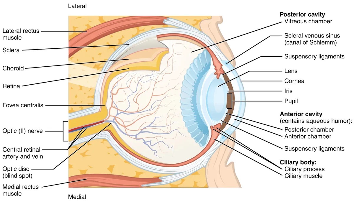
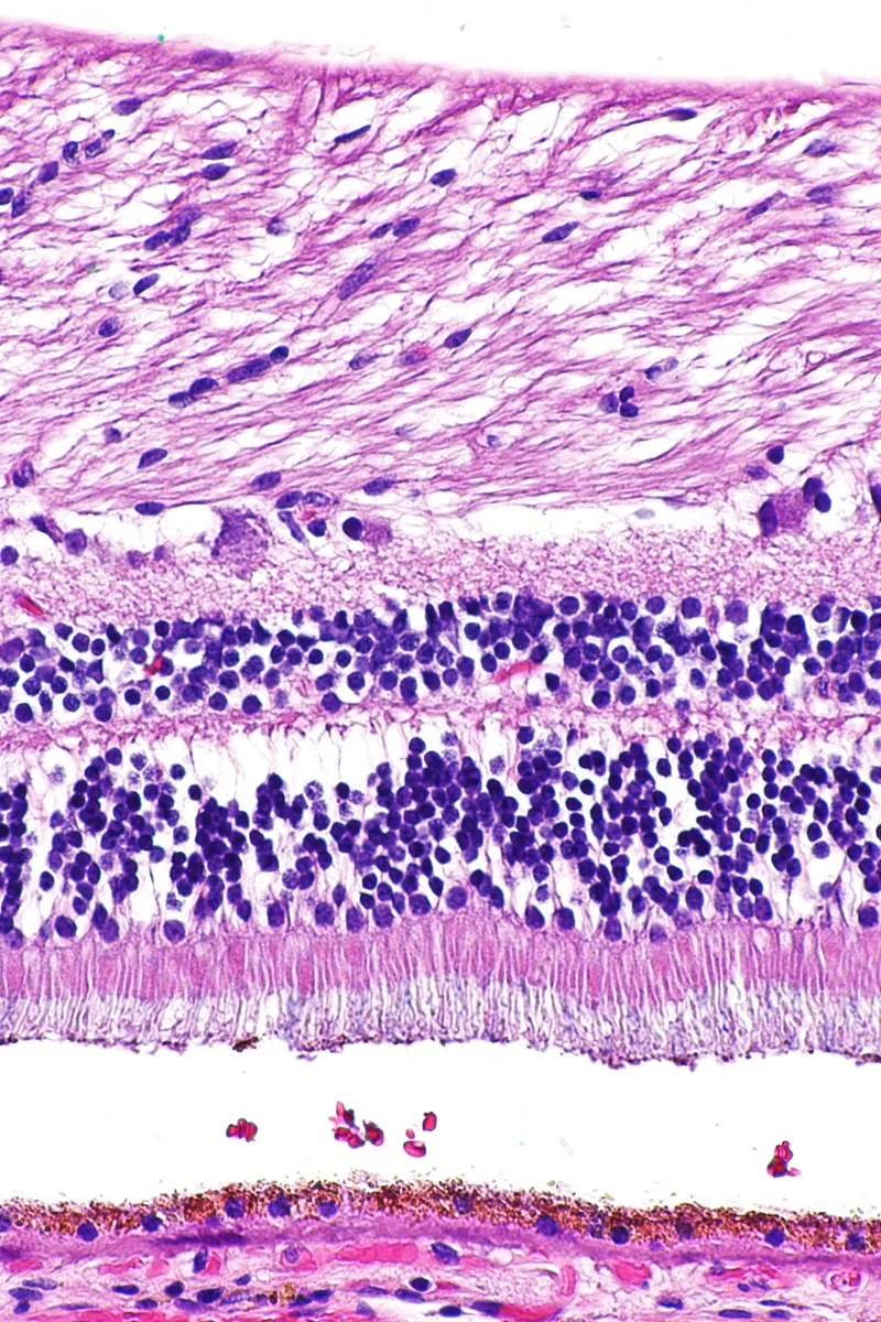
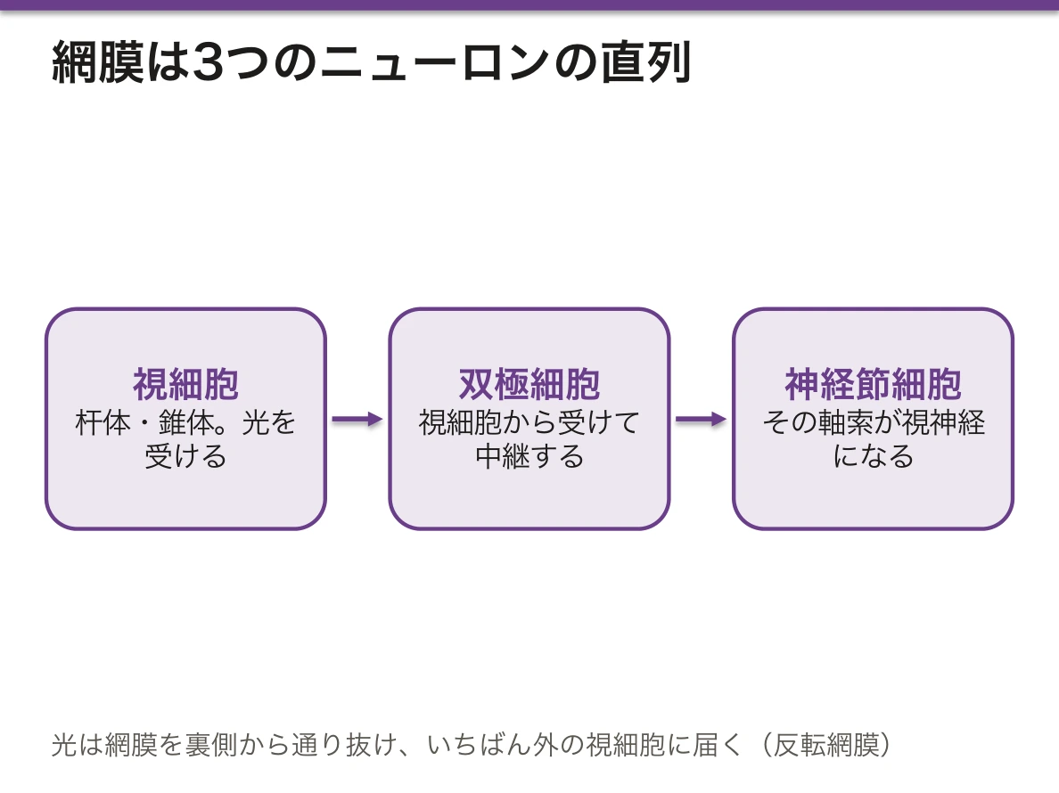
眼球壁の最内層。組織学的に10層に分けるが、要は3つのニューロンの直列である。
- 視細胞(杆体・錐体)→ 双極細胞 → 神経節細胞(その軸索が視神経になる)
- 横のつながりとして水平細胞・アマクリン細胞
- 支持するのはMüller 細胞(グリア)
- 最外層に網膜色素上皮
光は網膜を裏側から通り抜けていちばん外の視細胞に届く(反転網膜)。
- 杆体——暗所・周辺。感度が高いが色を見ない
- 錐体——明所・中心窩。色覚と鋭敏な視力
中心窩では内側の層が横へどけられ、錐体だけが露出して密に並ぶ。光を邪魔するものを物理的に取り除いた場所であり、だから最も視力が良い。同時に血管も無いので、栄養は脈絡膜からの拡散に頼る。中心窩が虚血や剥離に弱く、視力を一気に失う原因になるのはこの独立性による。
視神経乳頭には視細胞が無いので生理的な盲点(Mariotte 盲点)になる。
変換の向きが特異である。暗いとき視細胞は脱分極していて、光が当たると過分極する。ロドプシンのレチナールが11-シスから全トランスへ異性化し、トランスデューシン→ホスホジエステラーゼ→cGMP 分解と進んで、cGMP依存性ナトリウムチャネルが閉じるためである。「光でスイッチが切れる」という設計を知らないと、網膜電図も夜盲も理解できない。
レチナールはビタミンA から作られるので、ビタミンA欠乏では真っ先に杆体が働かなくなり夜盲になる。感度の高い杆体のほうが在庫不足に敏感に反応する。
網膜剥離は「網膜が眼球からはがれる」のではなく、網膜色素上皮と神経網膜のあいだが開く。この2層は発生学的に眼杯の外葉と内葉で、もともと癒合しない潜在的な隙間である。だからここだけが剥がれる。発生の名残が、そのまま病気の起きる場所を決めている。
色素上皮は視細胞外節を貪食して処理し、ビタミンAを再生し、網膜下の水を汲み出し、血液網膜関門を作る。この掃除係が老廃物を処理しきれなくなりドルーゼンが溜まるのが加齢黄斑変性である。糖尿病網膜症では毛細血管の周皮細胞が失われて瘤・出血・虚血が進み、血管内皮増殖因子により新生血管が出る。抗VEGF薬が両者に効くのはこの共通経路のためである。
English version
Core
The innermost layer of the wall of the eye. Histologically divided into ten layers, but in essence three neurones in series.
- Photoreceptor (rod, cone) → bipolar cell → ganglion cell (whose axons become the optic nerve)
- Lateral connections by horizontal and amacrine cells
- Supported by Müller cells (glia)
- Retinal pigment epithelium outermost
Light passes through the retina from behind to reach the outermost photoreceptors — the inverted retina.
- Rods — dim light, periphery. High sensitivity, no colour.
- Cones — bright light, fovea. Colour vision and fine acuity.
Going further
At the fovea the inner layers are swept aside and the cones alone are exposed and densely packed. It is a place where whatever obstructs light has been physically removed, and so it has the best acuity. At the same time it has no vessels, and depends on diffusion from the choroid. This independence is why the fovea is vulnerable to ischaemia and detachment and why vision can be lost at a stroke. The optic disc has no photoreceptors and is the physiological blind spot (of Mariotte).
The direction of the transduction is peculiar. In the dark the photoreceptor is depolarised, and light hyperpolarises it. The retinal of rhodopsin isomerises from 11-cis to all-trans, and through transducin → phosphodiesterase → breakdown of cGMP, the cGMP-gated sodium channels close. Without knowing this design — light switches it off — neither the electroretinogram nor night blindness can be understood.
Retinal is made from vitamin A, so in vitamin A deficiency the rods fail first and night blindness results — the more sensitive rods respond more sharply to a shortage of stock.
In the clinic
Retinal detachment is not the retina peeling off the eyeball — it is a separation between the retinal pigment epithelium and the neural retina. Developmentally these two are the outer and inner layers of the optic cup and never fused: a potential space. So it is here alone that separation occurs — a remnant of development deciding where the disease happens.
The pigment epithelium phagocytoses and processes the outer segments, regenerates vitamin A, pumps water out of the subretinal space and forms the blood–retinal barrier. Age-related macular degeneration is this cleaner failing to keep up, and drusen accumulating.
In diabetic retinopathy the capillary pericytes are lost, and microaneurysms, haemorrhage and ischaemia progress until vascular endothelial growth factor drives new vessels. Anti-VEGF agents work in both conditions because of this shared pathway.
網膜の視部と盲部 — 鋸状縁を境に、光を感じる網膜が終わるPars optica retinae
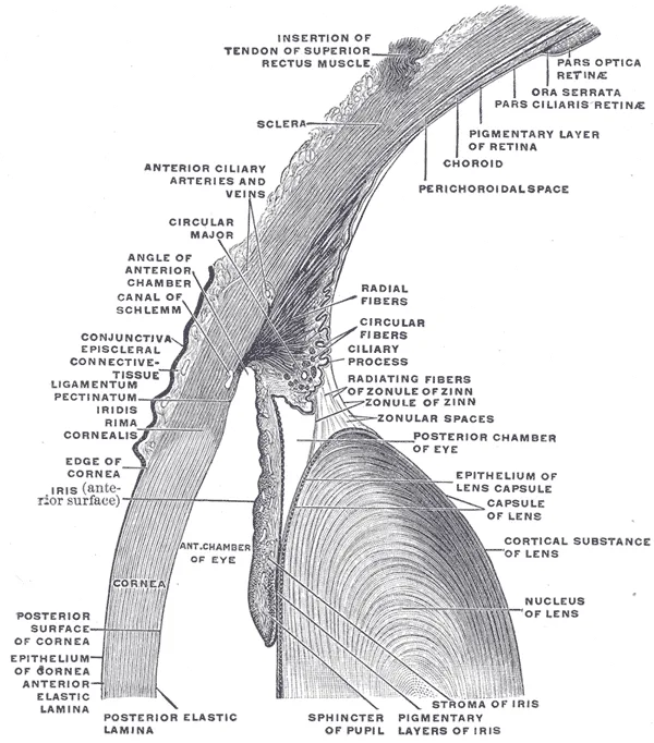
眼杯の内層は、眼球の内面を前の端まで覆う。そのうち光を感じるのは後ろの大部分だけで、境目を鋸状縁(ora serrata)と呼ぶ。
| 部分 | 場所 | 組織 |
|---|---|---|
| 網膜視部(pars optica) | 眼底から鋸状縁まで | 10層。色素上皮+神経網膜。光を感じる |
| 網膜盲部(pars caeca)・毛様体部 | 毛様体の内面 | 2層の立方上皮だけ。外層は有色素(色素上皮の続き)、内層は無色素(神経網膜の続き)で房水を作る |
| 網膜盲部・虹彩部 | 虹彩の後面 | 2層とも有色素。外層の細胞が瞳孔散大筋(筋上皮)になる |
鋸状縁で、何層もあった神経網膜が急に1層の無色素上皮に変わる。色素上皮の層は途切れずに前へ続く。2層が眼杯の内層と外層だった、という発生の名残がそのまま見える。
網膜の発生: 眼杯の内層の細胞が増えて、神経節細胞が最初に分化し、その軸索が視神経を作る。続いて水平細胞・錐体・アマクリン細胞、最後に杆体・双極細胞・Müller 細胞ができる。中心窩は生まれた後も成熟が続き、内側の層が横へどき、錐体が密に詰まっていく。視力が大人の水準に近づくのに幼児期までかかるのはこのためである。
網膜の血管は胎生期に視神経乳頭から周辺へ向かって伸び、鼻側の鋸状縁に届くのが約36週、耳側は出生のころである。
組織生理の要点を1つ加える。杆体の外節は先端から少しずつ捨てられ、根元で新しい円板が作られて、約10日で入れ替わる。捨てられた先端は色素上皮が毎日食べて処理する。
未熟児網膜症は、網膜血管が周辺まで届く前に生まれたために起きる。周辺の網膜は血管が無いまま残り、そこが低酸素になると VEGF が出て、異常な新生血管が硝子体へ伸び、牽引で網膜剥離に至る。早産児で眼底検査を繰り返すのは、まさに血管が鋸状縁へ伸びていく時期を見守るためである。治療はレーザー光凝固や抗 VEGF 薬である。
眼内に注射したり手術の器具を入れたりするときは、角膜の縁から数 mm 後方の毛様体扁平部を通す。そこは網膜盲部で、光を感じる網膜を傷つけないからである。
小児の白色瞳孔では網膜芽細胞腫(RB1 遺伝子)を考える。未分化な網膜の細胞の腫瘍である。
English version
Core
The inner layer of the optic cup lines the inside of the eyeball up to its front edge. Only the large posterior part senses light. The border is the ora serrata.
| Part | Site | Tissue |
|---|---|---|
| Pars optica | from the fundus to the ora serrata | 10 layers; pigment epithelium + neural retina; senses light |
| Pars caeca, ciliary part | inner surface of the ciliary body | only two layers of cuboidal epithelium: outer pigmented (continuing the pigment epithelium), inner non-pigmented (continuing the neural retina), which makes aqueous humour |
| Pars caeca, iridial part | back surface of the iris | both layers pigmented; outer-layer cells become the dilator pupillae (myoepithelium) |
At the ora serrata the many-layered neural retina suddenly becomes one layer of non-pigmented epithelium. The pigment layer continues forward without a break. The two layers still show their origin as the inner and outer layers of the optic cup.
Going further
Development of the retina: cells of the inner layer of the cup multiply, and ganglion cells differentiate first; their axons form the optic nerve. Horizontal cells, cones and amacrine cells follow, and rods, bipolar cells and Müller cells come last. The fovea keeps maturing after birth: the inner layers move aside and cones pack tightly. This is why visual acuity takes until early childhood to approach adult level.
Retinal vessels grow from the optic disc toward the periphery in fetal life. They reach the nasal ora serrata at about 36 weeks and the temporal side around birth.
One point of histophysiology: rod outer segments are shed bit by bit from the tip, and new discs are made at the base, so they are renewed in about ten days. The pigment epithelium eats and processes the shed tips every day.
In the clinic
Retinopathy of prematurity happens because the baby was born before retinal vessels reached the periphery. The peripheral retina stays avascular; when it becomes hypoxic, VEGF is released, abnormal new vessels grow into the vitreous, and traction can detach the retina. Repeated eye examinations in preterm babies watch exactly the period when vessels grow toward the ora serrata. Treatment is laser photocoagulation or anti-VEGF drugs.
Intraocular injections and surgical instruments enter through the pars plana of the ciliary body, a few millimetres behind the edge of the cornea. This is pars caeca, so no light-sensing retina is damaged.
White pupil (leukocoria) in a child suggests retinoblastoma (RB1 gene), a tumour of immature retinal cells.
角膜と水晶体 — 透明であるための組織Cornea
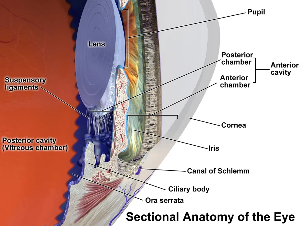
角膜は5層。
- 上皮(重層扁平・非角化)——再生する。知覚が体で最も鋭敏(三叉神経眼神経)
- Bowman 層——再生しない
- 実質——厚さの90%。規則正しく並んだI型コラーゲン
- Descemet 膜——内皮の基底膜
- 内皮——単層扁平。再生しない
血管が無い。栄養は房水と涙液から。
水晶体は上皮由来の線維が同心円状に重なり、生涯増え続ける。
透明性は「作ったら終わり」ではなく、絶えず維持されている状態である。
角膜実質のコラーゲン線維は直径も間隔も光の波長より小さく揃っているため、散乱が打ち消し合って光が素通りする。この配列を保つには実質が乾いた状態でなければならず、それを担うのが内皮のナトリウム・カリウムポンプである。内皮は水を実質から房水側へ汲み出し続けている。
したがって内皮が減ると実質が浮腫んで濁る。内皮は再生しないので一方通行で、白内障手術での機械的損傷や Fuchs 角膜内皮ジストロフィで細胞密度が閾値を下回ると水疱性角膜症となり、角膜移植以外に手がなくなる。「透明さはポンプが作っている」——これが角膜内皮を数える理由である。
角膜移植の拒絶が起きにくいのは、血管もリンパ管も無く、免疫細胞が到達しにくいためである(免疫特権)。逆に新生血管が入った角膜では拒絶率が跳ね上がる。無血管であることが治療の成否を決めている。
白内障は水晶体の透明性が失われる病態である。水晶体線維は核も細胞小器官も失い、クリスタリンという蛋白だけで満たされているので、入れ替えができない。生涯にわたり中心へ古い線維が押し込められて硬く(核硬化)、蛋白が変性・凝集して濁る。加齢で必発なのは捨てられない蛋白を一生使い続けるという構造の帰結である。
糖尿病白内障では、余ったグルコースがアルドース還元酵素でソルビトールに変えられ、細胞外へ出られず溜まって浸透圧性に水を引き込み混濁を早める。ステロイドでは後嚢下白内障が特徴的である。
English version
Core
The cornea has five layers.
- Epithelium (stratified squamous, non-keratinised) — regenerates. The most sensitive tissue in the body (ophthalmic division of the trigeminal).
- Bowman’s layer — does not regenerate.
- Stroma — 90 per cent of the thickness; regularly arranged type I collagen.
- Descemet’s membrane — the basal lamina of the endothelium.
- Endothelium — simple squamous. Does not regenerate.
It has no blood vessels; nutrition comes from aqueous humour and tears.
The lens is built of epithelially derived fibres laid concentrically, added to throughout life.
Going further
Transparency is not something made once but a state continuously maintained. The collagen fibrils of the stroma are smaller in diameter and spacing than the wavelength of light and uniformly arranged, so scattering cancels out and light passes through. Maintaining that arrangement requires the stroma to be kept dry, and that is the work of the sodium–potassium pump of the endothelium, which pumps water continuously from stroma to aqueous.
So when the endothelium is depleted, the stroma swells and clouds. The endothelium does not regenerate, so the process is one-way: when mechanical injury at cataract surgery or Fuchs’ endothelial dystrophy takes the cell density below the threshold, bullous keratopathy follows and nothing remains but corneal grafting. Transparency is made by a pump — this is why corneal endothelial cells are counted.
Corneal grafts are seldom rejected because there are neither blood nor lymphatic vessels and immune cells reach it poorly (immune privilege). Conversely, in a cornea that new vessels have entered, the rejection rate leaps. Being avascular decides the success of the treatment.
In the clinic
Cataract is loss of transparency of the lens. The lens fibres lose both nucleus and organelles and are filled with nothing but crystallins, so they cannot be replaced. Throughout life old fibres are compressed towards the centre and harden (nuclear sclerosis), and the proteins denature, aggregate and cloud. That it is inevitable with age is the consequence of a structure that must use, for a lifetime, proteins it cannot discard.
In diabetic cataract, surplus glucose is converted by aldose reductase to sorbitol, which cannot leave the cell, accumulates and draws in water osmotically, hastening the opacity. Steroids characteristically give posterior subcapsular cataract.
眼の発生と硝子体 — 脳の突起と表皮が出会ってできる器官Corpus vitreum
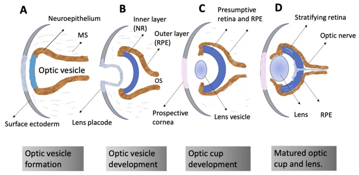
眼は脳(間脳)の壁が外へ伸びた突起と、体表の外胚葉と、周りの間葉の3つから作られる。
- 胎生4週ころ、前脳から眼胞が伸び出して表面外胚葉に触れる
- 触れられた表面外胚葉が厚くなって水晶体板になり、くぼんで水晶体胞として離れる
- 眼胞は内側へ折れ込んで2層の眼杯になる。外層が網膜色素上皮、内層が神経網膜になる
- 眼杯の下側の溝(眼杯裂)を硝子体動脈が通り、7週ころに閉じる
- 眼胞の柄(眼茎)を神経節細胞の軸索が通って視神経になる
| 由来 | できるもの |
|---|---|
| 神経外胚葉(眼杯) | 網膜、毛様体と虹彩の上皮、瞳孔括約筋・瞳孔散大筋、視神経 |
| 表面外胚葉 | 水晶体、角膜上皮、眼瞼の皮膚、結膜 |
| 間葉(神経堤と中胚葉) | 強膜、角膜実質と内皮、脈絡膜、虹彩と毛様体の間質 |
光は角膜・房水・水晶体・硝子体の4つの屈折媒体を通って網膜に届く。屈折力の約3分の2は角膜が担い、残りを水晶体が担う。水晶体だけが厚さを変えてピントを合わせられる(調節)。
硝子体は眼球の容積の大部分を占める透明なゲルである。約99%が水で、II型コラーゲンの細い線維の網とヒアルロン酸が水を抱えている。表層にわずかな細胞(硝子体細胞、マクロファージの仲間)がいる。網膜とは、鋸状縁をまたぐ硝子体基底部・視神経乳頭の縁・黄斑で強く付着する。胎生期に硝子体動脈が通った跡が、中を前後に走る細い管(硝子体管、Cloquet 管)として残る。
硝子体は一度失われても作り直されない。加齢でゲルが液化して縮むと、網膜から剥がれる(後部硝子体剥離、多くは50〜60代)。
後部硝子体剥離では、目の前に浮く影(飛蚊症)が急に増える。剥がれるときに強く付着した部分の網膜を引っ張ると網膜裂孔ができ、そこから水が入って網膜剥離になる。光が走って見える(光視症)とともに飛蚊症が急に増えたら、すぐに眼底を診る理由である。
硝子体は作り直されないので、手術(硝子体切除)で取り除いても、生理食塩水やガスで置き換えればよい。逆に、閉じた空間であることを利用して、抗 VEGF 薬を硝子体に注射する。
発生の誤りもこの順序で読める。眼杯裂が閉じないとコロボーマ(虹彩の下方が鍵穴形に欠ける)になる。水晶体胞ができる時期に母体が風疹にかかると先天白内障になり、難聴・動脈管開存と並ぶ先天性風疹症候群の3徴の1つになる。
English version
Core
The eye is built from three sources: an outgrowth of the brain wall (diencephalon), surface ectoderm, and the surrounding mesenchyme.
- At about week 4, the optic vesicle grows out from the forebrain and touches the surface ectoderm.
- The touched ectoderm thickens into the lens placode, sinks in, and separates as the lens vesicle.
- The optic vesicle folds in to form the two-layered optic cup. Its outer layer becomes the retinal pigment epithelium; its inner layer becomes the neural retina.
- The hyaloid artery runs in a groove on the underside of the cup (the choroid fissure), which closes at about week 7.
- Axons of ganglion cells grow through the stalk of the vesicle and become the optic nerve.
| Origin | What it forms |
|---|---|
| Neuroectoderm (optic cup) | retina, epithelium of ciliary body and iris, sphincter and dilator pupillae, optic nerve |
| Surface ectoderm | lens, corneal epithelium, eyelid skin, conjunctiva |
| Mesenchyme (neural crest and mesoderm) | sclera, corneal stroma and endothelium, choroid, stroma of iris and ciliary body |
Going further
Light reaches the retina through four refractive media: cornea, aqueous humour, lens and vitreous. About two-thirds of the refractive power is in the cornea and the rest in the lens. Only the lens can change its thickness to focus (accommodation).
The vitreous body is a clear gel that fills most of the eyeball. It is about 99% water, held by a net of fine type II collagen fibrils and hyaluronan. A few cells (hyalocytes, related to macrophages) lie in its surface. It is firmly attached to the retina at the vitreous base across the ora serrata, the edge of the optic disc, and the macula. The path of the fetal hyaloid artery remains as a thin canal running front to back (hyaloid or Cloquet canal).
The vitreous is not rebuilt once lost. With age the gel liquefies and shrinks, and separates from the retina (posterior vitreous detachment, often in the 50s–60s).
In the clinic
In posterior vitreous detachment, floating shadows (floaters) suddenly increase. If the vitreous pulls on a firmly attached spot as it separates, a retinal tear forms, fluid enters, and retinal detachment follows. That is why flashes of light with a sudden increase in floaters need a fundus examination at once.
Because the vitreous is not rebuilt, surgery can remove it (vitrectomy) and replace it with saline or gas. On the other hand, its closed space is used for intravitreal injection of anti-VEGF drugs.
Developmental errors follow the same sequence. If the choroid fissure does not close, a coloboma results (a keyhole-shaped gap in the lower iris). If the mother has rubella while the lens vesicle is forming, congenital cataract results — one of the classic triad of congenital rubella with deafness and patent ductus arteriosus.
隅角と房水 — 作る場所と、出ていく場所Angulus iridocornealis
房水は毛様体の無色素上皮が作り、後房 → 瞳孔 → 前房 → 隅角から出ていく。
毛様体上皮は2層で、向かい合っている——外側が色素上皮、内側(房水側)が無色素上皮。分泌するのは無色素上皮になる。
出口は2つ。
| 経路 | 道筋 | 割合 |
|---|---|---|
| 主経路(線維柱帯路) | 線維柱帯 → Schlemm 管 → 上強膜静脈 | 約 80〜90% |
| 副経路(ぶどう膜強膜路) | 毛様体筋の隙間 → 強膜 | 約 10〜20% |
ぶどう膜は3つ——虹彩・毛様体・脈絡膜。血管とメラノサイトに富む一続きの層になる。
眼圧の正常は 10〜21 mmHg。産生と流出の釣り合いで決まる。
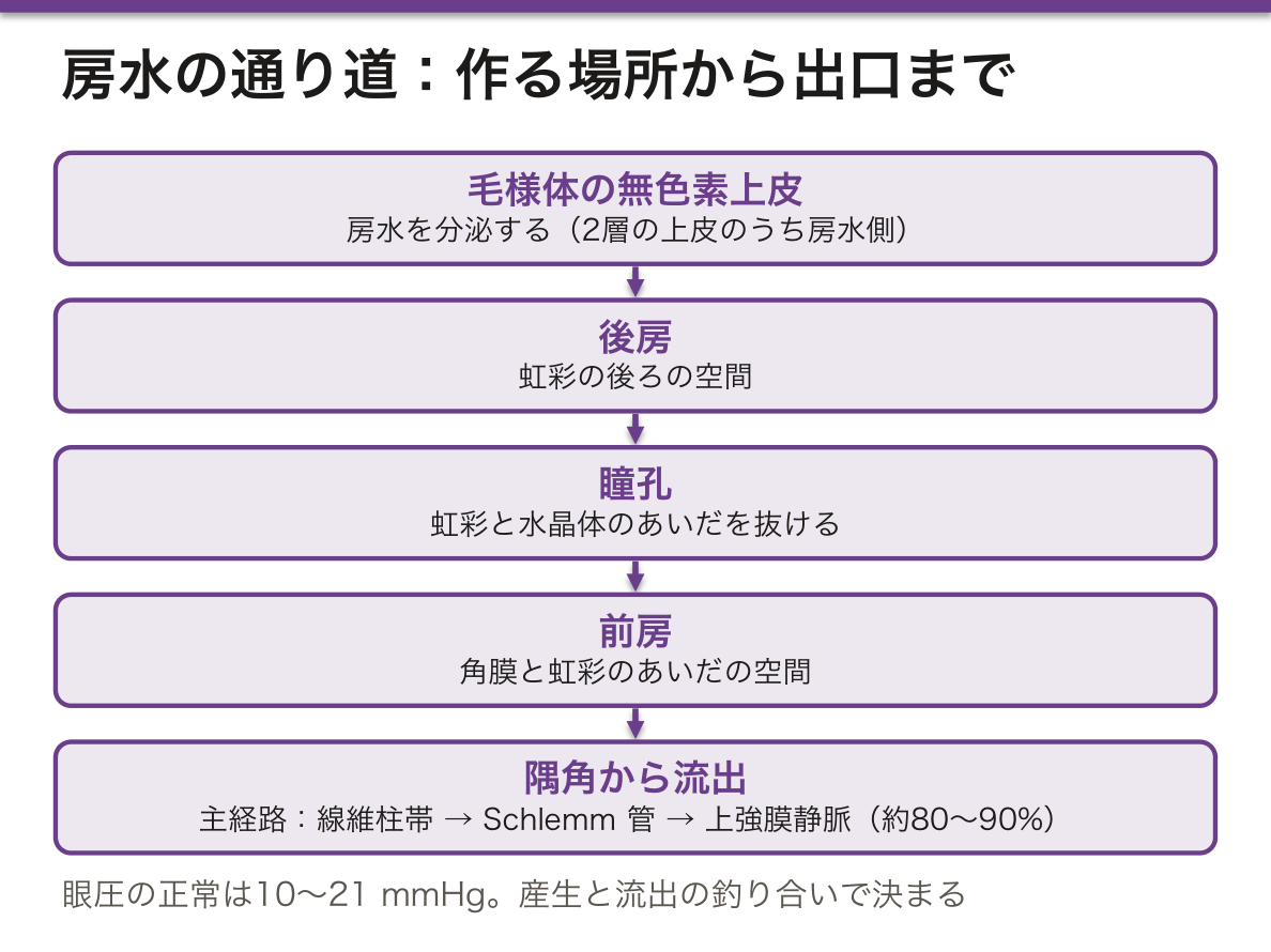
眼圧は「作る量」ではなく、ほぼ「出る抵抗」で決まっている。だから緑内障の大半は、出口の病気になる——「蛇口ではなく、排水口の話」。
出口の詰まり方が2つあることが、そのまま病型を分けている。
| 開放隅角 | 閉塞隅角 | |
|---|---|---|
| 詰まる場所 | 線維柱帯そのもの | 隅角が虹彩で塞がれる |
| 経過 | 無症状で進む | 急激(発作) |
| 症状 | 視野が欠ける(気づかない) | 眼痛・頭痛・嘔吐・虹視・充血 |
| 治療 | 点眼で眼圧を下げる | レーザー虹彩切開で、後房から前房への近道を作る |
(緑内障の章)——「急性緑内障発作が嘔吐を伴うので、消化器疾患と間違われる」。
薬の効き方も、この図の上に置ける。プロスタグランジン関連薬は副経路を広げ、β遮断薬と炭酸脱水酵素阻害薬は産生を減らし、縮瞳薬は線維柱帯を開く(自律神経作用薬の章)——「どの点眼薬も、産生か流出のどちらかに置ける」。
そして抗コリン作用のある薬が禁忌になるのは、閉塞隅角のほうだけになる。散瞳して虹彩が隅角に寄る(薬の副作用の章)——「『緑内障に禁忌』と書かれた薬は、正確には閉塞隅角緑内障に禁忌である」。
作る場所と出る場所の関係が、緑内障の型を決める。
開放隅角緑内障 — 隅角は開いているが、線維柱帯・Schlemm 管での流出抵抗が高い。慢性に進行し、視野が周辺から欠けるが自覚が遅れる。日本人に多い正常眼圧緑内障もこの型である。
閉塞隅角緑内障(急性発作) — 隅角が物理的に塞がる。眼痛・充血・霧視・虹輪視に、頭痛と嘔吐を伴うため消化器疾患や脳疾患と間違われる。眼球は硬く、瞳孔は中等度散大で対光反射が鈍い。抗コリン薬・散瞳薬が誘発するので高齢者への処方で常に意識する。
治療は「作るのを減らす」か「出るのを増やす」かに整理できる。β遮断薬・炭酸脱水酵素阻害薬は産生を抑え、プロスタグランジン関連薬はぶどう膜強膜流出路を広げる。
English version
Core
Aqueous humour is made by the non-pigmented epithelium of the ciliary body and leaves by posterior chamber → pupil → anterior chamber → the angle.
The ciliary epithelium is two layers facing each other — pigmented on the outside, non-pigmented within (on the aqueous side). It is the non-pigmented layer that secretes.
There are two exits.
| Route | Path | Share |
|---|---|---|
| Conventional (trabecular) | Trabecular meshwork → Schlemm’s canal → episcleral veins | About 80–90% |
| Unconventional (uveoscleral) | Between the bundles of ciliary muscle → sclera | About 10–20% |
The uvea is three parts — iris, ciliary body, choroid: one continuous layer rich in vessels and melanocytes.
Normal intraocular pressure is 10–21 mmHg, set by the balance of production and outflow.
Going further
Intraocular pressure is set almost entirely by the resistance to outflow, not by the amount produced. So most glaucoma is a disease of the exit — a question of the drain, not of the tap.
That there are two ways for the exit to block divides the disease into its two types.
| Open angle | Closed angle | |
|---|---|---|
| Site of block | The trabecular meshwork itself | The angle is occluded by the iris |
| Course | Progresses without symptoms | Abrupt (an attack) |
| Symptoms | The field is lost (unnoticed) | Ocular pain, headache, vomiting, haloes, redness |
| Treatment | Drops to lower the pressure | Laser iridotomy, making a short cut from posterior to anterior chamber |
An acute attack of glaucoma is accompanied by vomiting, and so is mistaken for a gastrointestinal illness.
The action of the drugs can be laid on the same diagram: prostaglandin analogues widen the unconventional route, beta blockers and carbonic anhydrase inhibitors reduce production, and miotics open the meshwork — every eye drop can be placed on either production or outflow.
And drugs with anticholinergic action are contraindicated only in the closed-angle type, since dilating the pupil crowds the iris into the angle — a drug labelled "contraindicated in glaucoma" is, strictly, contraindicated in closed-angle glaucoma.
In the clinic
The relation between where it is made and where it leaves decides the type of glaucoma.
Open-angle glaucoma — the angle is open but resistance at the trabecular meshwork and Schlemm’s canal is high. It progresses chronically, the field is lost from the periphery, and awareness comes late. Normal-tension glaucoma, common in Japan, is of this type.
Closed-angle glaucoma (acute attack) — the angle is physically occluded. Ocular pain, redness, blurring and haloes, with headache and vomiting, so it is mistaken for gastrointestinal or intracranial disease. The globe is hard and the pupil is mid-dilated with a sluggish light reflex. Anticholinergics and mydriatics precipitate it — always keep this in mind when prescribing for the elderly.
Treatment sorts into reducing production or increasing outflow. Beta blockers and carbonic anhydrase inhibitors suppress production; prostaglandin analogues widen the uveoscleral route.
眼瞼と涙器 — 表面を守る3層の膜Glandula lacrimalis
眼瞼は外から内へ——皮膚(体で最も薄い)・皮下組織・眼輪筋・瞼板(密性結合組織)・瞼結膜。
3種類の腺がある。
| 腺 | 種類 | 出すもの | 詰まると |
|---|---|---|---|
| Meibom 腺(瞼板腺) | 脂腺(ホロクリン) | 油層 | 霰粒腫(無菌性の肉芽腫) |
| Zeis 腺 | 脂腺(睫毛に付属) | 脂 | 外麦粒腫 |
| Moll 腺 | アポクリン汗腺 | 汗 | 外麦粒腫 |
涙液は3層構造になる——外から油層(Meibom 腺)・水層(涙腺)・粘液層(結膜の杯細胞)。
涙の流れ——涙腺 → 眼表面 → 涙点 → 涙小管 → 涙嚢 → 鼻涙管 → 下鼻道。
涙が3層であることが、ドライアイの分類をそのまま作っている。水が足りない型(涙腺の問題)と、蒸発しやすい型(油層の問題)に分かれる(乾く眼の章)——「涙の量が正常でも、蓋が壊れていれば乾く」。
だから Meibom 腺機能不全が、ドライアイの最も多い原因になる——「温罨法と眼瞼の清拭が治療になるのは、脂の腺の詰まりだから」。
水が足りない型では、全身疾患を探す。Sjögren 症候群では涙腺と唾液腺が同時に障害される(膠原病の章)——「目と口が同時に乾くという訴えを、1つの病気として聞く」。
鼻涙管が下鼻道へ開くことは、日常の現象を説明する。泣くと鼻水が出るのは、涙がそのまま鼻へ流れているから——「点眼薬が全身に効くのも、この道から鼻粘膜で吸収されるため」。
だから点眼後に涙点を押さえると、全身への移行が減る(眼の薬の章)——「β遮断薬の点眼で喘息が悪化することがある」。
新生児の鼻涙管閉塞は、この道の下端(Hasner 弁)が開通していない状態になる(子どもの眼の章)——「生後まもない流涙と眼脂の大半が、これで説明できる」。
腺の種類で、詰まったときの病名が変わる。
Meibom 腺(瞼板腺・脂腺) — 詰まると霰粒腫(無痛性の硬い結節)、感染すると内麦粒腫(痛い)。「痛くない=霰粒腫、痛い=麦粒腫」で分ける。
Zeis 腺(脂腺)・Moll 腺(アポクリン汗腺)の感染が外麦粒腫である。
涙液は3層——油層(Meibom 腺)・水層(涙腺)・ムチン層(結膜杯細胞)。どの層が足りないかでドライアイの型が変わる。Meibom 腺機能不全では油が足りず涙が蒸発しやすい——温罨法と眼瞼清拭が治療になる。
涙は上外側から出て、内眼角の涙点へ流れる。鼻涙管が閉塞すると流涙になる。新生児の鼻涙管閉塞は多くが自然に開通する。点眼薬が全身に吸収されるのもこの経路による——点眼後に涙点を押さえると全身移行を減らせる。
English version
Core
The eyelid, from outside in — skin (the thinnest in the body), subcutaneous tissue, orbicularis oculi, tarsal plate (dense connective tissue), palpebral conjunctiva.
Three kinds of gland.
| Gland | Type | Secretes | When blocked |
|---|---|---|---|
| Meibomian (tarsal) | Sebaceous (holocrine) | The oil layer | Chalazion (a sterile granuloma) |
| Gland of Zeis | Sebaceous, attached to a lash | Sebum | External hordeolum |
| Gland of Moll | Apocrine sweat gland | Sweat | External hordeolum |
The tear film is three layers — from outside: oil (meibomian), aqueous (lacrimal gland), mucin (conjunctival goblet cells).
The flow of tears — lacrimal gland → ocular surface → punctum → canaliculus → lacrimal sac → nasolacrimal duct → inferior meatus.
Going further
That the tear film has three layers creates the classification of dry eye directly: an aqueous-deficient type (a problem of the gland) and an evaporative type (a problem of the oil layer) — even with a normal volume of tears, a broken lid lets the eye dry.
So meibomian gland dysfunction is the commonest cause of dry eye — warm compresses and lid hygiene are the treatment because it is a blocked oil gland.
In the aqueous-deficient type, look for systemic disease. In Sjögren’s syndrome the lacrimal and salivary glands are affected together — hear the complaint of dry eyes and dry mouth together as one disease.
That the nasolacrimal duct opens into the inferior meatus explains an everyday phenomenon. Crying makes the nose run because the tears flow straight into it — and eye drops act systemically because they are absorbed from the nasal mucosa by the same route. So pressing on the punctum after instillation reduces systemic absorption — beta blocker drops can worsen asthma.
Nasolacrimal duct obstruction in the newborn is failure of the lower end of this passage (the valve of Hasner) to open — most watering and discharge in the first weeks of life is explained by it.
In the clinic
The type of gland changes the name of the disease when it blocks.
Meibomian gland (tarsal, sebaceous) — blocked, it gives a chalazion (a painless firm nodule); infected, an internal hordeolum (painful). Divide them as painless = chalazion, painful = hordeolum. Infection of the glands of Zeis (sebaceous) and Moll (apocrine sweat) is an external hordeolum.
The tear film has three layers — oil (meibomian), aqueous (lacrimal gland), mucin (conjunctival goblet cells). Which layer is lacking changes the type of dry eye. In meibomian gland dysfunction the oil is deficient and tears evaporate readily — warm compresses and lid hygiene are the treatment.
Tears arise superolaterally and flow to the punctum at the medial canthus. Obstruction of the nasolacrimal duct causes watering; in the newborn it usually opens spontaneously. Systemic absorption of eye drops takes the same route — pressing the punctum after instillation reduces it.
篩状板 — 視神経が眼球を出るときに通る、弱い場所Lamina cribrosa
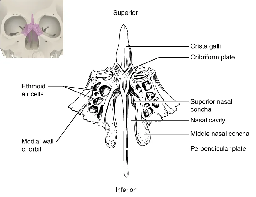
強膜は丈夫な膜だが、視神経が出る場所だけは網の目状に穴が開いている。そこを 100 万本を超える神経線維が束になって通る。
- 構造的に弱い——強膜の中で唯一、コラーゲンが連続していない
- 圧の境目になる——ここを境に、眼圧と頭蓋内圧が接している
- 血流も乏しい
だから圧が上がると、真っ先にここが変形する。眼圧が高いと篩状板が後方へ押し出され、通っている軸索が圧迫されて軸索輸送が止まり、神経節細胞が死ぬ。これが緑内障で視野が失われる機序である。
眼底で見えるのは、この変形の結果である。
| 陥凹の拡大 | 視神経乳頭の凹みが広がる | C/D 比で表す。左右差 0.2 以上も異常 |
| リムの菲薄化 | 神経線維が集まる縁が薄くなる | 下→上→鼻側→耳側の順に薄くなる(ISNT の規則が崩れる) |
| 乳頭出血 | 縁の線状出血 | 進行を示す徴候 |
視野の欠け方も、線維の走り方で決まっている。網膜の耳側の線維は黄斑を避けて弓状に迂回するので、障害されると弓状の暗点ができ、水平の縫線を越えないため、鼻側の階段状の欠損になる。中心視力は最後まで保たれるので、患者は末期まで自覚しない。
⚠️ 眼圧が正常でも緑内障は起こる。日本では正常眼圧緑内障のほうが多い——篩状板の構造の弱さや、乳頭の血流が関わると考えられている。「眼圧が正常だから大丈夫」とは言えないので、眼底(乳頭)と視野で判定する。
反対に、頭蓋内圧が上がると軸索輸送が乳頭で滞ってうっ血乳頭になる。同じ場所が、圧の向きによって陥凹にも隆起にもなる——篩状板が2つの圧の境界にあることの帰結である。
緑内障は失われた視野が戻らない。自覚症状が出る前に見つけるため、健診での眼底検査に意味がある。
English version
Core
The sclera is a tough coat, but at the place where the optic nerve leaves it is perforated like a net, and more than a million nerve fibres pass through in bundles.
- Structurally weak — the only place in the sclera where the collagen is not continuous.
- A boundary of pressures — intraocular pressure and intracranial pressure meet across it.
- The blood supply is also sparse.
So when pressure rises it is the first thing to deform. A high intraocular pressure pushes the lamina cribrosa backwards, the axons passing through it are compressed, axonal transport stops, and the ganglion cells die. This is the mechanism by which the visual field is lost in glaucoma.
Going further
What is seen at the fundus is the result of that deformation.
| Sign | What it is | How it is read |
|---|---|---|
| Enlarged cup | The hollow of the disc widens | Expressed as the C/D ratio. A difference of 0.2 between the eyes is also abnormal |
| Thinning of the rim | The margin where the fibres gather thins | Thins in the order inferior → superior → nasal → temporal (the ISNT rule breaks down) |
| Disc haemorrhage | A linear haemorrhage at the margin | A sign of progression |
The way the field is lost is likewise decided by the course of the fibres. The temporal fibres arch around the macula to avoid it, so damage produces an arcuate scotoma, and because they do not cross the horizontal raphe, a nasal step. Central acuity is preserved to the last, so the patient notices nothing until the end.
⚠️ Glaucoma occurs at a normal intraocular pressure. In Japan normal-tension glaucoma is the commoner form — structural weakness of the lamina cribrosa and the blood supply of the disc are thought to be involved. "The pressure is normal, so it is safe" cannot be said: judge by the disc and the visual field.
Conversely, a rise in intracranial pressure stalls axonal transport at the disc and gives papilloedema. The same place becomes either a cup or a swelling according to the direction of the pressure — the consequence of the lamina cribrosa lying at the boundary of two pressures.
In the clinic
In glaucoma the visual field that has been lost does not return. Fundus examination at a health check has value because it finds the disease before symptoms appear.
感覚器の分類と分析器 — 受容細胞が神経か上皮かで分けるOrgana sensuum
感覚器に共通する性質は、決まった種類の刺激(適刺激)だけを受け取り、それを電気の信号に変える(変換)ことである。強さが一定の刺激が続くと反応が弱まる順応もある。多くの感覚器では、受容細胞のほかに刺激を集めて整える補助装置(眼の屈折系、耳小骨など)が付く。
ヨーロッパの組織学の教科書では、受容細胞の種類で感覚器を3つに分ける。
| 型 | 受容細胞 | 例 |
|---|---|---|
| 一次感覚(神経感覚性) | 受容細胞そのものがニューロン。自分の突起で信号を中枢側へ送る | 嗅覚(嗅細胞)、視覚(杆体・錐体) |
| 二次感覚(感覚上皮性) | 受容細胞は上皮細胞で軸索を持たない。感覚ニューロンの終末にシナプスで渡す | 味覚(味細胞)、聴覚・平衡覚(有毛細胞) |
| 独立した器官を持たないもの | 感覚ニューロンの終末(自由終末・被包終末) | 皮膚・筋・関節・内臓の一般感覚 |
日本の生理学の教科書では、特殊感覚(視・聴・平衡・嗅・味)と体性感覚・内臓感覚に分け、受容器を刺激の種類(機械・化学・光・温度・侵害)で分けるのが一般的である。並べて覚えておくとよい。
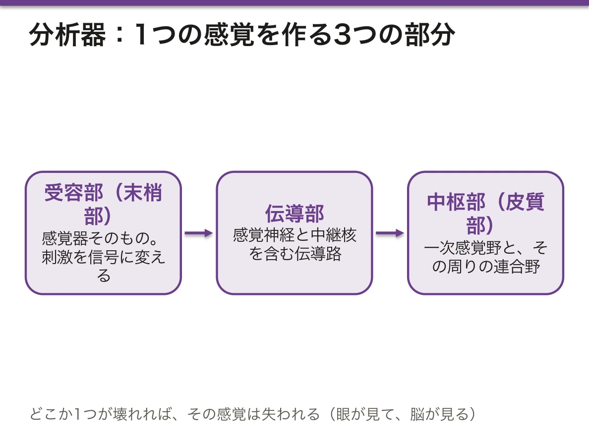
分析器(analyser)は、ロシアの生理学者 Pavlov に由来する考え方で、ヨーロッパの組織学でよく使われる。1つの感覚は、次の3つがそろって初めて成り立つ。
- 受容部(末梢部)——感覚器そのもの。刺激を信号に変える
- 伝導部——感覚神経と、途中の中継核を含む伝導路
- 中枢部(皮質部)——大脳皮質の一次感覚野と、その周りの連合野
日本語の教科書の「受容器 → 伝導路 → 感覚中枢」と同じ内容である。「眼が見て、脳が見る」——どこか1つが壊れれば、その感覚は失われる。
型の違いは再生の違いになる。嗅細胞はニューロンでありながら、基底細胞から約1〜2か月ごとに入れ替わる数少ない例である。味細胞も上皮として約10日で入れ替わる。ところが哺乳類の蝸牛の有毛細胞は一度失われると再生しない。
分析器の3部のどこが壊れたかで、同じ「見えない」でも所見が違う。網膜や視神経(受容部・伝導部の前半)の障害では対光反射が弱まるが、後頭葉(中枢部)の障害による皮質盲では対光反射が保たれる。対光反射の線維は、外側膝状体より手前で視蓋前域へ分かれるからである。
ウイルス感染後の嗅覚障害が数か月で戻ることが多いのは、嗅上皮が再生するからである。頭部外傷で篩板を通る嗅糸が切れると戻りにくい。一方、騒音やアミノグリコシド系抗菌薬で失われた有毛細胞は戻らないので、感音難聴は不可逆になる。亜鉛欠乏で味覚障害が起きるのは、入れ替わりの速い味細胞の再生が追いつかなくなるためと考えられている。
English version
Core
All sense organs share two properties: they receive one kind of stimulus (the adequate stimulus), and they turn it into an electrical signal (transduction). Their response fades if a constant stimulus continues (adaptation). Many sense organs also have accessory structures that collect and shape the stimulus (the optics of the eye, the ossicles).
European histology textbooks divide sense organs into three groups by the type of receptor cell.
| Type | Receptor cell | Examples |
|---|---|---|
| Primary sensory (neurosensory) | the receptor is itself a neuron and sends the signal on with its own process | smell (olfactory cells), vision (rods and cones) |
| Secondary sensory (sensory epithelial) | the receptor is an epithelial cell without an axon; it passes the signal by synapse to a sensory neuron | taste (taste cells), hearing and balance (hair cells) |
| No separate organ | endings of sensory neurons (free and encapsulated) | general sensation of skin, muscle, joints and viscera |
Japanese physiology textbooks usually separate special senses (vision, hearing, balance, smell, taste) from somatic and visceral senses, and classify receptors by stimulus (mechanical, chemical, light, thermal, noxious). It helps to keep both views side by side.
Going further
The analyser is an idea from the Russian physiologist Pavlov and is common in European histology. A sense works only when all three parts are present:
- Receptor (peripheral) part — the sense organ itself; it turns the stimulus into a signal.
- Conducting part — the sensory nerve and the pathway with its relay nuclei.
- Central (cortical) part — the primary sensory cortex and nearby association areas.
This matches "receptor → pathway → sensory centre" in Japanese textbooks. "The eye looks, the brain sees" — if any one part fails, the sense is lost.
The types also differ in regeneration. Olfactory cells are neurons, yet they are among the few replaced from basal cells, roughly every one to two months. Taste cells, as epithelium, are replaced in about ten days. But mammalian cochlear hair cells do not regenerate once lost.
In the clinic
"Cannot see" looks different depending on which of the three parts failed. Damage to the retina or optic nerve weakens the pupillary light reflex, but in cortical blindness from the occipital lobe, the light reflex is kept, because reflex fibres leave for the pretectal area before the lateral geniculate body.
Loss of smell after a viral infection often returns within months because the olfactory epithelium regenerates. If a head injury tears the olfactory fibres through the cribriform plate, recovery is poor. Hair cells lost to noise or aminoglycosides do not return, so sensorineural hearing loss is irreversible. Zinc deficiency is thought to cause taste disorders because renewal of the fast-turning taste cells cannot keep up.
視覚の分析器 — 網膜から後頭葉まで、どこで切れたかで視野が変わるChiasma opticum
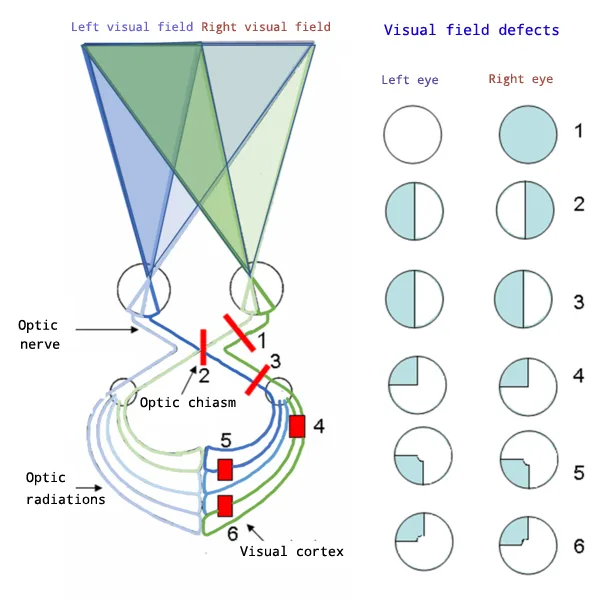
視覚の分析器を3部に分けて追う。
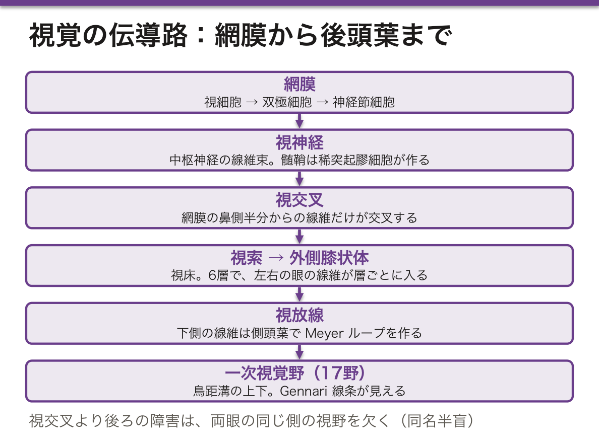
受容部——網膜。視細胞(第1ニューロン)→ 双極細胞(第2)→ 神経節細胞(第3)の3つが網膜の中で直列につながる。
伝導部——神経節細胞の軸索の道。
- 視神経——中枢神経の線維束である。髄鞘は稀突起膠細胞が作り、周りを髄膜と脳脊髄液が包む
- 視交叉——網膜の鼻側半分(耳側の視野)からの線維だけが交叉する。耳側の網膜からの線維は同じ側を進む
- 視索 → 外側膝状体(視床。6層で、左右の眼からの線維が層ごとに分かれて入る)
- 視放線——下側の線維(上の視野)は側頭葉を前へ回り込んで(Meyer ループ)から後ろへ向かう
中枢部——後頭葉の一次視覚野(Brodmann 17野、鳥距溝の上下)。第IV層が厚く、その中を走る有髄線維が肉眼でも白い線として見える(Gennari 線条)。このため有線野とも呼ぶ。黄斑からの入力が後頭極の広い範囲を占める。周りの18・19野が連合野である。
一部の線維は外側膝状体へ行かずに分かれる。視蓋前域へ行く線維は対光反射を、上丘へ行く線維は眼の動きの反射を、視交叉上核へ行く線維(メラノプシンを持つ神経節細胞)は体内時計の調整を担う。
交叉が半分だけなのは、左右の眼が見た同じ側の視野を、1つの大脳半球に集めるためである。右の視野は両眼とも左半球へ届く。だから視交叉より後ろの障害は、両眼の同じ側の視野を欠く(同名半盲)。
| 障害部位 | 視野 | 代表的な原因 |
|---|---|---|
| 視神経 | 片眼の視力・視野の障害 | 視神経炎(多発性硬化症)、虚血 |
| 視交叉の中央 | 両耳側半盲 | 下垂体腺腫、頭蓋咽頭腫 |
| 視索 | 反対側の同名半盲 | — |
| Meyer ループ(側頭葉) | 反対側の上四分盲 | 側頭葉の腫瘍・手術 |
| 頭頂葉の視放線 | 反対側の下四分盲 | — |
| 後頭葉 | 黄斑回避を伴う同名半盲 | 後大脳動脈の梗塞 |
後頭葉の梗塞で中心の視野が残る(黄斑回避)のは、黄斑の領域である後頭極が後大脳動脈と中大脳動脈の両方から血を受けるためと説明される。視神経の障害では、光を当てた眼の入力が弱いことが瞳孔の反応で分かる(相対的入力瞳孔反射異常)。視神経は中枢神経なので、多発性硬化症の最初の症状として視神経炎が起こりうる。
English version
Core
Follow the visual analyser in three parts.
Receptor part — the retina. Photoreceptors (1st neuron) → bipolar cells (2nd) → ganglion cells (3rd) are linked in series inside the retina.
Conducting part — the path of ganglion-cell axons.
- Optic nerve — a tract of the central nervous system. Its myelin is made by oligodendrocytes, and it is wrapped by meninges and CSF.
- Optic chiasm — only fibres from the nasal half of the retina (temporal visual field) cross. Fibres from the temporal retina stay on the same side.
- Optic tract → lateral geniculate body (thalamus; six layers, each receiving from one eye).
- Optic radiation — the lower fibres (upper visual field) loop forward through the temporal lobe (Meyer loop) before running back.
Central part — the primary visual cortex of the occipital lobe (Brodmann area 17, above and below the calcarine sulcus). Layer IV is thick, and the myelinated fibres in it are visible to the naked eye as a white line (stria of Gennari); hence the name striate cortex. Input from the macula takes up a large area at the occipital pole. Areas 18 and 19 around it are association areas.
Going further
Some fibres leave before the lateral geniculate body. Fibres to the pretectal area serve the pupillary light reflex; fibres to the superior colliculus serve reflex eye movements; fibres to the suprachiasmatic nucleus (from melanopsin-containing ganglion cells) set the body clock.
Crossing is only half so that the same side of the visual field, seen by both eyes, is gathered into one hemisphere. The right visual field from both eyes reaches the left hemisphere. So damage behind the chiasm removes the same side of the field in both eyes (homonymous hemianopia).
In the clinic
| Site | Visual field | Typical cause |
|---|---|---|
| Optic nerve | loss in one eye | optic neuritis (multiple sclerosis), ischaemia |
| Centre of the chiasm | bitemporal hemianopia | pituitary adenoma, craniopharyngioma |
| Optic tract | contralateral homonymous hemianopia | — |
| Meyer loop (temporal lobe) | contralateral upper quadrantanopia | temporal tumour or surgery |
| Parietal optic radiation | contralateral lower quadrantanopia | — |
| Occipital lobe | homonymous hemianopia with macular sparing | posterior cerebral artery infarct |
Central vision survives an occipital infarct (macular sparing) because, it is explained, the occipital pole that holds the macula receives blood from both the posterior and middle cerebral arteries. In optic nerve damage, the weak input from the lit eye shows in the pupil response (relative afferent pupillary defect). Because the optic nerve is central nervous tissue, optic neuritis can be the first sign of multiple sclerosis.
まとめ
| 網膜 | 眼球壁の最内層。組織学的に10層に分けるが、要は3つのニューロンの直列である。 |
|---|---|
| 網膜の視部と盲部 — 鋸状縁を境に、光を感じる網膜が終わる | 眼杯の内層は、眼球の内面を前の端まで覆う。 |
| 角膜と水晶体 — 透明であるための組織 | 角膜は5層。上皮(重層扁平・非角化)——再生する。 |
| 眼の発生と硝子体 — 脳の突起と表皮が出会ってできる器官 | 眼は脳(間脳)の壁が外へ伸びた突起と、体表の外胚葉と、周りの間葉の3つから作られる。 |
| 隅角と房水 — 作る場所と、出ていく場所 | 房水は毛様体の無色素上皮が作り、後房 → 瞳孔 → 前房 → 隅角から出ていく。 |
| 眼瞼と涙器 — 表面を守る3層の膜 | 眼瞼は外から内へ——皮膚(体で最も薄い)・皮下組織・眼輪筋・瞼板(密性結合組織)・瞼結膜。 |
| 篩状板 — 視神経が眼球を出るときに通る、弱い場所 | 強膜は丈夫な膜だが、視神経が出る場所だけは網の目状に穴が開いている。 |
| 感覚器の分類と分析器 — 受容細胞が神経か上皮かで分ける | 感覚器に共通する性質は、決まった種類の刺激(適刺激)だけを受け取り、それを電気の信号に変える(変換)ことである。 |
| 視覚の分析器 — 網膜から後頭葉まで、どこで切れたかで視野が変わる | 視覚の分析器を3部に分けて追う。受容部——網膜。 |
このノートは医学生が授業の内容を自分の言葉でまとめたもので、診療の判断に使うものではありません。誤りに気づいたらお知らせください。