消化腺は導管を持つ外分泌腺で、口の中から始まる。漿液性・粘液性・混合性という分け方は、細胞の染まり方と核の位置でそのまま見分けられる。
大唾液腺・小唾液腺の項目はまだ書かれていない。3つの大唾液腺の見分けは腺上皮の章にある。
- 膵外分泌部 — 自分を溶かさないための工夫
膵外分泌部 — 自分を溶かさないための工夫Pancreas
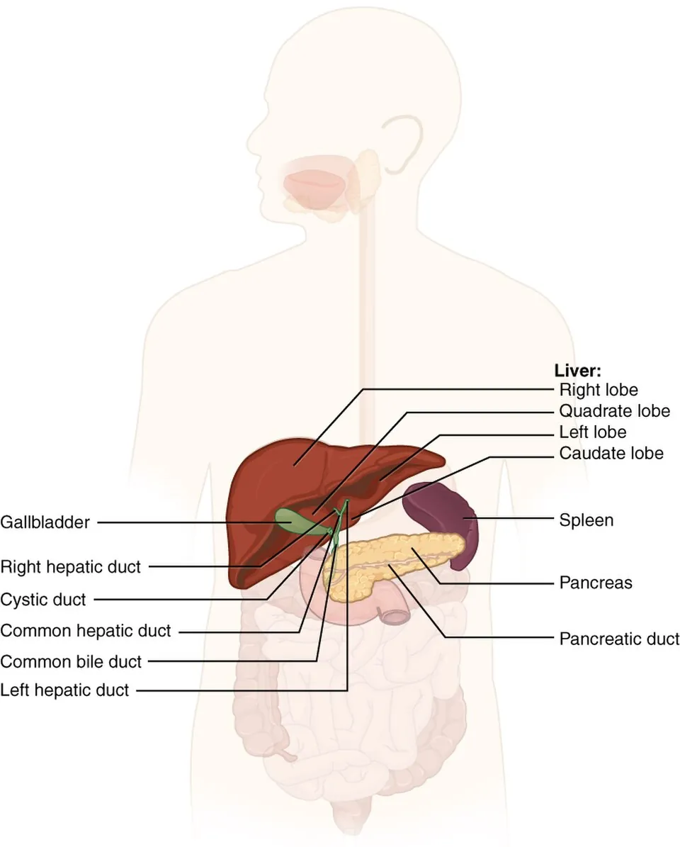
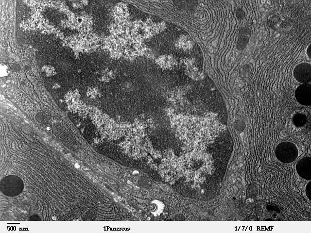
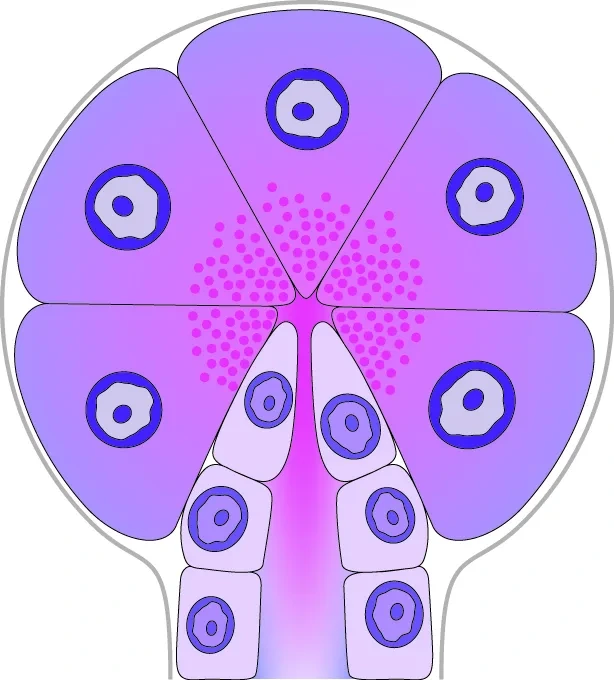
複合管状胞状腺。膵臓の98%以上を占める。
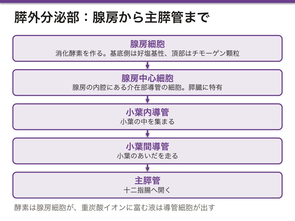
- 腺房細胞——錐体形。基底側は粗面小胞体で濃く染まり、頂部はチモーゲン顆粒で好酸性。1個の細胞の中で色が二分される
- 腺房中心細胞——腺房の内腔にある介在部導管の細胞。膵臓に特有
- 導管——介在部 → 小葉内 → 小葉間 → 主膵管
酵素は腺房細胞が、重炭酸イオンに富む液は導管細胞が出す。
膵臓は蛋白分解酵素を作りながら自分は溶けないという難題を、三重の仕掛けで解いている。
- 不活性な前駆体(チモーゲン)として作る——トリプシノーゲンのまま出す
- 膜で包んで運ぶ——顆粒の中に閉じ込め、細胞質と接触させない
- 阻害物質を同梱する——顆粒の中に膵分泌性トリプシンインヒビター(PSTI/SPINK1)を一緒に入れておく
活性化は十二指腸に着いてからで、刷子縁のエンテロキナーゼがトリプシノーゲンをトリプシンに変え、トリプシンが他の全部を活性化する。スイッチは膵臓の外に置いてある。
急性膵炎はこの仕掛けが膵内で破れた状態である。だから重症化の理屈も一貫している——トリプシンが連鎖的に他の酵素を起こし、ホスホリパーゼが膜を、エラスターゼが血管壁を壊す。脂肪壊死で遊離脂肪酸がカルシウムと結合するため低カルシウム血症になる。
遺伝性膵炎では、この三重の防御のうち SPINK1 またはトリプシン自身を分解する仕組み(PRSS1)が壊れている。防御の設計図を知っていれば、どの遺伝子が壊れると膵炎になるかが読める。
膵液が漏れると周囲が溶けるという性質が、そのまま合併症の形になる。仮性嚢胞、被包化壊死、周囲血管の仮性動脈瘤と出血。手術や内視鏡処置の後の膵液瘻が恐れられるのも同じ理由である。
慢性膵炎では腺房が失われて線維に置き換わり、外分泌不全(脂肪便・体重減少)が出る。膵島は比較的あとまで残るので、糖尿病は遅れて現れる。失われる順序が症状の順序になる。
外分泌不全の治療は高力価の消化酵素薬を食事とともに。胃酸で失活するため腸溶性製剤を使い、必要なら酸分泌抑制薬を併用する——活性化の場所が十二指腸だという組織学が、そのまま処方の根拠になる。
English version
Core
A compound tubulo-acinar gland making up over 98 per cent of the pancreas.
- Acinar cell — pyramidal. The basal pole stains darkly with rough endoplasmic reticulum and the apex is eosinophilic with zymogen granules. The colour of a single cell is divided in two.
- Centroacinar cell — the cell of the intercalated duct lying within the lumen of the acinus. Unique to the pancreas.
- Ducts — intercalated → intralobular → interlobular → main pancreatic duct.
The enzymes come from the acinar cells; the bicarbonate-rich fluid from the duct cells.
Going further
The pancreas solves the problem of making proteolytic enzymes without digesting itself by a triple device.
- Made as an inactive precursor (zymogen) — released as trypsinogen.
- Carried wrapped in membrane — shut inside a granule, never in contact with the cytoplasm.
- An inhibitor is packed with it — pancreatic secretory trypsin inhibitor (PSTI / SPINK1) inside the same granule.
Activation occurs only after arrival in the duodenum: brush-border enterokinase converts trypsinogen to trypsin, and trypsin activates all the rest. The switch is placed outside the pancreas.
Acute pancreatitis is that device breaking inside the gland, and the logic of severe disease follows from it — trypsin triggers the other enzymes in a chain, phospholipase destroys membranes and elastase the vessel walls. Free fatty acids from fat necrosis bind calcium, hence hypocalcaemia.
In hereditary pancreatitis, the broken part of that triple defence is SPINK1 or the mechanism that degrades trypsin itself (PRSS1). Knowing the design of the defence tells you which gene, when broken, causes pancreatitis.
In the clinic
That leaking pancreatic juice dissolves what surrounds it becomes directly the shape of the complications — pseudocyst, walled-off necrosis, pseudoaneurysm of the adjacent vessels and haemorrhage. The same reason makes a pancreatic fistula after surgery or endoscopy so feared.
In chronic pancreatitis the acini are lost and replaced by fibrous tissue, giving exocrine insufficiency (steatorrhoea, weight loss). The islets survive comparatively longer, so diabetes appears later — the order in which they are lost becomes the order of the symptoms.
Exocrine insufficiency is treated with high-potency pancreatic enzymes taken with food. They are inactivated by gastric acid, so enteric-coated preparations are used, with acid suppression if needed — the histological fact that activation occurs in the duodenum becomes directly the basis of the prescription.
まとめ
| 膵外分泌部 — 自分を溶かさないための工夫 | 複合管状胞状腺。膵臓の98%以上を占める。 |
|---|
このノートは医学生が授業の内容を自分の言葉でまとめたもので、診療の判断に使うものではありません。誤りに気づいたらお知らせください。