西洋医学はいつの時代も自分を「科学的」だと考えてきたが、その意味は変わってきた。ヒポクラテス派にとっての科学は、筋の通った理屈と経験だった。近世から、経験(experience)は実験(experiment)を含むようになり、その場所が実験室(laboratory, 「働く場所」の意)になった。
Bynum の区分の最後の型、実験室の医学の目標は、病気を理解すること——仕組みと原因を明らかにすることである。19世紀、主にドイツとフランスの大学・研究所で、細胞病理学・細菌学・実験生理学の3つが立ち上がり、外科は麻酔と無菌法で一変した。
病気の単位は、体全体(体液)→臓器(モルガーニ)→組織(ビシャ)と小さくなってきた。この章でそれは細胞になり、さらに外から来る病原体という新しい原因が加わる。
- 実験の伝統と顕微鏡 — 量れるものは知ることができる
- 細胞説と細胞病理学 —「すべての細胞は細胞から」
- パスツール — 発酵・自然発生の否定・狂犬病ワクチン
- コッホと「コッホの原則」— 病原体を特定する方法
- 麻酔と、消毒から無菌へ — 外科を変えた2つの革命
- クロード・ベルナールと「内部環境」— 実験医学の方法
- 生理学の研究所・ホルモン・動物実験のルール
実験の伝統と顕微鏡 — 量れるものは知ることができる
実験室の医学は19世紀に突然生まれたのではない。初期の実験室は裕福な人の自宅の一室で、その典型は錬金術師の部屋——炉・蒸留器・試薬・天秤・フラスコ——だった。医学でも量を測る実験の伝統は長い。
イタリアのサントーリオ(1561–1636)は自分の体重を量れる椅子を作り、飲食した量と排泄した量を記録して、その差を「不感蒸泄」と呼んだ。ベルギーのファン・ヘルモントは鉢植えの木を5年間水だけで育て、土の重さがほとんど減らないのに木が大きく重くなったことを確かめた。スイスのハラー(1708–1777)は生きた動物の実験で、筋肉の被刺激性と神経の感受性を区別した。
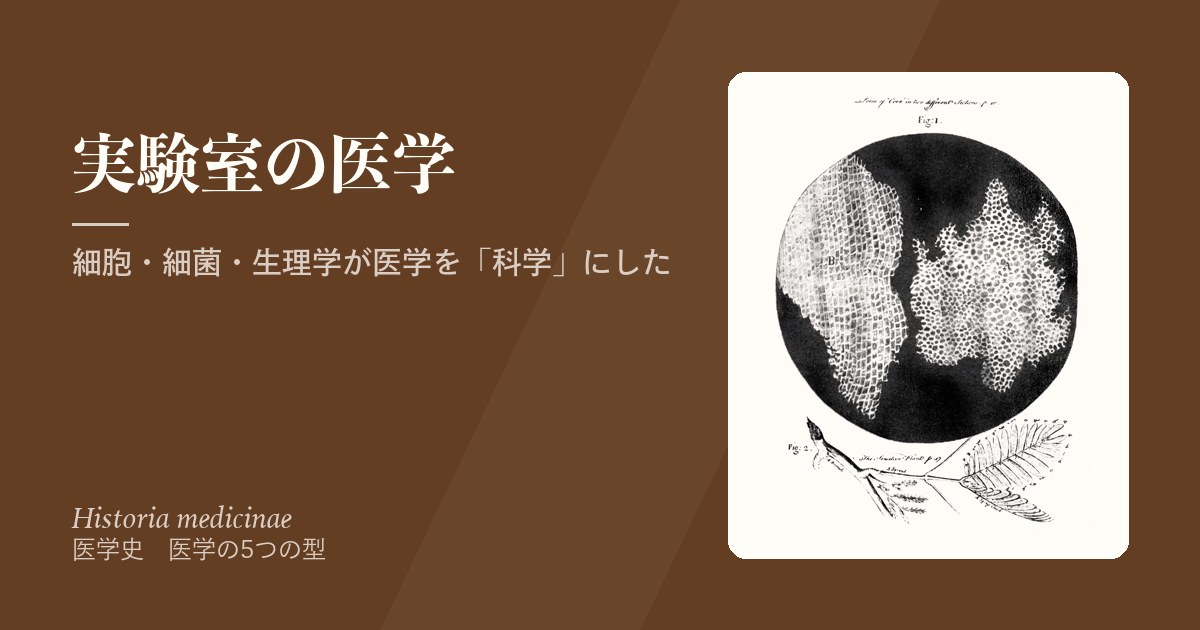
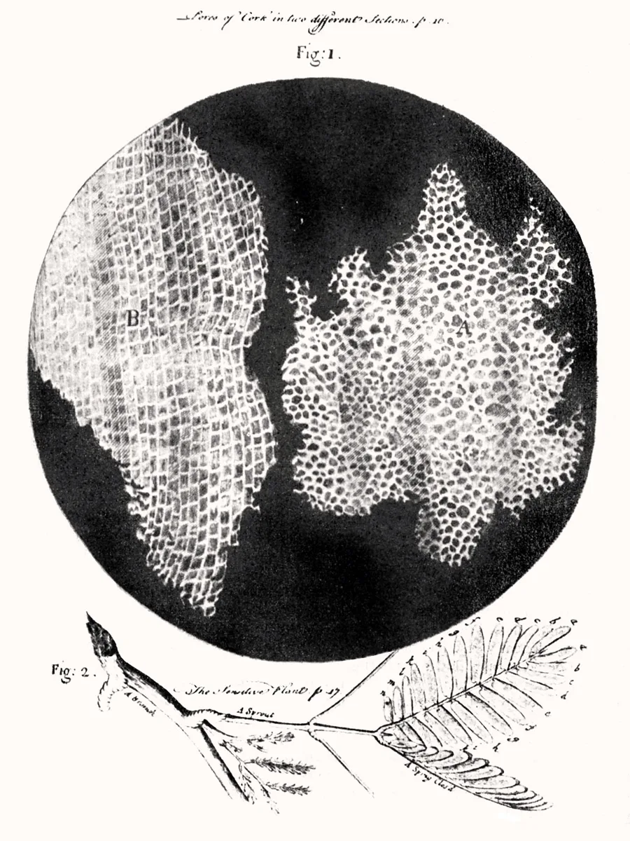
こうした実験の道具の一つが顕微鏡である。初期の顕微鏡には像のゆがみがあり、19世紀以前の顕微鏡学は金持ちの道楽と軽く見られることもあった。しかし実際には17世紀から重要な議論に使われていた。オランダの織物商で独学のレーウェンフック(1632–1723)は自作のレンズで微小な生き物を観察し、イギリスのフック(1635–1703)は1665年の『ミクログラフィア』でコルクの小部屋をcell(細胞)と名づけた。
なぜ欠点のある道具が受け入れられたのか。新しい世界が見えるという可能性が、技術的な不便を上回ったからである。レンズの改良が進んだ19世紀には、顕微鏡は医学研究者の象徴になった——ちょうど聴診器が進歩的な臨床医の象徴だったように。
臨床では:「不感蒸泄」は今も輸液の計算で使う言葉で、皮膚と呼気から失われる水分を見積もる。病理診断・血液像・グラム染色など、顕微鏡で見ることは今も診断の中心にあり、それは17世紀の「見えないものを見る」試みの延長である。
English version
Core
Laboratory medicine did not suddenly appear in the 19th century. Early laboratories were rooms in the homes of rich people; the typical one was the alchemist’s room, with furnace, still, reagents, balance and flasks. Medicine, too, had a long tradition of experiments that measure.
In Italy, Santorio (1561–1636) built a chair in which he could weigh himself. He recorded what he ate and drank and what he excreted, and called the difference “insensible perspiration”. In Belgium, van Helmont grew a tree in a pot for five years with only water, and found that the soil hardly lost weight while the tree grew much heavier. In Switzerland, Haller (1708–1777) used experiments on living animals to separate the irritability of muscle from the sensibility of nerve.
Going further
One tool of such experiments was the microscope. Early microscopes distorted images, and microscopy before the 19th century was sometimes dismissed as a hobby of the rich. In fact it was used in serious debates from the 17th century. The self-taught Dutch cloth merchant Leeuwenhoek (1632–1723) saw tiny living things through his own lenses, and the Englishman Hooke (1635–1703) named the little rooms in cork “cells” in his Micrographia (1665).
Why was an imperfect tool accepted? Because the chance to see a new world outweighed the technical trouble. With better lenses in the 19th century, the microscope turned into the emblem of anyone doing medical research — just as the stethoscope was the symbol of the progressive clinician.
In the clinic
“Insensible perspiration” (insensible water loss) is still used in fluid calculations, to estimate water lost through the skin and breath. Pathology, blood films, Gram staining — looking through a microscope is still at the centre of diagnosis, continuing the 17th-century effort to see the invisible.
細胞説と細胞病理学 —「すべての細胞は細胞から」Omnis cellula e cellula
1830年代末、ドイツのシュライデン(植物, 1838年)とシュワン(動物, 1839年)が、細胞が植物と動物の体の基本単位であると唱えた。2人ともドイツ人なのは偶然ではない。近代の生命医学研究の多くはドイツの大学制度から生まれた。シュワンは、新しい細胞は「芽体(blastema)」という無定形の液から結晶のようにできると考えた。
この細胞説を医学に持ち込んだのがルドルフ・ウィルヒョウである。彼は芽体説を否定し、細胞はつねに細胞の分裂から生じると主張した。標語「すべての細胞は細胞から」(Omnis cellula e cellula)は彼の発案ではないが、それを科学界に納得させたのは彼である。1858年の講義録『細胞病理学』で、細胞が正常な働きと病気の両方の基本単位であることを示した。
ウィルヒョウは、急性・慢性の炎症、がんの増殖と転移、刺激や圧迫への体の反応といった日常の臨床の出来事を、細胞の言葉で説明できることを示した。静脈炎・塞栓症・がん・アミロイドーシスなどについても重要な観察を残し、ベルリンの研究所からは次の世代の病理学者が多数育った。彼が創刊した病理学の雑誌は今もVirchows Archivとして刊行されている。
なぜ細胞説はそれほど重要なのか。病気の単位が、体液→臓器→組織→細胞と小さくなるほど、説明が具体的になり、確かめられるようになるからである。「体液のつり合いが崩れた」は検証のしようがないが、「この細胞が増えている」は顕微鏡で誰でも確かめられる。ミクロトームで薄く切り、染色で核や細胞質を染め分ける技術がこれを支えた。教え子に「顕微鏡で見ることを学べ」と説いた彼は、一方で細菌説には最後まで全面的には賛成しなかった。
臨床では:病理学総論の「ウィルヒョウの三徴」(血流のうっ滞・血管内皮の障害・血液凝固能の亢進)は、今も深部静脈血栓症の危険因子を考える枠組みである。がんが一つの細胞の異常な増殖から始まるという今の理解も、「すべての細胞は細胞から」の延長にある。
English version
Core
In the late 1830s two Germans, Schleiden (plants, 1838) and Schwann (animals, 1839), proposed that the cell is the basic unit of plant and animal bodies. It is no accident that both were German: much of modern biomedical research grew out of the German university system. Schwann thought new cells formed like crystals from a shapeless fluid, the “blastema”.
Rudolf Virchow brought cell theory into medicine. He rejected the blastema idea and argued that cells always arise from the division of cells. The slogan “every cell from a cell” (Omnis cellula e cellula) was not his invention, but he convinced the scientific world of it. In his 1858 lectures, Cellular Pathology, he showed that the cell is the basic unit of both normal function and disease.
Going further
Virchow showed that everyday clinical events — acute and chronic inflammation, the growth and spread of cancer, the body’s reaction to irritation or pressure — can be explained in terms of cells. He made important observations on phlebitis, embolism, cancer, amyloidosis and more, and his Berlin institute trained many of the next generation of pathologists. The pathology journal he founded is still published as Virchows Archiv.
Why is cell theory so important? As the unit of disease became smaller — fluids, organs, tissues, cells — explanations became more concrete and could be tested. “The balance of the humours is broken” cannot be tested, but “these cells are increasing” can be checked by anyone with a microscope. The microtome, for thin sections, and stains that colour the nucleus and cytoplasm made this possible. He told students to “learn to see microscopically”, yet he never fully accepted germ theory.
In the clinic
“Virchow’s triad” in general pathology — stasis of blood flow, damage to the vessel wall and increased clotting — is still the framework for thinking about the risk factors of deep vein thrombosis. Today’s understanding that cancer begins from the abnormal growth of one cell also extends “every cell from a cell”.
パスツール — 発酵・自然発生の否定・狂犬病ワクチン
ルイ・パスツールは医師ではなく、物理と化学を学んだ科学者だった。そのこと自体が、医学にとって科学がどれほど重要になったかを示している。
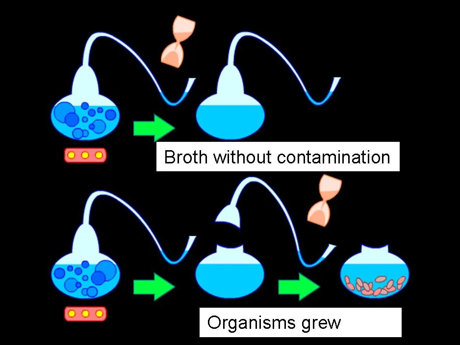
彼の研究は一つの流れでつながっている。酒石酸の結晶の研究で、生物が作った結晶だけが偏光を回転させることに気づき、生物の特別な働きに関心をもった。そこから発酵(ビール・ワイン・牛乳の酸敗)が化学反応ではなく微生物の働きであることを示し、加熱で腐敗を遅らせる低温殺菌(pasteurization)を広めた。1860年代には首の長い白鳥の首フラスコで、煮沸した液に空気中のものが入らなければ微生物は自然には発生しないことを示した。カイコの病気の原因を突き止めたあと、彼は人と動物の病気へ進んだ。
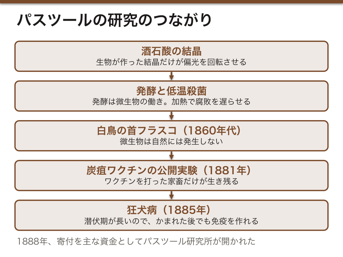
まず炭疽(anthrax)で、病原菌を弱毒化して免疫を作るワクチンを作った。1881年、記者たちを招いて家畜に公開実験を行い、ワクチンを打った動物だけが生き残ることを見せた。彼はジェンナーをたたえて、vaccine という言葉をあらゆる予防接種に広げた。
最後の大仕事が狂犬病(rabies)である。原因のウイルスは当時見ることも培養することもできなかったが、彼は症状から神経系の感染と見て、ウサギの脊髄で病原体を継代し、毒力を調節した。なぜかまれた後でも間に合うのか。かまれてから発症までの潜伏期が長いので、その間に免疫を作れるからである。1885年、少年ジョゼフ・マイスターが助かると、国際的な称賛が集まり、1888年には寄付を主な資金としてパスツール研究所が開かれた。
ただし後世の研究ノートの分析では、自然発生の実験で都合の悪い結果を公表しなかったことも分かっている。熱に強い芽胞をもつ枯草菌では、煮沸しても菌が生き残るからである。
臨床では:狂犬病の曝露後ワクチン接種は、パスツールの「潜伏期のうちに免疫を作る」原理そのものである。発症すればほぼ100%死亡するので、流行地でかまれたら直ちに接種する。日本では国内での感染は1950年代以降ないが、海外でかまれて帰国後に発症した例がある。また、彼のノートの問題は、都合の悪いデータも記録し報告する研究公正の大切さを教えている。
English version
Core
Louis Pasteur was not a doctor but a scientist trained in physics and chemistry. That fact alone shows how important science had become for medicine.
His research forms one connected line. Studying crystals of tartaric acid, he noticed that only crystals made by living things rotated polarised light, and became interested in what living things can do. He then showed that fermentation (of beer and wine, the souring of milk) is the work of micro-organisms, not just a chemical reaction, and he spread pasteurisation — heating to slow spoiling. In the 1860s, with his swan-necked flasks, he showed that if nothing from the air enters a boiled liquid, micro-organisms do not appear by themselves. After finding the cause of a silkworm disease, he moved on to diseases of animals and humans.
Going further
First, for anthrax, he made a vaccine by attenuating (weakening) the bacterium so that it gave immunity. In 1881 he invited journalists to a public test on farm animals: only the vaccinated animals survived. Honouring Jenner, he extended the word vaccine to all such preventive inoculations.
His last great work was rabies. Its virus could not be seen or grown at the time, but from the symptoms he judged it to be an infection of the nervous system, and he passed the agent through rabbit spinal cords to control its strength. Why can treatment work even after a bite? Because the incubation period between the bite and illness is long, so there is time to build immunity. When the boy Joseph Meister survived in 1885, praise came from around the world, and in 1888 the Pasteur Institute opened, paid for mainly by public donations.
Later study of his notebooks showed, however, that in his experiments on spontaneous generation he did not publish results that did not suit him. With hay bacillus, whose spores resist heat, bacteria could survive boiling.
In the clinic
Post-exposure rabies vaccination is exactly Pasteur’s principle of building immunity during the incubation period. Once symptoms start, rabies is almost always fatal, so anyone bitten in an endemic area must be vaccinated at once. There has been no infection inside Japan since the 1950s, but people bitten abroad have fallen ill after returning. The problem in his notebooks also teaches the importance of research integrity — recording and reporting inconvenient data too.
コッホと「コッホの原則」— 病原体を特定する方法
ロベルト・コッホ(1843–1910)はドイツの開業医から出発した医学細菌学者である。最初の仕事で炭疽菌の生活環——土の中で何年も生き延びる芽胞の時期があること——を明らかにした。
パスツールより一世代若い彼は、技術がはるかに精密だった。顕微鏡写真で客観的な証拠を示し、汚染の少ない寒天の固形培地で細菌を育て、滅菌の装置を広め、弟子のペトリはペトリ皿を作った。そして1882年に結核菌、1883〜84年にコレラ菌を特定した。結核菌は増えるのが遅く染まりにくく、しかも結核は体質や環境の病気と考えられていたので、細菌が原因とは誰も思っていなかった。
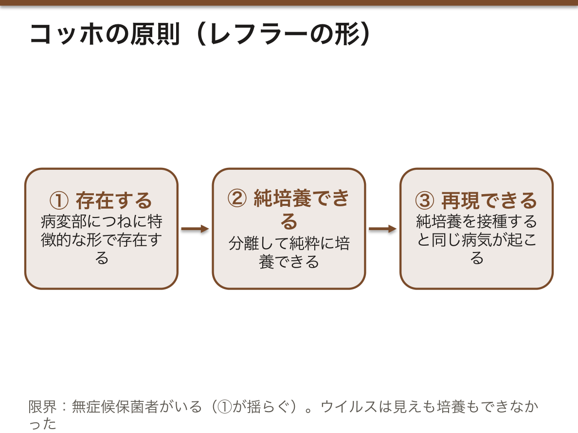
病原体を特定する基準として知られるのがコッホの原則である。弟子のレフラーがジフテリアについて最も簡潔に書いた形では、①その微生物が病変部につねに特徴的な形で存在する、②それを分離して純粋に培養できる、③純培養を接種すると同じ病気が起こる、の3つである。
しかしこの基準は、多くの病気で満たすのが難しかった。健康な人の皮膚や腸からも同じ菌が見つかる。コッホ自身が見つけた無症候保菌者(腸チフスのメアリーの例)は、原則の①を揺るがした。ウイルスは見えも培養もできなかった。有名な反証の試みもある。衛生学者ペッテンコーファーは、病気は菌だけでなく土地や体の条件が重なって起こると考え、コレラ菌の培養液を公衆の前で飲み、軽い下痢で済んだ。
それでも細菌説は医学を変えた。病気の原因が患者の外にあると分かったことで、診断の客観的な基準を作りやすくなった。また病気ごとに原因が違う(特異性)ことがはっきりし、「熱」は病気そのものではなく病気の徴候になった。
臨床では:コッホが結核菌を発表した3月24日は世界結核デーである。彼が治療薬として出したツベルクリンは治療には失敗したが、ツベルクリン反応として診断に生き残った。日本からは北里柴三郎がコッホに学び、破傷風菌の純培養と血清療法を、志賀潔が赤痢菌(Shigella の名の由来)を発見した。今の分子生物学では、遺伝子で原因を確かめる「分子版コッホの原則」も使われる。
English version
Core
Robert Koch (1843–1910) began as a German country doctor and became a medical bacteriologist. In his first work he showed the life cycle of the anthrax bacterium, including a spore stage that can survive in soil for years.
A generation younger than Pasteur, he had far more precise techniques. He used photomicrographs as objective evidence, grew bacteria on solid agar media that reduced contamination, spread sterilising equipment, and his assistant Petri made the Petri dish. He then identified the tubercle bacillus in 1882 and the cholera bacterium in 1883–84. The tubercle bacillus grows slowly and stains poorly, and tuberculosis was thought to be a disease of constitution and environment, so few expected a bacterial cause.
Going further
The famous standard for identifying a cause is Koch’s postulates. In the clearest form, written by his pupil Löffler about diphtheria, there are three: (1) the microbe is found every time in the diseased tissue, with a typical appearance; (2) it can be separated out and grown on its own; (3) when that culture is given to an animal, the same disease appears.
But many diseases could not meet this standard. The same bacteria were found on the skin and in the gut of healthy people. The healthy carrier, which Koch himself described (as in Typhoid Mary), shook postulate (1). Viruses could be neither seen nor grown. There was also a famous attempt to disprove him: the hygienist Pettenkofer, who believed disease needed local and bodily conditions as well as the germ, publicly drank a culture of cholera bacteria and had only mild diarrhoea.
Still, germ theory changed medicine. Knowing that the cause of disease lies outside the patient made it easier to set objective standards for diagnosis. It also made clear that each disease has its own cause (specificity), and “fever” became a sign of disease, not a disease itself.
In the clinic
March 24, the day Koch announced the tubercle bacillus, is World Tuberculosis Day. Tuberculin, which he offered as a cure, failed as treatment, but survived in diagnosis as the tuberculin skin test. From Japan, Kitasato Shibasaburō studied under Koch and achieved the pure culture of the tetanus bacillus and serum therapy, and Shiga Kiyoshi discovered the dysentery bacillus (the origin of the name Shigella). Modern molecular biology also uses “molecular Koch’s postulates”, which test causes with genes.
麻酔と、消毒から無菌へ — 外科を変えた2つの革命
近代外科は2つの柱の上に立つ。一つは麻酔である。1846年10月16日、ボストンのマサチューセッツ総合病院でエーテル麻酔による手術が初めて公開され、知らせは船でヨーロッパへ広まった。翌年にはクロロホルムが続いた。どちらも化学研究の産物で、実験室が臨床を変えた例である。痛みが取れたことで外科医は時間をかけて組織を温存できるようになったが、傷が長く開いているぶん術後感染も増え、手術の範囲は広がっても患者の生存率は上がらなかった。
もう一つの柱が感染を防ぐ方法である。ウィーンの産科医ゼンメルワイスは1847年、医師と学生が担当する病棟で産褥熱(febris puerperalis)の死亡が助産師の病棟より多いことに気づき、剖検室から来る医師の手が原因と考えてさらし粉(塩素水)での手洗いを命じ、死亡を激減させた。しかし彼の考えは受け入れられなかった。
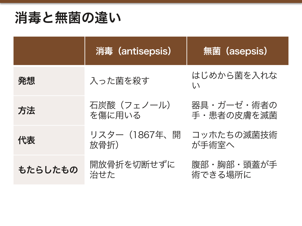
イギリスの外科医ジョゼフ・リスター(1827–1912)は、パスツールの発酵と腐敗の研究に触発され、下水の消毒に使われていた石炭酸(フェノール)を傷に用いた。1867年、開放骨折——折れた骨が皮膚を破って外に出た骨折——を石炭酸の包帯で治療し、切断せずに治せることを示した。当時、開放骨折はほとんど切断しか手がなかった。普仏戦争では、彼の方法を取り入れたドイツの外科医の成績がフランスより良かった。
やがて消毒(antisepsis)は無菌(asepsis)に取って代わられた。違いは発想にある。消毒は入った菌を殺す、無菌ははじめから菌を入れない。器具・ガーゼ・術者の手・患者の皮膚を滅菌すれば、もともと無菌の体の組織は膿まずに治る(一次治癒)。コッホたちが実験室で作った滅菌の技術が手術室に移り、腹部・胸部・頭蓋の3つの体腔がはじめて手術できる場所になった。ただし、出血や輸血・抗菌薬の欠如といった支えが追いつかず、必要のない手術も多く行われた。
臨床では:日本では1804年、華岡青洲が自作の麻酔薬「通仙散」で全身麻酔をかけて乳がんの手術を行い、エーテルより40年以上早い。今のWHO の手指衛生の5つのタイミングはゼンメルワイスの直系である。開放骨折は今も整形外科の緊急疾患で、早期の抗菌薬投与・洗浄・デブリードマンが感染を防ぐ鍵になる。
English version
Core
Modern surgery stands on two pillars. One is anaesthesia. On 16 October 1846, at Massachusetts General Hospital in Boston, an operation under ether was shown in public for the first time, and the news crossed to Europe by ship. Chloroform followed the next year. Both were products of chemical research — the laboratory changing the clinic. Without pain, surgeons could take time and save tissue, but wounds stayed open longer, so post-operative infection rose; the range of operations grew, but patient survival did not.
The other pillar was preventing infection. In 1847 the Viennese obstetrician Semmelweis noticed that deaths from puerperal fever (febris puerperalis) were higher on the ward run by doctors and students than on the midwives’ ward. He blamed doctors’ hands coming from the autopsy room, ordered hand-washing in chlorinated lime, and deaths fell sharply. But his idea was not accepted.
Going further
The British surgeon Joseph Lister (1827–1912), inspired by Pasteur’s work on fermentation and decay, used carbolic acid (phenol), then used to disinfect sewage, on wounds. In 1867 he treated compound (open) fractures — where the broken bone breaks through the skin — with carbolic dressings, and showed they could heal without amputation. At the time, amputation was almost the only option for such fractures. In the Franco-Prussian War, German surgeons who used his method did better than the French.
Later, antisepsis was replaced by asepsis. The difference is the idea: antisepsis kills germs that got in; asepsis keeps germs out from the start. If everything that touches the wound — tools, gauze, the operator’s hands, the skin at the incision — is sterile, body tissue, which is germ-free to begin with, heals without pus (first intention). Sterilising methods from Koch’s laboratory moved into the operating theatre, and the three body cavities — abdomen, chest and skull — could be operated on for the first time. But support such as transfusion and antibiotics lagged behind, and many unnecessary operations were done.
In the clinic
In Japan in 1804, Hanaoka Seishū operated on breast cancer under general anaesthesia with his own drug, tsūsensan — more than 40 years before ether. Today’s WHO “five moments for hand hygiene” descend directly from Semmelweis. Open fractures are still orthopaedic emergencies, and early antibiotics, irrigation and debridement are the keys to preventing infection.
クロード・ベルナールと「内部環境」— 実験医学の方法
19世紀最大の生理学者は、フランスのクロード・ベルナール(1813–1878)である。パリの医学校で学んだ彼は、病院の臨床観察だけでは病気の仕組みや新しい治療にはたどり着けないと考えた。実験室での手術の腕にすぐれ、肝臓が糖を作り蓄える働き(グリコーゲン)、膵臓の消化での役割、末梢神経の働き、一酸化炭素中毒の仕組みを明らかにし、脳の一部を傷つけて一種の糖尿病を起こすことも示した。
彼の最も大きな概念が内部環境(milieu intérieur)である。生物は、体温・血中の塩類・血糖などを狭い範囲に保つことで、外の環境が変わっても生きていける。この考えはのちにアメリカのキャノンによってホメオスタシス(恒常性)と名づけられた。
1865年の『実験医学序説』で、彼は医学研究の方法を論じた。実験室こそ医学の聖域である。病院では患者の世話が優先され、条件が多すぎて観察は断片的になる。実験室でのみ条件を一定に保ち、変化をはっきり捉えられる。
その方法は今でいう仮説演繹法である。仮説を立て、そこから何が起こるはずかを導き、実験で確かめる。ベルナールはこれを帽子にたとえた。実験中は帽子(考え)を掛けておき、予想にとらわれずに観察する。実験室を出るときは帽子をかぶり直して、何を見たかを考える。偶然の観察も大切にした。ふつうアルカリ性で濁ったウサギの尿が、絶食させると酸性で澄むことに気づき、ウサギが自分の組織を燃やしていると推論して、食物の消化の研究へ進んだ。彼にとって実験医学の3本柱は、正常を扱う生理学・異常を扱う病理学・治療法を探す治療学だった。
臨床では:血糖・電解質・酸塩基平衡・体温の調節を「ずれ」と「戻す仕組み」で考えるのは、ベルナールの内部環境の考え方である。輸液や電解質補正は、崩れた内部環境を外から戻す治療と言える。仮説を立て、予測し、データで確かめるという手順は、研究だけでなく日々の鑑別診断の進め方でもある。
English version
Core
The greatest physiologist of the 19th century was the Frenchman Claude Bernard (1813–1878). Trained in the Paris medical school, he believed that clinical observation in hospitals alone could not reach the mechanisms of disease or new treatments. A skilled surgeon in the laboratory, he showed how the liver makes and stores sugar (glycogen), the role of the pancreas in digestion, the functions of peripheral nerves and how carbon monoxide poisons, and he caused a kind of diabetes by damaging part of the brain.
His greatest idea was the internal environment (milieu intérieur). Living things survive changes in the outside world by keeping body temperature, blood salts, blood sugar and more within narrow limits. The American Cannon later called this homeostasis.
Going further
In his Introduction to the Study of Experimental Medicine (1865), he wrote about the method of medical research. The laboratory is the true sanctuary of medicine. In a hospital, caring for patients comes first, there are too many variables, and observations are only partial. Only in the laboratory can conditions be kept constant so that changes are clear.
His method is what we now call the hypothetico-deductive method: form a hypothesis, work out what should happen, and test it by experiment. Bernard compared this to a hat. During the experiment, hang up your hat (your ideas) and observe without expectations; when leaving the lab, put it back on and think about what you saw. He also valued chance observations. Rabbit urine is normally alkaline and cloudy; when he saw that the urine of fasting rabbits was acidic and clear, he reasoned that they were burning their own tissues, and moved on to study digestion. For him the three pillars of experimental medicine were physiology (normal function), pathology (abnormal function) and therapeutics (finding remedies).
In the clinic
Thinking about blood sugar, electrolytes, acid–base balance and body temperature as “a shift” and “a mechanism that brings it back” is Bernard’s idea of the internal environment. Fluids and electrolyte correction can be seen as treatment that restores a broken internal environment from outside. And forming a hypothesis, predicting and checking with data is not only research — it is how we work through a differential diagnosis every day.
生理学の研究所・ホルモン・動物実験のルール
19世紀のドイツは、医学のあらゆる基礎科学に国の資金で研究所を作った。生理学で最も有名なのは、ライプツィヒ大学のカール・ルートヴィヒ(1816–1895)の研究所で、世界中から学生が集まった。ルートヴィヒを含む若い生理学者4人は1840年代後半、生理学の問題はすべて物理学と化学で解けるという立場を打ち出した。その一人ヘルムホルツは感覚器の生理学を研究し、検眼鏡を発明した。ルートヴィヒは脈拍や筋収縮を連続して記録するキモグラフを作った。生体の変化を波形として記録するやり方は、以後の研究と臨床の特徴になった。この時代、医学の国際語はドイツ語だった。
生理学の研究は多くの専門科を生んだ。大脳の機能局在の研究は神経内科を、心臓の収縮と拍動の調節の研究は循環器を支えた。イギリスのベイリスとスターリングは1902年に十二指腸から出て膵液の分泌を促すセクレチンを発見し、血液で運ばれて遠くの臓器に働く化学の使者——ホルモン——という考えを確立した。内分泌学はここから始まる。
一方、実験生理学は生きた動物を手術するため、とくにイギリスで激しい動物実験反対運動を引き起こした。その結果、1876年にイギリスで動物虐待防止法ができ、動物実験が法律で規制された。研究者は最初これを恐れたが、実際には研究を個人の自宅から大学や公的機関へ移し、制度化する働きをした。麻酔は痛みを防ぐだけでなく実験を容易にし、無菌法も生理学に役立った——臨床と実験科学が互いを強め合った例である。
臨床では:検眼鏡で眼底を見ることは、今も高血圧・糖尿病・頭蓋内圧亢進の評価に使われる。心電図・脳波・動脈圧の連続モニターは、キモグラフの「波形で記録する」発想の子孫である。動物実験には今、3R(代替 Replacement・削減 Reduction・苦痛の軽減 Refinement)の原則があり、日本の動物愛護管理法にも書き込まれ、各機関の委員会が計画を審査している。
English version
Core
19th-century Germany built state-funded institutes for every basic medical science. The most famous in physiology was the institute of Carl Ludwig (1816–1895) at Leipzig, where students came from around the world. In the late 1840s, four young physiologists including Ludwig declared that all problems of physiology can be solved by physics and chemistry. One of them, Helmholtz, studied the physiology of the senses and invented the ophthalmoscope. Ludwig built the kymograph, which records the pulse or muscle contractions continuously. Recording changes in the living body as waveforms became a mark of later research and clinical medicine. In this era, German was the international language of medicine.
Going further
Physiological research gave rise to many specialties. Work on localisation in the brain supported neurology; work on the contraction and rhythm of the heart supported cardiology. In 1902 the British Bayliss and Starling discovered secretin, released from the duodenum to stimulate pancreatic secretion, and established the idea of chemical messengers carried in the blood to act on distant organs — hormones. Endocrinology starts here.
But experimental physiology operated on living animals, and so, especially in Britain, it provoked a strong anti-vivisection movement. As a result, Britain passed the Cruelty to Animals Act in 1876, which regulated animal experiments by law. Researchers feared it at first, but in fact it moved research from private homes into universities and public institutions and made it an institution. Anaesthesia not only prevented pain but made experiments easier, and asepsis helped physiology too — clinical medicine and experimental science strengthening each other.
In the clinic
Looking at the fundus with an ophthalmoscope is still used to assess hypertension, diabetes and raised intracranial pressure. ECG, EEG and continuous arterial pressure monitoring descend from the kymograph’s idea of recording waveforms. Animal research now follows the 3Rs — Replacement, Reduction and Refinement. They are written into Japan’s animal welfare law, and committees in each institution review research plans.
まとめ
| 実験の伝統と顕微鏡 — 量れるものは知ることができる | 実験室の医学は19世紀に突然生まれたのではない。 |
|---|---|
| 細胞説と細胞病理学 —「すべての細胞は細胞から」 | 1830年代末、ドイツのシュライデン(植物, 1838年)とシュワン(動物, 1839年)が、細胞が植物と動物の体の基本単位であると唱えた。 |
| パスツール — 発酵・自然発生の否定・狂犬病ワクチン | ルイ・パスツールは医師ではなく、物理と化学を学んだ科学者だった。 |
| コッホと「コッホの原則」— 病原体を特定する方法 | ロベルト・コッホ(1843–1910)はドイツの開業医から出発した医学細菌学者である。 |
| 麻酔と、消毒から無菌へ — 外科を変えた2つの革命 | 近代外科は2つの柱の上に立つ。一つは麻酔である。 |
| クロード・ベルナールと「内部環境」— 実験医学の方法 | 19世紀最大の生理学者は、フランスのクロード・ベルナール(1813–1878)である。 |
| 生理学の研究所・ホルモン・動物実験のルール | 19世紀のドイツは、医学のあらゆる基礎科学に国の資金で研究所を作った。 |
このノートは医学生が授業の内容を自分の言葉でまとめたもので、診療の判断に使うものではありません。誤りに気づいたらお知らせください。