眼はよく「カメラ」に例えられるが、解剖で押さえるべきは3枚の膜と、その内側の中身という作りである。外から線維膜(形を保つ)、血管膜(栄養を運び、光の量とピントを調節する)、内膜=網膜(光を受ける)。その内側に房水・水晶体・硝子体が詰まっている。どの構造も、この5つのどれに属するかを先に決めると、名前の多さに振り回されない。
眼球のまわりには、守る装置(眼瞼・結膜・涙器)と動かす装置(外眼筋)がある。さらに網膜から後頭葉までの視覚路と、途中で中脳へ分かれる対光反射・近見反応の経路は、神経診察でいちばん基本の道具になる。組織の細部は視覚器(組織学)、外眼筋の起始・停止は頭部の筋、視交叉から先の脳の構造は中脳・間脳にある。この章では、それらを「どこに何があり、なぜそう働くか」でつなぐ。
- 眼球 — 外形・3本の軸・3枚の膜
- 線維膜 — 角膜と強膜
- 血管膜(ぶどう膜)— 脈絡膜・毛様体・虹彩
- 網膜と視神経 — 見える部分・見えない部分・出口
- 水晶体・硝子体・眼房 — 眼の中身と調節
- 眼の付属器 — 眼瞼・結膜・涙器
- 外眼筋と眼窩の血管・神経 — 作用は眼窩の向きから導く
- 視覚路と瞳孔の反射 — 対光反射と近見反応
- 眼球 — 外形・3本の軸・3枚の膜Bulbus oculi
- 線維膜 — 角膜と強膜Tunica fibrosa bulbi
- 血管膜(ぶどう膜)— 脈絡膜・毛様体・虹彩Tunica vasculosa bulbi
- 網膜と視神経 — 見える部分・見えない部分・出口Retina et nervus opticus
- 水晶体・硝子体・眼房 — 眼の中身と調節Lens et corpus vitreum
- 眼の付属器 — 眼瞼・結膜・涙器Palpebrae et apparatus lacrimalis
- 外眼筋と眼窩の血管・神経 — 作用は眼窩の向きから導くMusculi bulbi; vasa et nervi orbitae
- 視覚路と瞳孔の反射 — 対光反射と近見反応Via optica
- まとめ
眼球 — 外形・3本の軸・3枚の膜Bulbus oculi
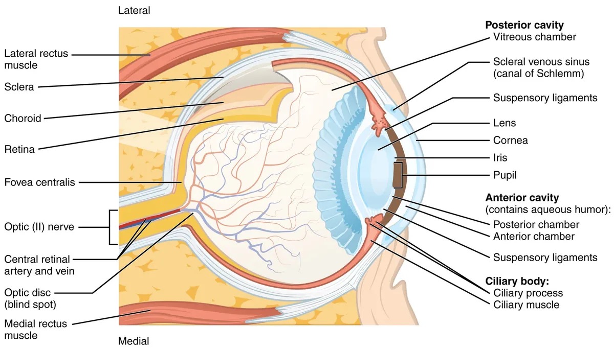
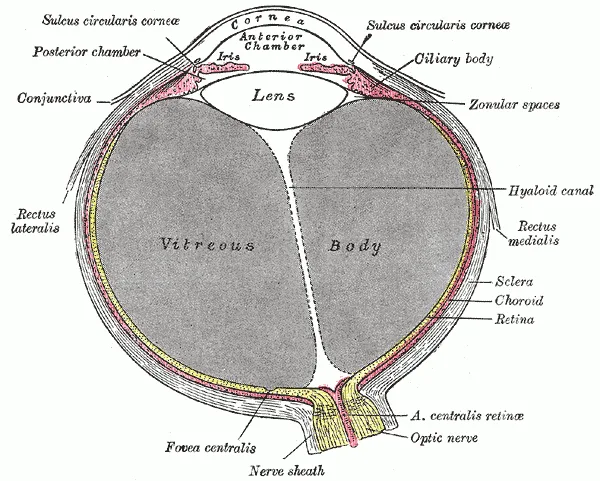
眼球は眼窩 orbita の前半分に収まる、重さ約7.5 g のほぼ球形の器官である。角膜の中心を前極 polus anterior、強膜の後ろの中心を後極 polus posterior、両極から等しい距離で一周する線を赤道 aequatorと呼ぶ。外眼筋の停止を「赤道より前か後か」で言い分けるのは、この線が基準になっている。
| 軸 | 結ぶもの | 長さの目安 |
|---|---|---|
| 外眼球軸 axis bulbi externus | 前極の外面 〜 後極の外面 | 約24 mm |
| 内眼球軸 axis bulbi internus | 角膜の内面 〜 網膜 | 約22 mm |
| 視軸 axis opticus | 前極 〜 黄斑(中心窩) | — |
壁は外から線維膜 tunica fibrosa・血管膜 tunica vasculosa・内膜 tunica interna(網膜)の3層。中身は房水 humor aquosus・水晶体 lens・硝子体 corpus vitreumの3つである。視神経が眼球を出る場所(視神経円板)は後極ではなく、黄斑から約4 mm 内側(鼻側)にずれている。像を結ぶ中心と、神経の出口を重ねない配置になっている。
なぜ眼軸の長さで近視・遠視が決まるのか。眼の屈折力は角膜と水晶体でほぼ決まり、焦点を結ぶ距離はおおむね一定である。その焦点がちょうど網膜の上に落ちるかどうかは、レンズの側ではなくスクリーン(網膜)までの距離=眼軸長で決まる。
眼軸が長すぎると焦点は網膜の手前に来る——近視 myopia。短すぎると網膜の後ろに来る——遠視 hypermetropia。目安として眼軸が1 mm 伸びると、約3 ディオプター近視の側へずれる。成長期に近視が進むのは、主に眼球が前後に伸びるためである(軸性近視)。近視は凹レンズで焦点を後ろへ、遠視は凸レンズで前へ動かして網膜に合わせる。
眼軸が大きく伸びた強度近視(病的近視)では、後極の網膜・脈絡膜・強膜が風船のように引き延ばされて薄くなる。そのため網膜剥離・近視性の黄斑の変性・緑内障が起こりやすく、日本では視覚障害の主な原因の一つになっている。「眼鏡で矯正できる」ことと「眼球が健康である」ことは別の話であり、強度近視の人には眼底の定期的な検査が勧められる。
English version
Core
The eyeball sits in the front half of the orbit. It weighs about 7.5 g and is almost a sphere. The centre of the cornea is the anterior pole, the centre of the back of the sclera is the posterior pole, and the line around the eye halfway between them is the equator. We describe the insertion of eye muscles as “in front of” or “behind” the equator.
| Axis | Joins | Length (approx.) |
|---|---|---|
| Axis bulbi externus | outer surface of anterior pole – outer surface of posterior pole | about 24 mm |
| Axis bulbi internus | inner surface of cornea – retina | about 22 mm |
| Axis opticus (visual axis) | anterior pole – macula (fovea) | — |
The wall has three layers: the fibrous layer, the vascular layer and the inner layer (retina). The contents are the aqueous humour, lens and vitreous body. The optic nerve does not leave at the posterior pole: the optic disc lies about 4 mm medial (nasal) to the macula. The centre of the image and the exit of the nerve are kept apart.
Going further
Why does the length of the eye decide myopia and hyperopia? The focusing power comes mostly from the cornea and lens, so the focal distance is fairly fixed. Whether the focus falls exactly on the retina depends on the distance to the screen — the axial length.
If the eye is too long, the focus falls in front of the retina: myopia. If it is too short, the focus falls behind it: hypermetropia. As a rule of thumb, 1 mm of extra length gives about 3 dioptres of myopia. Myopia increases in childhood mainly because the eye grows longer (axial myopia). A concave lens moves the focus back; a convex lens moves it forward.
In the clinic
In high (pathological) myopia the long eye stretches the retina, choroid and sclera at the back, and they become thin. Retinal detachment, myopic macular degeneration and glaucoma become more common, and in Japan it is one of the main causes of visual impairment. Being corrected by glasses is not the same as having a healthy eye, so regular fundus examination is advised.
線維膜 — 角膜と強膜Tunica fibrosa bulbi
眼球のいちばん外の膜で、前の約1/6 が角膜 cornea、残りが強膜 scleraである。両者の境目が角膜輪部 limbus corneaeで、その外面の浅い溝を強膜溝 sulcus sclerae、その深部を輪状に走る静脈の管を強膜静脈洞 sinus venosus sclerae(Schlemm 管)と呼ぶ。房水の主な出口である。
角膜は透明で前へ凸、血管を持たない。栄養は房水と涙液、周辺は輪部の血管から受ける。結膜には覆われない。一方で知覚神経はきわめて密で、三叉神経の第1枝(鼻毛様体神経から出る長毛様体神経)が担う。
強膜は白く不透明な丈夫な膜で、厚さは1 mm 前後。前方は眼球結膜に覆われ、6つの外眼筋が付く。後方では視神経が網の目状の篩状板 lamina cribrosa scleraeを貫いて出ていき、視神経の外鞘(硬膜の続き)が強膜に溶け込む。篩状板の弱さと緑内障の関係は篩状板(組織学)、角膜の5層は角膜と水晶体にある。
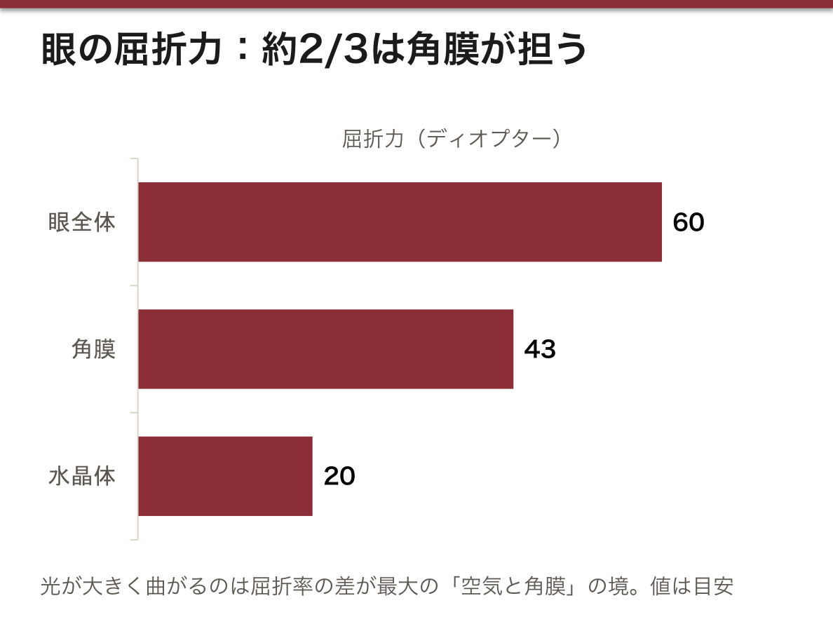
なぜ角膜が眼の屈折の主役なのか。眼全体の屈折力は約60 ディオプターで、そのうち約2/3(約43 D)を角膜が担い、水晶体は約20 D にすぎない。光が大きく曲がるのは屈折率の差が大きい境目であり、眼の中で差が最大なのは「空気と角膜」の境だからである。水晶体は房水と硝子体という、屈折率の近い液体に挟まれているので、それ自体は強いレンズでも効きは小さい。水中で物がぼやけるのは、角膜の前が空気でなく水になって角膜の屈折がほとんど消えるからで、ゴーグルで空気の層を戻すとまた見える。近視の屈折矯正手術が水晶体ではなく角膜の形を変えるのも、この理由による。
角膜反射 corneal reflex:角膜に触れると両眼が閉じる。求心路は三叉神経(V1)、遠心路は顔面神経(眼輪筋)である。両側に出る反射なので、片眼ずつ刺激して左右の瞬目を比べると壊れている側が分かる。右を触れても左右どちらも閉じなければ右の V1、どちらを触れても右だけ閉じなければ右の VII が障害されている。
角膜反射は意識障害の患者で脳幹の働きを確かめる反射の一つで、脳死判定の項目にも入っている。強膜は I 型コラーゲンでできているため、骨形成不全症では強膜が薄くなり、下の脈絡膜の色が透けて青色強膜になる。骨と強膜が同じ材料でできている、という解剖の事実がそのまま所見になっている。
English version
Core
The outer coat of the eye. The front sixth is the cornea and the rest is the sclera. Their border is the limbus. On the surface there is a shallow groove (sulcus sclerae), and deep to it runs a ring-shaped venous channel, the sinus venosus sclerae (canal of Schlemm) — the main exit for aqueous humour.
The cornea is transparent, convex and has no blood vessels. It is nourished by the aqueous humour and tears, and at the edge by limbal vessels. It is not covered by conjunctiva. Its sensory nerves are very dense and come from the ophthalmic nerve (V1, via the long ciliary nerves).
The sclera is the tough white coat, about 1 mm thick. The bulbar conjunctiva covers its front part, and the six extraocular muscles insert on it. At the back the optic nerve leaves through the sieve-like lamina cribrosa, and the outer sheath of the nerve (a continuation of the dura) blends with the sclera.
Going further
Why is the cornea the main lens of the eye? The whole eye has about 60 dioptres of power; the cornea gives about two-thirds (about 43 D) and the lens only about 20 D. Light bends most where the difference in refractive index is largest, and in the eye that is the air–cornea boundary. The lens sits between fluids with similar indices, so its effect is smaller. Under water the cornea loses most of its power, so we see blurred; goggles bring back the air layer. This is also why refractive surgery reshapes the cornea, not the lens.
Corneal reflex: touching the cornea closes both eyes. The afferent limb is the trigeminal nerve (V1); the efferent limb is the facial nerve (orbicularis oculi). Because the response is bilateral, you can find the damaged side: if touching the right eye closes neither eye, the right V1 is affected; if the right eye never closes whichever side is touched, the right VII is affected.
In the clinic
The corneal reflex is used to test brainstem function in unconscious patients and is one of the items in brain-death testing. The sclera is made of type I collagen, so in osteogenesis imperfecta it becomes thin and the dark choroid shows through as a blue sclera. Bone and sclera share the same material, and the sign follows directly from that.
血管膜(ぶどう膜)— 脈絡膜・毛様体・虹彩Tunica vasculosa bulbi
血管と色素細胞と平滑筋に富む中間の膜で、後ろから脈絡膜・毛様体・虹彩の3部が一続きになっている。
脈絡膜 choroideaは強膜と網膜の間の最大の部分で、網膜の外層(視細胞)を養う。写真の「赤目」は、この血液に富む層で反射した光である。毛様体 corpus ciliareは脈絡膜の前の厚い輪で、断面は三角形。後ろの平らな毛様体輪 orbiculus ciliaris(pars plana)と、前の毛様体冠 corona ciliaris(pars plicata)に分かれ、毛様体冠から約70〜80本の毛様体突起 processus ciliaresが出る。突起の上皮が房水を作り、突起から水晶体へ毛様体小帯が張る。土台は平滑筋の毛様体筋 m. ciliaris(経線・放線・輪状の線維)である。虹彩 irisは前頭面に張った円板で、中央の穴が瞳孔 pupilla(直径およそ1〜8 mm)。外縁を毛様体縁 margo ciliaris、瞳孔を囲む内縁を瞳孔縁 margo pupillaris という。色はメラニンの量で決まり、根元と瞳孔縁の近くに大・小虹彩動脈輪がある。
| 平滑筋 | 線維の向き | 働き | 支配 |
|---|---|---|---|
| 瞳孔括約筋 m. sphincter pupillae | 瞳孔縁を輪状 | 縮瞳 | 副交感:動眼神経 → 毛様体神経節 → 短毛様体神経 |
| 瞳孔散大筋 m. dilatator pupillae | 放射状 | 散瞳 | 交感:上頸神経節 → 内頸動脈神経叢 → 長毛様体神経 |
| 毛様体筋 m. ciliaris | 経線・放線・輪状 | 調節(近くに合わせる) | 副交感(瞳孔括約筋と同じ経路) |
血液は眼動脈の枝から来る。短後毛様体動脈が脈絡膜へ、長後毛様体動脈と前毛様体動脈が大虹彩動脈輪をつくる。静脈は渦静脈 vv. vorticosae(ふつう4本)から眼静脈へ出る。
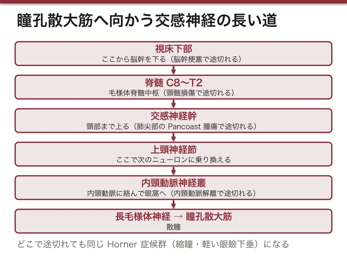
瞳孔の左右差は「どちらが異常か」を明暗で決める。縮瞳は副交感、散瞳は交感が担うので、片側だけ異常なら明るさを変えて比べる。暗い所で左右差が大きくなるなら、散大できない小さいほうが異常(交感の障害=Horner 症候群)。明るい所で差が大きくなるなら、縮瞳できない大きいほうが異常(副交感の障害=動眼神経麻痺)である。
交感神経の道は長い。視床下部から脳幹を下り、脊髄の C8〜T2(毛様体脊髄中枢)を出て、交感神経幹を頸部まで上り、上頸神経節から内頸動脈に絡んで眼窩へ入る。途中のどこが壊れても Horner 症候群になるので、脳幹梗塞・頸髄損傷・肺尖部の腫瘍(Pancoast 腫瘍)・内頸動脈解離が同じ瞳孔所見を出す。
毛様体輪(pars plana)の内面には光を受ける網膜が無い。そのため硝子体手術の器具は、角膜輪部から約3.5〜4 mm 後ろのこの部分から眼内に入れる。
動眼神経麻痺で瞳孔を見る理由。瞳孔へ向かう副交感線維は動眼神経の表面(上内側)を走る。外から押す病変——とくに内頸動脈・後交通動脈分岐部の動脈瘤——では早くから散瞳が出る。糖尿病などによる神経の内部の虚血では中心部が傷み、表面の線維が残って瞳孔は保たれやすい。「散瞳を伴う動眼神経麻痺は、動脈瘤を除外するまで緊急」とされるのはこのためである。
脈絡膜・毛様体・虹彩は一続きの層なので、炎症も広がる(ぶどう膜炎)。日本ではサルコイドーシス・Behçet 病・Vogt-小柳-原田病が三大原因とされ、全身疾患を探す入口になる。
English version
Core
The middle coat, rich in vessels, pigment cells and smooth muscle. From back to front it has three continuous parts: choroid, ciliary body and iris.
The choroid lies between sclera and retina and feeds the outer retina (photoreceptors). The red eye in photos is light reflected from this blood-rich layer. The ciliary body is a thick ring in front of it, triangular in section. Its flat back part is the orbiculus ciliaris (pars plana); its front part is the corona ciliaris (pars plicata), which carries about 70–80 ciliary processes. Their epithelium makes aqueous humour, and zonular fibres run from them to the lens. Its base is the smooth ciliary muscle (meridional, radial and circular fibres). The iris is a disc in the frontal plane with a central hole, the pupil (about 1–8 mm). Its colour depends on melanin. It has a greater and a lesser arterial circle.
| Smooth muscle | Fibres | Action | Nerve supply |
|---|---|---|---|
| M. sphincter pupillae | circular, at pupil margin | constricts pupil | parasympathetic: III → ciliary ganglion → short ciliary nerves |
| M. dilatator pupillae | radial | dilates pupil | sympathetic: superior cervical ganglion → internal carotid plexus → long ciliary nerves |
| M. ciliaris | meridional, radial, circular | accommodation (near focus) | parasympathetic (same route as sphincter) |
Blood comes from branches of the ophthalmic artery: short posterior ciliary arteries to the choroid; long posterior and anterior ciliary arteries to the greater arterial circle of the iris. Veins leave through the vorticose veins (usually four) to the ophthalmic veins.
Going further
Use light and dark to decide which pupil is abnormal. Constriction is parasympathetic and dilation is sympathetic. If the difference grows in the dark, the smaller pupil is abnormal — it cannot dilate (sympathetic lesion, Horner syndrome). If it grows in bright light, the larger pupil is abnormal — it cannot constrict (parasympathetic lesion, oculomotor palsy).
The sympathetic route is long: from the hypothalamus down the brainstem to C8–T2, out to the sympathetic trunk, up to the superior cervical ganglion, and along the internal carotid artery to the orbit. A lesion anywhere on it gives Horner syndrome, so brainstem stroke, cervical cord injury, an apical lung tumour (Pancoast) and carotid dissection can all give the same pupil.
The pars plana has no light-sensitive retina on its inner surface. For this reason, vitrectomy instruments enter the eye here, about 3.5–4 mm behind the limbus.
In the clinic
Why check the pupil in oculomotor palsy. The parasympathetic pupil fibres run on the surface of the nerve. Compression from outside — especially an aneurysm at the internal carotid–posterior communicating junction — dilates the pupil early. Ischaemia inside the nerve, as in diabetes, damages the core and often spares the pupil. So an oculomotor palsy with a dilated pupil is treated as an emergency until an aneurysm is excluded.
Because choroid, ciliary body and iris form one continuous layer, inflammation spreads through it (uveitis). In Japan the three main causes are sarcoidosis, Behçet disease and Vogt–Koyanagi–Harada disease, so uveitis is a door to systemic disease.
網膜と視神経 — 見える部分・見えない部分・出口Retina et nervus opticus
網膜は、発生のときに間脳の壁が眼杯として外へ張り出したもので、脳の一部である。鋸状縁 ora serrataを境に、光を受ける網膜視部 pars optica retinaeと、視細胞を持たない網膜盲部 pars caeca retinaeに分かれる。盲部は毛様体の内面を覆う毛様体部 pars ciliaris と、虹彩の裏を覆う虹彩部 pars iridica からなる。
| 目印 | 場所と中身 |
|---|---|
| 視神経円板 discus nervi optici | 神経節細胞の軸索が集まって眼球を出る所。視細胞が無いので盲点になる。中央のくぼみ excavatio disci から網膜中心動静脈が出入りする |
| 黄斑 macula lutea | 円板の約4 mm 外側(耳側)。錐体が最も密な領域 |
| 中心窩 fovea centralis | 黄斑の中央のくぼみ。錐体だけが並び、視力が最も高い |
視細胞は杆体(約1億個、暗い所・明暗)と錐体(約600万個、明るい所・色。光の波長の好みが違う3種類)である。10層の構造と3つのニューロンの並びは網膜(組織学)にある。
視神経 n. opticusは眼球から視交叉まで約4.5〜5 cm。眼窩の中をゆるくたわんで走り、視神経管を眼動脈と一緒に通って頭蓋内へ入る。外鞘は硬膜の続き、内鞘はくも膜・軟膜の続きで、その間には脳のくも膜下腔がそのまま続いている。網膜中心動脈 a. centralis retinae は眼動脈の枝で、眼球の後ろ約1 cm で視神経に入り、その中心を通って円板から網膜に広がる。
視神経は「末梢神経」ではない。脳の一部が伸びたものなので、髄鞘を作るのはオリゴデンドロサイトで、切れると再生しない。多発性硬化症や視神経脊髄炎で視神経炎が起こるのも、中枢神経の病気が中枢神経の一部を襲っているからである。
網膜中心動脈は終動脈で、詰まると網膜の内層が白くむくむ。中心窩には内層が無いので、その部分だけ下の脈絡膜の赤が透けて見える——桜実紅斑 cherry-red spotである。網膜の内層は網膜中心動脈、外層(視細胞)は脈絡膜、という二重の血液供給がこの所見を作っている。毛様体の血管から黄斑へ枝(毛様網膜動脈)を持つ人では、閉塞しても中心の視力が残ることがある。
うっ血乳頭:頭蓋内圧が上がると、視神経のまわりのくも膜下腔を通って圧が眼球の後ろまで伝わり、軸索の流れと静脈の戻りが円板で滞って乳頭が腫れる。両側に出て、初めのうちは視力が保たれる。眼底を見るだけで頭蓋内圧の上昇が分かるのは、視神経鞘が脳の膜の続きだからである。網膜中心動脈閉塞症は突然の無痛性の視力低下で、数時間で網膜が回復しなくなるため、眼科の緊急疾患として扱う。
English version
Core
The retina develops from the wall of the diencephalon that grows out as the optic cup, so it is part of the brain. At the ora serrata it divides into the light-sensitive pars optica and the pars caeca, which has no photoreceptors. The pars caeca covers the ciliary body (pars ciliaris) and the back of the iris (pars iridica).
| Landmark | Position and content |
|---|---|
| Optic disc (discus nervi optici) | where ganglion cell axons leave the eye. No photoreceptors, so it is the blind spot. Central retinal vessels enter and leave at its central cup |
| Macula lutea | about 4 mm lateral (temporal) to the disc. The densest area of cones |
| Fovea centralis | the pit in the centre of the macula. Only cones; the sharpest vision |
Photoreceptors are rods (about 100 million; dim light, black and white) and cones (about 6 million; bright light and colour; three types).
The optic nerve runs about 4.5–5 cm from the eye to the chiasm. It is slack in the orbit and passes through the optic canal with the ophthalmic artery. Its outer sheath continues the dura, its inner sheath the arachnoid and pia, and between them the subarachnoid space of the brain continues. The central retinal artery, a branch of the ophthalmic artery, enters the nerve about 1 cm behind the eye and reaches the retina at the disc.
Going further
The optic nerve is not a peripheral nerve. It is an extension of the brain: oligodendrocytes make its myelin, and it does not regenerate. Optic neuritis in multiple sclerosis and neuromyelitis optica is a CNS disease attacking a part of the CNS.
The central retinal artery is an end artery. When it is blocked, the inner retina becomes white and swollen. The fovea has no inner layers, so the red choroid shows through there — the cherry-red spot. The inner retina is fed by the central retinal artery and the outer retina by the choroid; this double supply creates the sign. People with a cilioretinal artery may keep central vision.
In the clinic
Papilloedema: raised intracranial pressure travels along the subarachnoid space around the optic nerve to the back of the eye. Axonal flow and venous return are blocked at the disc, and the disc swells — on both sides, with vision kept at first. We can see raised intracranial pressure in the fundus because the nerve sheath is a continuation of the meninges. Central retinal artery occlusion causes sudden painless loss of vision and is an eye emergency, because the retina cannot recover after a few hours.
水晶体・硝子体・眼房 — 眼の中身と調節Lens et corpus vitreum
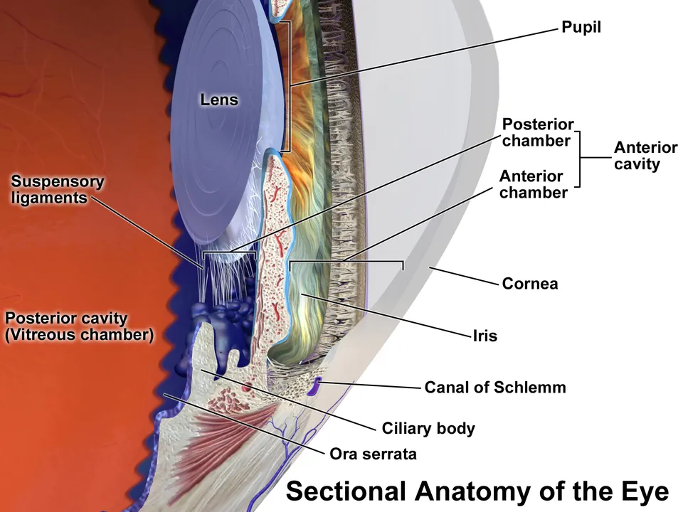
水晶体 lensは虹彩の後ろ、硝子体の前にある両凸のレンズで、前極・後極・その間の水晶体軸・まわりの赤道を区別する。血管も神経も無い。弾力のある水晶体包 capsula lentisに包まれ、水晶体上皮は前面にしか無い。上皮は赤道で新しい水晶体線維を一生作り続け、古い線維は中心へ押し込まれて硬い水晶体核 nucleus lentisとなり、まわりを柔らかい皮質 cortex lentis が囲む。水晶体は毛様体小帯 zonula ciliaris(Zinn 小帯)で毛様体突起から吊られている。小帯は細い小帯線維 fibrae zonulares の集まりで、線維の間の隙間を小帯隙 spatia zonularia という。
硝子体 corpus vitreumは眼球の後ろ約4/5 を満たすゼリーで、99% が水、残りはコラーゲン線維とヒアルロン酸である。硝子体膜に包まれ、前面の硝子体窩 fossa hyaloidea に水晶体がはまる。中を前後に走る硝子体管 canalis hyaloideus は、胎児期の硝子体動脈の通り道の名残である。
眼房は2つ。前眼房 camera anteriorは角膜と虹彩の間、後眼房 camera posteriorは虹彩と水晶体・毛様体小帯の間である。房水は毛様体突起 → 後眼房 → 瞳孔 → 前眼房 → 虹彩角膜角(隅角)angulus iridocornealis → 強膜静脈洞 → 眼の静脈、と流れ、角膜と水晶体を養い、眼圧で眼球の形を保つ。作る細胞と緑内障の型は隅角と房水にある。
調節は「筋が縮むとレンズが解放される」仕組みで、直感と逆である。近くを見るとき毛様体筋が収縮すると、毛様体の輪が小さくなって水晶体に近づき、小帯がゆるむ。引っ張りが消えた水晶体は、包の弾力で自ら丸くなり、屈折力が増す。遠くを見るときは筋がゆるみ、輪が広がって小帯が張り、水晶体は平たく引かれる。
老視はこの仕組みから説明できる。水晶体線維は捨てられずに中心へたまり続けるので、年齢とともに水晶体が硬くなる。筋が縮んでも丸くなれず、40歳代から近くにピントが合いにくくなる。
瞳孔は房水の唯一の通り道である。虹彩が水晶体に張りつくと(虹彩後癒着、あるいは前房の浅い眼での瞳孔ブロック)房水は後眼房に閉じこめられ、虹彩を前へ押してふくらませる(膨隆虹彩 iris bombé)。ふくらんだ虹彩の根元が隅角を塞ぎ、閉塞隅角緑内障になる。レーザーで虹彩に小さな穴を開けると後眼房から前眼房への近道ができ、発作が防げるのはこのためである。
白内障手術は水晶体包を「袋」として使う。前面の包を丸く切り取り、中身を超音波で砕いて吸い出し、残した包の中に眼内レンズを入れる。小帯が袋の吊り紐として残るので、レンズが正しい位置に保たれる。数か月〜数年後に残った上皮が後面の包で増えて再び濁ること(後発白内障)があり、レーザーで後面の包に穴を開けて治す。
硝子体は加齢で液化して縮み、網膜から離れる(後部硝子体剥離)。このとき見える浮遊物が飛蚊症である。網膜と強く付いた場所で網膜が引き裂かれると網膜裂孔となり、そこから網膜剥離へ進むことがある。飛蚊症が急に増えたり光が走って見えたりしたら、眼底の検査が必要になる。
English version
Core
The lens is a biconvex lens behind the iris and in front of the vitreous. We name its anterior and posterior poles, its axis and its equator. It has no vessels or nerves. It is enclosed in an elastic capsule, and the lens epithelium is only on the front surface. At the equator the epithelium makes new lens fibres all through life; old fibres are pushed to the centre and form the hard nucleus, surrounded by the softer cortex. The lens hangs from the ciliary processes by the ciliary zonule (zonule of Zinn), made of fine zonular fibres with spaces between them.
The vitreous body fills the back four-fifths of the eye. It is a gel of 99% water with collagen fibres and hyaluronic acid, inside a thin membrane. The lens sits in the hyaloid fossa at its front. The hyaloid canal through its centre is the path of the fetal hyaloid artery.
There are two chambers: the anterior chamber between cornea and iris, and the posterior chamber between iris and lens/zonule. Aqueous humour flows: ciliary processes → posterior chamber → pupil → anterior chamber → iridocorneal angle → canal of Schlemm → veins. It feeds the cornea and lens, and its pressure keeps the shape of the eye.
Going further
In accommodation, contracting the muscle releases the lens — the opposite of what you expect. For near vision the ciliary muscle contracts, the ring of the ciliary body becomes smaller, and the zonule relaxes. Free from tension, the lens becomes rounder by the elasticity of its capsule, and its power increases. For distance the muscle relaxes, the ring widens, the zonule tightens and the lens is pulled flat.
Presbyopia follows from this. Lens fibres are never removed and collect in the centre, so the lens hardens with age. Even when the muscle contracts, the lens cannot round up, and near focus becomes difficult from the forties.
The pupil is the only path for aqueous humour. If the iris sticks to the lens (posterior synechiae, or pupillary block in an eye with a shallow anterior chamber), fluid is trapped in the posterior chamber and pushes the iris forward (iris bombé). The root of the iris then closes the angle, causing angle-closure glaucoma. A small laser hole in the iris makes a short cut from the posterior to the anterior chamber; that is why laser iridotomy prevents attacks.
In the clinic
Cataract surgery uses the capsule as a bag. The surgeon opens the front of the capsule in a circle, breaks up and removes the lens contents with ultrasound, and places an artificial lens inside the remaining capsule. The zonule still holds the bag in place. Months or years later, remaining epithelial cells may grow on the back of the capsule and cloud it again (posterior capsule opacification); a laser opening in the posterior capsule treats it.
With age the vitreous liquefies, shrinks and separates from the retina (posterior vitreous detachment); the floating shadows are floaters. Where the vitreous is firmly attached, it can tear the retina, and a retinal tear can lead to detachment. A sudden increase in floaters or flashes of light needs a fundus examination.
眼の付属器 — 眼瞼・結膜・涙器Palpebrae et apparatus lacrimalis
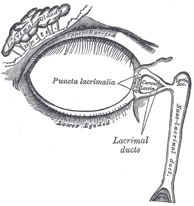
付属器 structurae oculi accessoriae は、眼球を守るものと動かすもの(外眼筋、次の項目)に分かれる。眼窩の中では、骨膜にあたる眼窩骨膜 periorbitaが壁を覆い、眼窩脂肪体 corpus adiposum orbitaeがすき間を埋める。眼球は眼球鞘 vagina bulbi(Tenon 嚢)という結合組織の袋に包まれ、脂肪から隔てられて、関節の受け皿のように滑らかに回る。
眼瞼 palpebraeは上下2枚で、間のすき間が眼瞼裂 rima palpebrarum、両端が内眼角・外眼角である。芯は密な結合組織の板瞼板 tarsusで、内側・外側眼瞼靱帯と眼窩隔膜 septum orbitaleで眼窩縁につながる。前から皮膚・眼輪筋・瞼板・瞼結膜と重なる。自由縁は、睫毛 cilia が2〜3列に生える前縁 limbus anterior と、瞼板腺の口が並ぶ後縁 limbus posterior に分かれる。上眼瞼を上げる筋は2つある。動眼神経が支配する骨格筋の上眼瞼挙筋 m. levator palpebrae superiorisと、その下に付く平滑筋で交感神経が支配する上瞼板筋 m. tarsalis superior(Müller 筋)である。
結膜 tunica conjunctivaは眼瞼の裏を覆う瞼結膜と、強膜の前面を角膜の縁まで覆う眼球結膜からなり、折り返しが上・下の結膜円蓋 fornix である。閉眼すると袋(結膜嚢)になる。内眼角には結膜半月ひだ plica semilunaris と涙丘 caruncula lacrimalis がある。
涙器 apparatus lacrimalis:涙腺 glandula lacrimalisは眼窩の上外側(前頭骨の涙腺窩)にあり、上眼瞼挙筋の腱膜で眼窩部と眼瞼部に分けられる。6〜12本の導管が上結膜円蓋の外側に開く。涙は眼の表面を内下方へ流れ、涙河 rivus lacrimalis → 涙湖 lacus lacrimalis → 涙乳頭の涙点(上下2つ)→ 涙小管(2本)→ 涙嚢(1つ)→ 鼻涙管(1本)→ 下鼻道へ出る。
涙は流れるのではなく、汲み出されている。眼輪筋の涙嚢部は涙嚢のまわりに付き、瞬きのたびに涙嚢を広げたり縮めたりして、涙点から涙を吸い込む。顔面神経麻痺で涙がこぼれる(流涙)のは涙が増えたからではなく、閉瞼とこのポンプが止まり、下眼瞼が外へ垂れて涙点が涙湖から離れるからである。
涙を出させる神経は顔面神経の副交感線維である。上唾液核 → 中間神経 → 膝神経節を素通り → 大錐体神経 → 翼突管神経 → 翼口蓋神経節(ここで乗り換え)→ 上顎神経の枝(頬骨神経)→ 涙腺神経との交通 → 涙腺、と大回りする。顔面神経麻痺で涙の量を測る(Schirmer 試験)と、障害が膝神経節より中枢側(涙が減る)か末梢側(涙は保たれる)かを推定できる。
眼瞼下垂は、どの筋が働かないかで程度が違う。動眼神経麻痺では上眼瞼挙筋が止まり、ほぼ完全な下垂になる(散瞳と外下方への偏位を伴う)。Horner 症候群では Müller 筋だけが止まるので、1〜2 mm の軽い下垂にとどまり、縮瞳を伴う。高齢者に多いのは挙筋の腱膜が瞼板から外れる腱膜性眼瞼下垂である。瞼板腺・睫毛腺の病気(霰粒腫・麦粒腫)と涙の3層、鼻涙管閉塞は眼瞼と涙器(組織学)にある。
English version
Core
The accessory structures either protect the eye or move it (the extraocular muscles, next item). In the orbit the periorbita lines the bony walls and the orbital fat body fills the space. The eyeball sits in a connective tissue sac, the fascial sheath (Tenon’s capsule), which separates it from the fat and lets it turn like a ball in a socket.
The eyelids are upper and lower; the gap between them is the palpebral fissure, with medial and lateral angles. The core of each lid is a dense connective tissue plate, the tarsus, attached to the orbital margin by the medial and lateral palpebral ligaments and the orbital septum. From front to back: skin, orbicularis oculi, tarsus, palpebral conjunctiva. The free margin has an anterior lip with 2–3 rows of eyelashes and a posterior lip with the openings of the tarsal glands. The upper lid is raised by the levator palpebrae superioris (oculomotor nerve) and by the smooth superior tarsal muscle (Müller’s muscle, sympathetic) below it.
The conjunctiva lines the back of the lids and covers the front of the sclera up to the cornea; the folds between are the upper and lower fornices. With the lids closed it forms the conjunctival sac. At the medial angle are the plica semilunaris and the lacrimal caruncle.
Lacrimal apparatus: the lacrimal gland lies in the upper lateral orbit (lacrimal fossa of the frontal bone) and is divided by the levator aponeurosis into orbital and palpebral parts. Its 6–12 ducts open into the lateral upper fornix. Tears flow medially and downward: lacrimal stream → lacrimal lake → puncta (2) → canaliculi (2) → lacrimal sac (1) → nasolacrimal duct (1) → inferior nasal meatus.
Going further
Tears are pumped, not just drained. The lacrimal part of orbicularis oculi lies around the lacrimal sac and opens and squeezes it with each blink, sucking tears into the puncta. In facial palsy tears overflow not because more tears are made, but because lid closure and this pump stop, and the lower lid falls away so the punctum leaves the tear lake.
The secretory nerve to the gland is parasympathetic from the facial nerve: superior salivatory nucleus → nervus intermedius → through the geniculate ganglion without synapse → greater petrosal nerve → nerve of the pterygoid canal → pterygopalatine ganglion (synapse) → zygomatic branch of the maxillary nerve → communication with the lacrimal nerve → gland. Measuring tears in facial palsy (Schirmer test) helps decide whether the lesion is proximal to the geniculate ganglion (less tears) or distal (tears preserved).
In the clinic
Ptosis depends on which muscle fails. In oculomotor palsy the levator stops and the ptosis is almost complete (with a dilated pupil and the eye turned down and out). In Horner syndrome only Müller’s muscle stops, so the ptosis is mild (1–2 mm) and the pupil is small. In older people the common type is aponeurotic ptosis, where the levator aponeurosis separates from the tarsus. Diseases of the lid glands, the three layers of the tear film and nasolacrimal obstruction are covered in the histology chapter.
外眼筋と眼窩の血管・神経 — 作用は眼窩の向きから導くMusculi bulbi; vasa et nervi orbitae
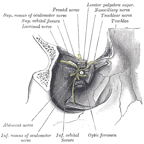
眼窩の筋は7つ:上眼瞼挙筋、直筋4つ(上・下・内側・外側)、斜筋2つ(上・下)。直筋4つは視神経管を囲む線維の輪総腱輪 anulus tendineus communisから起こり、前へ広がって視神経を包む筋円錐をつくり、角膜輪部から数 mm 後ろ(赤道より前)の強膜に付く。上斜筋は視神経管の上内側(蝶形骨体)から前へ進み、軟骨の滑車 trochleaで後外側へ向きを変えて赤道の後ろに付く。下斜筋だけは眼窩底の前内側(上顎骨)から起こる。支配は「外側直筋=外転神経 VI、上斜筋=滑車神経 IV、残りは動眼神経 III」。1つずつの起始・停止は上直筋から下斜筋までの項目にある。
血管:眼動脈 a. ophthalmicaは内頸動脈の枝で、視神経管を視神経と一緒に通り、網膜中心動脈・毛様体動脈・涙腺動脈・筋枝・眼窩上動脈・篩骨動脈などを出す。静脈は上・下眼静脈に集まり、上眼窩裂を通って海綿静脈洞へ注ぐ。眼静脈には弁が無く、顔の眼角静脈ともつながる。
| 通り道 | 通るもの |
|---|---|
| 視神経管 canalis opticus | 視神経 II・眼動脈 |
| 上眼窩裂のうち総腱輪の内側 | 動眼神経 III(上枝・下枝)・外転神経 VI・鼻毛様体神経(V1) |
| 上眼窩裂のうち総腱輪の外側 | 滑車神経 IV・前頭神経(V1)・涙腺神経(V1)・上眼静脈 |
視神経の外側には、瞳孔括約筋と毛様体筋へ行く副交感線維の乗り換え所毛様体神経節 ganglion ciliareがある。骨の孔そのものは上眼窩裂を参照。
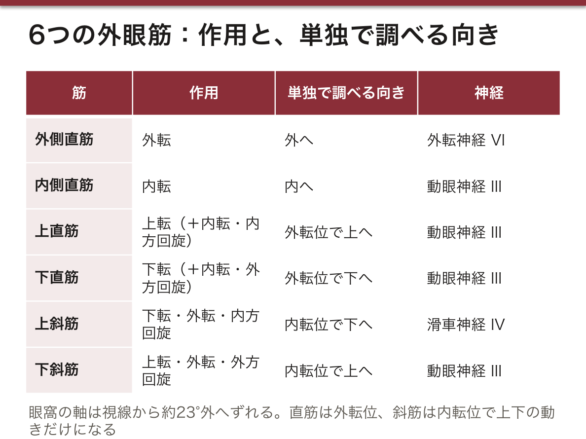
外眼筋の作用は、眼窩の向きと視線のずれから導ける。眼窩の軸は前外側へ開いていて、まっすぐ前を見たときの視線と約23°ずれている。上直筋・下直筋は眼窩の奥(内側寄り)から眼窩の軸に沿って引くので、上げる・下げるに加えて内転と回旋が混じる。眼を約23°外転させると筋の引く向きと視線がそろい、純粋な上転・下転になる。
斜筋は前内側(滑車と眼窩底の前内側)から、赤道より後ろの強膜を引く。眼球の後ろ半分が引かれると、前の角膜は反対へ傾く。上斜筋は後ろの上部を前内側へ引くので角膜は下・外を向き(下転・外転・内方回旋)、下斜筋は上・外を向かせる(上転・外転・外方回旋)。眼を内転させると斜筋の引く向きと視線がそろうので、斜筋は内転位で上下の動きを調べる。診察で「H の字」に眼を動かしてもらうのは、6つの筋がそれぞれ単独で働く方向を順に確かめるためである。ヨーロッパの教科書では、上直筋・下直筋が「角膜を内側へも向ける」ことを、付着が眼球の垂直軸より内側にあるため、と説明している。
総腱輪の内か外かが、麻酔と病変の出方を決める。眼の手術で筋円錐の中に麻酔薬を入れる(球後麻酔)と、輪の内側を通る III と VI が効いて眼球が止まるが、輪の外を通る滑車神経は効きにくく、上斜筋の動きが残ることがある。
反対に、上眼窩裂や海綿静脈洞の病変(腫瘍・炎症・血栓)では III・IV・VI・V1 がまとめて障害され、眼球がほとんど動かなくなり、額と角膜の感覚が落ちる。顔の中央(いわゆる危険三角)の感染が眼角静脈 → 眼静脈 → 海綿静脈洞へ逆流して海綿静脈洞血栓症を起こしうるのは、眼静脈に弁が無いためである。
English version
Core
There are seven muscles in the orbit: levator palpebrae superioris, four recti (superior, inferior, medial, lateral) and two obliques. The four recti arise from a fibrous ring around the optic canal, the common tendinous ring, spread forward as a muscle cone around the optic nerve, and insert on the sclera a few millimetres behind the limbus (in front of the equator). The superior oblique runs forward from above and medial to the optic canal, turns back and laterally through the cartilage trochlea, and inserts behind the equator. Only the inferior oblique arises from the front of the orbital floor (maxilla). Innervation: lateral rectus = abducens (VI), superior oblique = trochlear (IV), the rest = oculomotor (III).
Vessels: the ophthalmic artery, a branch of the internal carotid, passes through the optic canal with the optic nerve and gives the central retinal, ciliary, lacrimal, muscular, supraorbital and ethmoidal arteries. Veins collect into the superior and inferior ophthalmic veins, pass through the superior orbital fissure and drain into the cavernous sinus. The ophthalmic veins have no valves and connect with the angular vein of the face.
| Passage | Contents |
|---|---|
| Optic canal | optic nerve (II), ophthalmic artery |
| Superior orbital fissure, inside the ring | oculomotor (III, both divisions), abducens (VI), nasociliary nerve (V1) |
| Superior orbital fissure, outside the ring | trochlear (IV), frontal and lacrimal nerves (V1), superior ophthalmic vein |
Lateral to the optic nerve lies the ciliary ganglion, where parasympathetic fibres to the sphincter pupillae and ciliary muscle synapse.
Going further
You can work out the actions from the angle between the orbit and the line of sight. The orbit opens forward and laterally; its axis is about 23° from the line of sight in straight-ahead gaze. The superior and inferior recti pull along the orbital axis from the back, so besides elevation and depression they also adduct and rotate the eye. When the eye is abducted about 23°, the pull and the line of sight match, and the action becomes pure elevation or depression.
The obliques pull from the front and medial side on the sclera behind the equator. When the back half is pulled, the cornea tips the other way. The superior oblique pulls the upper back part forward and medially, so the cornea turns down and out (depression, abduction, intorsion). The inferior oblique turns it up and out (elevation, abduction, extorsion). In adduction the obliques line up with the line of sight, so they are tested in adduction. The “H” pattern of eye movements in the clinic tests each muscle in the direction where it works alone. European textbooks explain that the superior and inferior recti also turn the cornea medially because they insert medial to the vertical axis of the eye.
In the clinic
Inside or outside the ring decides how anaesthesia and lesions show. In retrobulbar anaesthesia, the drug in the muscle cone blocks III and VI, which run inside the ring, and the eye stops moving; the trochlear nerve runs outside the ring, so superior oblique movement may remain.
In contrast, lesions of the superior orbital fissure or cavernous sinus (tumour, inflammation, thrombosis) damage III, IV, VI and V1 together: the eye hardly moves, and sensation of the forehead and cornea is lost. Infection in the central face (the danger triangle) can spread via the angular vein and ophthalmic veins to the cavernous sinus because these veins have no valves.
視覚路と瞳孔の反射 — 対光反射と近見反応Via optica
視覚路はニューロンを4つ継ぐと数える(ヨーロッパの教科書の数え方。日本の本では網膜の中の2つをまとめて数えることもあるが、乗り換えの場所は同じ)。
| ニューロン | 場所 | 軸索の行き先 |
|---|---|---|
| 1 | 視細胞(杆体・錐体) | 双極細胞 |
| 2 | 双極細胞 | 神経節細胞 |
| 3 | 網膜神経節細胞 | 視神経 → 視交叉 → 視索 → 外側膝状体 |
| 4 | 外側膝状体(視床) | 視放線 → 鳥距溝の両岸(一次視覚野、17野) |
視交叉では鼻側の網膜からの線維だけが交叉し、耳側の線維は同じ側を進む。そのため視索より後ろは、左右の眼の「同じ側の視野」を運ぶ。視放線は内包後脚の後部を通って後頭葉へ向かう。
視索の線維の一部は外側膝状体に入らず、上丘腕を通って中脳の視蓋前域 area pretectalisと上丘へ行く。ここが反射の入口である。上丘から出る視蓋脊髄路は中脳被蓋で交叉して頸髄の前角へ下り、急に視野に入ったものへ頭と眼を向ける反射を担う。
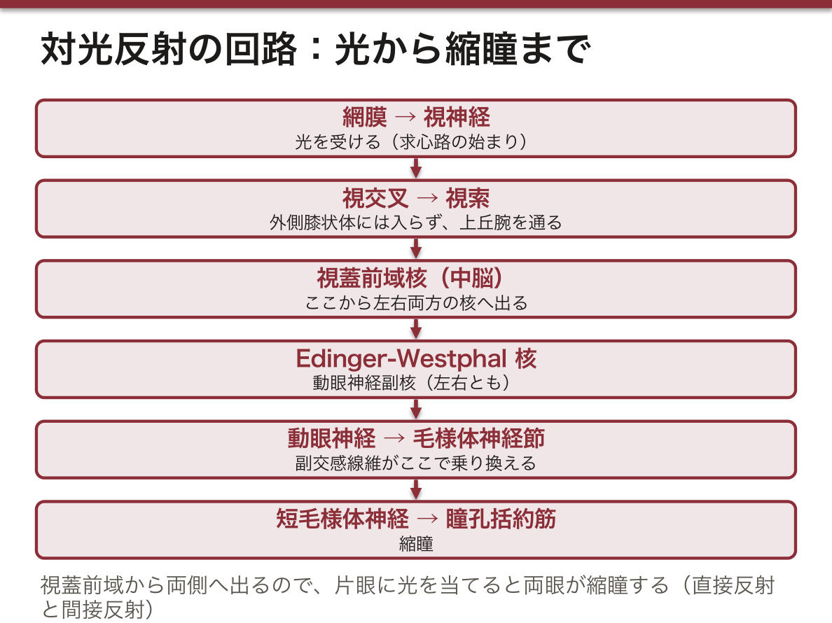
対光反射の回路:網膜 → 視神経 → 視交叉 → 視索 →(上丘腕)→ 視蓋前域核 → 左右両方の Edinger-Westphal 核(動眼神経副核)→ 動眼神経 → 毛様体神経節(乗り換え)→ 短毛様体神経 → 瞳孔括約筋。視蓋前域から両側へ出るので、片眼に光を当てると両眼が縮瞳する(直接反射と間接反射)。ヨーロッパの教科書には中継を上丘として書くものもあるが、現在の標準は視蓋前域である。
この回路から所見が読める。片眼の視神経が傷むと、その眼に光を当てても両眼とも縮瞳が弱く、反対の眼に当てると両眼とも縮瞳する。光を左右交互に当てると、傷んだ眼に移った瞬間に両眼の瞳孔が開く(相対的瞳孔求心路障害 RAPD、Marcus Gunn 瞳孔)。動眼神経が傷んだ眼は、どちらから光を当てても縮瞳しない。入力の障害は左右の瞳孔の大きさを変えず、出力の障害は変えるのである。
近見反応:近くを見ると、輻輳(両側の内側直筋)・調節(毛様体筋)・縮瞳の3つが同時に起こる。こちらは視覚野で「ぼやけている」と判断してから中脳の核へ下りる経路を使い、視覚野を通らない対光反射とは道が違う。
視放線のうち下方の網膜(=上方の視野)からの線維は、側頭葉で側脳室下角の前へ大きく回り込む(Meyer ループ)。側頭葉の病変では反対側の上1/4が、頭頂葉側の線維の病変では下1/4が欠ける。
English version
Core
The visual pathway is counted as a chain of four neurons (the European way; some Japanese books count the two retinal neurons together, but the relay sites are the same).
| Neuron | Location | Axons go to |
|---|---|---|
| 1 | photoreceptors (rods, cones) | bipolar cells |
| 2 | bipolar cells | ganglion cells |
| 3 | retinal ganglion cells | optic nerve → chiasm → optic tract → lateral geniculate body |
| 4 | lateral geniculate body (thalamus) | optic radiation → banks of the calcarine sulcus (primary visual cortex, area 17) |
At the chiasm only fibres from the nasal retina cross; temporal fibres stay on the same side. So behind the chiasm each optic tract carries the same half of the visual field from both eyes. The optic radiation passes through the back of the posterior limb of the internal capsule to the occipital lobe.
Some optic tract fibres do not enter the lateral geniculate body but go through the brachium of the superior colliculus to the pretectal area and superior colliculus of the midbrain — the entry to the reflexes. The tectospinal tract from the superior colliculus crosses in the midbrain tegmentum and runs to the anterior horn of the cervical cord; it turns the head and eyes toward something that suddenly appears.
Going further
Pupillary light reflex: retina → optic nerve → chiasm → optic tract → (brachium of superior colliculus) → pretectal nucleus → Edinger–Westphal nuclei on both sides → oculomotor nerve → ciliary ganglion (synapse) → short ciliary nerves → sphincter pupillae. Because the pretectal area projects to both sides, light in one eye makes both pupils constrict (direct and consensual reflex). Some European textbooks give the superior colliculus as the relay; the current standard is the pretectal area.
Findings follow from this circuit. If one optic nerve is damaged, light in that eye gives a weak response in both eyes, and light in the other eye constricts both. In the swinging-flashlight test both pupils dilate when the light moves to the damaged eye (relative afferent pupillary defect, Marcus Gunn pupil). An eye with oculomotor damage does not constrict whichever eye is lit. Afferent lesions do not make the pupils unequal; efferent lesions do.
Near response: looking at something near causes convergence (both medial recti), accommodation (ciliary muscle) and miosis together. It uses a route through the visual cortex, which judges that the image is blurred, and then down to the midbrain nuclei. The light reflex does not pass through the cortex.
Optic radiation fibres from the lower retina (upper visual field) loop forward around the temporal horn in the temporal lobe (Meyer’s loop). A temporal lobe lesion causes loss of the opposite upper quadrant; a lesion of the parietal fibres causes loss of the lower quadrant.
In the clinic
The two separate routes explain light–near dissociation. The Argyll Robertson pupil of neurosyphilis is small, does not react to light, but constricts for near vision; the lesion is thought to be near the dorsal pretectal area, sparing the more ventral near pathway. In contrast, in cortical blindness from damage to both occipital lobes the patient cannot see, but the light reflex is kept. In unconscious patients the pupils show the level of brainstem damage, and a unilateral dilated pupil is the first sign of uncal herniation compressing the oculomotor nerve.
まとめ
| 眼球 — 外形・3本の軸・3枚の膜 | 眼球は眼窩 orbita の前半分に収まる、重さ約7.5 g のほぼ球形の器官である。 |
|---|---|
| 線維膜 — 角膜と強膜 | 眼球のいちばん外の膜で、前の約1/6 が角膜 cornea、残りが強膜 scleraである。 |
| 血管膜(ぶどう膜)— 脈絡膜・毛様体・虹彩 | 血管と色素細胞と平滑筋に富む中間の膜で、後ろから脈絡膜・毛様体・虹彩の3部が一続きになっている。 |
| 網膜と視神経 — 見える部分・見えない部分・出口 | 網膜は、発生のときに間脳の壁が眼杯として外へ張り出したもので、脳の一部である。 |
| 水晶体・硝子体・眼房 — 眼の中身と調節 | 水晶体 lensは虹彩の後ろ、硝子体の前にある両凸のレンズで、前極・後極・その間の水晶体軸・まわりの赤道を区別する。 |
| 眼の付属器 — 眼瞼・結膜・涙器 | 付属器 structurae oculi accessoriae は、眼球を守るものと動かすもの(外眼筋、次の項目)に分かれる。 |
| 外眼筋と眼窩の血管・神経 — 作用は眼窩の向きから導く | 眼窩の筋は7つ:上眼瞼挙筋、直筋4つ(上・下・内側・外側)、斜筋2つ(上・下)。 |
| 視覚路と瞳孔の反射 — 対光反射と近見反応 | 視覚路はニューロンを4つ継ぐと数える(ヨーロッパの教科書の数え方。 |
このノートは医学生が授業の内容を自分の言葉でまとめたもので、診療の判断に使うものではありません。誤りに気づいたらお知らせください。