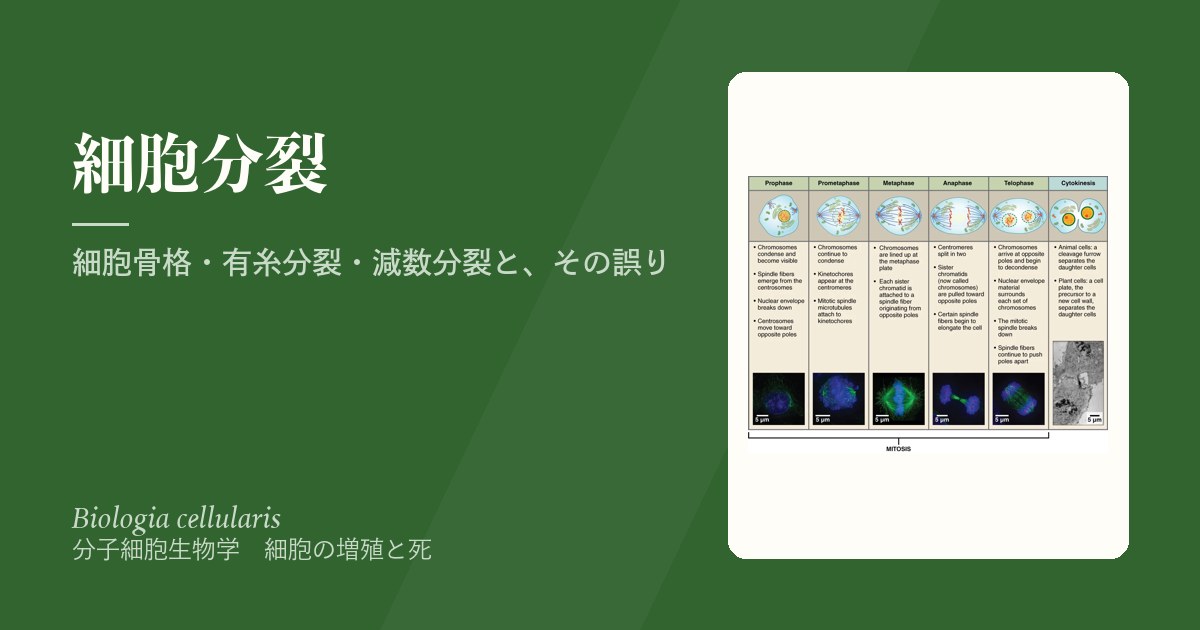
細胞分裂は、複製したDNAを2つの細胞に間違いなく分ける作業である。そのために細胞は、細胞骨格という「線路と綱」を組み替え、リン酸化という「スイッチ」で順番を守らせる。体細胞の有糸分裂は同じ細胞を2つ作り、生殖細胞の減数分裂は染色体の数を半分にしながら組み合わせを混ぜる。
この章では、細胞骨格とモータータンパク質から始めて、有糸分裂の各期と細胞質分裂、減数分裂の第一分裂前期の細かい段階までを追い、最後に分配の誤りが何を起こすか(トリソミー・モザイク・片親性ダイソミー・癌のゲノム不安定性)を見る。各期の出来事を暗記する前に、「なぜその順番でないといけないのか」を押さえると、全体が1本の筋でつながる。
- 細胞骨格の3種類 — 太さと材料で役割が分かれる
- モータータンパク質 — 線路の上を、決まった向きに歩く
- 有糸分裂の調節 — CDK・Plk・Aurora・APC/Cのリレー
- 前期と前中期 — 染色体を固め、核膜をほどき、紡錘体で捕まえる
- 中期・後期・終期 — 全員がそろうまで待ち、そろったら一斉に引く
- 細胞質分裂 — 動物はくびれ、植物は内側から壁を築く
- 減数分裂 — 複製1回・分裂2回で、数を半分にしつつ混ぜる
- 分裂の誤りと病気 — トリソミー・モザイク・片親性ダイソミー・癌
細胞骨格の3種類 — 太さと材料で役割が分かれる
細胞骨格は、細胞の形を保ち、動くときには形を変え、小器官の位置を決め、変形に耐え、荷物を運ぶ。3種類の線維からなる。
| 微小管 | 中間径フィラメント | アクチンフィラメント(微小線維) | |
|---|---|---|---|
| 太さ | 約25 nm(最も太い) | 約10 nm | 約7〜8 nm(最も細い) |
| 材料 | α・βチューブリン | 細胞の種類ごとに異なるタンパク質(ヒトで約70種) | アクチン |
| 特徴 | 中空の管。伸び縮みが速い | 丈夫で安定。動物細胞にある | 筋に特に多い。収縮・細胞質分裂 |
微小管は、αとβのチューブリンが縦につながったプロトフィラメントが、哺乳類では13本集まって筒をつくる。チューブリンが付けば伸び、外れれば縮む。微小管のマイナス端は微小管形成中心(MTOC)につなぎとめられ、その中心が中心体である。中心体は2個の中心小体と、その周りの100種以上のタンパク質からなる中心小体周辺物質でできており、S期に複製される。
なぜ微小管は、わざわざ伸び縮みを繰り返すのか。伸びては急に縮む性質(動的不安定性)のおかげで、微小管は細胞の中をあちこち「探る」ことができる。分裂のとき、伸びてきた微小管の先がたまたま染色体の動原体に当たると、そこで捕まえられて安定になる。当たらなかったものは縮んで、また別の方向へ伸びる。固定された線路では、どこにあるかわからない染色体を46本すべて捕まえることはできない。
中間径フィラメントは、上皮のケラチン、筋のデスミン、神経のニューロフィラメントのように、細胞の種類ごとに材料がちがう。核の内側のラミンも中間径フィラメントの仲間である。
中間径フィラメントが細胞の種類ごとに決まっていることは、腫瘍の由来を見分ける免疫染色に使われる。サイトケラチンが陽性なら上皮由来(癌腫)、デスミンなら筋由来、GFAPならグリア由来が疑われる。腫瘍の形だけでは区別できないとき、骨格の材料が「出身地」を教えてくれる。
English version
Core
The cytoskeleton keeps cell shape, changes shape during movement, places organelles, resists deformation and carries cargo. It is made of three kinds of filament.
| Microtubules | Intermediate filaments | Actin filaments (microfilaments) | |
|---|---|---|---|
| Diameter | About 25 nm (thickest) | About 10 nm | About 7–8 nm (thinnest) |
| Material | α- and β-tubulin | Proteins that differ by cell type (about 70 in humans) | Actin |
| Features | Hollow tube; grows and shrinks quickly | Strong and stable; found in animal cells | Very abundant in muscle; contraction, cytokinesis |
In a microtubule, α- and β-tubulin join end to end into protofilaments, and in mammals 13 of them form a tube. It grows when tubulin is added and shrinks when tubulin leaves. The minus end is anchored in a microtubule organising centre (MTOC); the main one is the centrosome. The centrosome is made of two centrioles and the pericentriolar material around them (over 100 proteins), and it is duplicated in S phase.
Going further
Why do microtubules keep growing and shrinking? Because they grow and then suddenly shrink (dynamic instability), they can “search” the cell. During division, if a growing tip happens to hit a chromosome’s kinetochore, it is caught and becomes stable. Tips that miss shrink and grow again in another direction. Fixed rails could never catch all 46 chromosomes when their positions are unknown.
Intermediate filaments are made of different materials in different cell types: keratin in epithelium, desmin in muscle, neurofilaments in neurons. The lamins inside the nucleus also belong to this family.
In the clinic
Because intermediate filaments depend on cell type, they are used in immunostaining to find where a tumour came from. Cytokeratin-positive suggests epithelial origin (carcinoma), desmin suggests muscle, GFAP suggests glia. When shape alone cannot tell, the skeleton’s material shows the tumour’s “home town”.
モータータンパク質 — 線路の上を、決まった向きに歩く
モータータンパク質は、ATPを分解して得たエネルギーで、細胞骨格の線維の上を動き、力を生む。
| モーター | 線路 | 主な向きと働き |
|---|---|---|
| キネシン | 微小管 | 多くはプラス端へ=細胞の周辺(細胞膜)へ小器官や小胞を運ぶ |
| ダイニン | 微小管 | マイナス端へ=核の方へ引き寄せる。微小管どうしを滑らせる |
| ミオシン | アクチン | 筋の収縮、細胞質分裂、エンドサイトーシス・エキソサイトーシス |
なぜモーターは向きを間違えないのか。微小管にもアクチンにも向き(極性)があるからである。チューブリンは常にαとβが同じ向きで並ぶので、管の両端は化学的にちがう。モーターの「足」はこの向きに合わせて一方向にしか歩けない。微小管のマイナス端は中心体(核のそば)に、プラス端は細胞の周辺に向いているので、キネシンで外へ、ダイニンで内へという往復の輸送ができる。
分裂のときには、これらのモーターが紡錘体の微小管を滑らせて極を押し広げ、染色体を引く。線毛や鞭毛の屈曲も、ダイニンが隣の微小管を滑らせることで生まれる。
微小管を狙う抗癌薬は2種類ある。タキサン系(パクリタキセルなど)は微小管を安定化させて縮めなくし、ビンカアルカロイド(ビンクリスチンなど)は重合を妨げる。どちらも紡錘体が働かなくなり、分裂期で細胞を止める。共通の副作用が末梢神経障害なのは、長い軸索の中の物質輸送が、同じ微小管とモーターに頼っているからである。痛風に使うコルヒチンも微小管の重合を妨げる薬である。
English version
Core
Motor proteins use energy from ATP breakdown to move along cytoskeletal filaments and produce force.
| Motor | Track | Main direction and job |
|---|---|---|
| Kinesin | Microtubules | Mostly toward the plus end = to the cell edge (membrane); carries organelles and vesicles |
| Dynein | Microtubules | Toward the minus end = pulls toward the nucleus; slides microtubules past each other |
| Myosin | Actin | Muscle contraction, cytokinesis, endocytosis and exocytosis |
Going further
Why do motors not go the wrong way? Because both microtubules and actin have a direction (polarity). Tubulin always lines up with α and β in the same orientation, so the two ends of the tube are chemically different. A motor’s “feet” can only walk one way along this direction. Microtubule minus ends sit at the centrosome (near the nucleus) and plus ends point to the cell edge, so transport goes both ways: out with kinesin, in with dynein.
During division, these motors slide spindle microtubules to push the poles apart and pull chromosomes. The bending of cilia and flagella also comes from dynein sliding neighbouring microtubules.
In the clinic
There are two kinds of anticancer drugs that target microtubules. Taxanes (such as paclitaxel) stabilise microtubules so they cannot shrink; vinca alkaloids (such as vincristine) block their assembly. Both stop the spindle from working and halt cells in mitosis. Their shared side effect is peripheral neuropathy, because transport inside long axons depends on the same microtubules and motors. Colchicine, used for gout, also blocks microtubule assembly.
有糸分裂の調節 — CDK・Plk・Aurora・APC/CのリレーMitosis
体細胞の分裂は、遺伝的に同じ2つの娘細胞をつくり、組織の成長・再生・置き換えを支える。分裂期(M期)は2段階からなる。有糸分裂(核分裂)は核の分裂だけを指し、前期・前中期・中期・後期・終期の5期に分けられる。続く細胞質分裂で細胞質と小器官が分けられ、2つの細胞が独立する。2つの段階は一部重なって進む。
進行の中心にいるのがサイクリン依存性キナーゼ(CDK)である。キナーゼは、セリン・トレオニン・チロシンにリン酸基を付ける酵素で、CDKはサイクリンと結合したときだけ働く。CDKの量はほぼ一定だが、サイクリンの量は周期に合わせて上下する。分裂期を動かすのはCdk1–サイクリンBである。これを助けるのがPlk(ポロ様キナーゼ)とAuroraキナーゼで、中心体の働き、紡錘体の形成、染色体の分配、細胞質分裂を受け持つ。
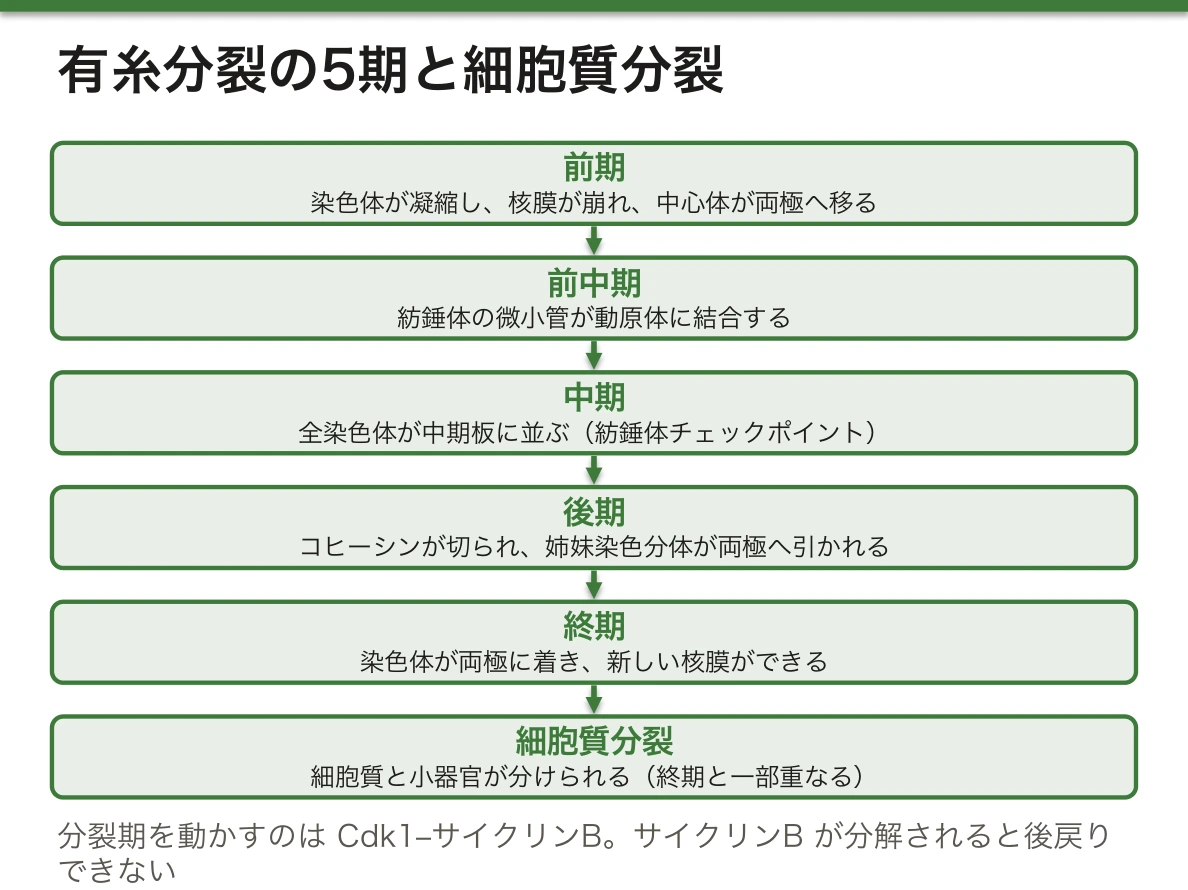
なぜ量の変わらないCDKではなく、量の変わるサイクリンで時計を刻むのか。酵素そのものを毎回作って壊すより、相棒の量を上下させるほうが、オンとオフをはっきり切り替えられるからである。サイクリンBは分裂期に向けて溜まってCdk1を働かせ、中期の終わりにAPC/C(後期促進複合体)によってユビキチンの目印を付けられ、まとめて分解される。分解は元に戻せないので、細胞は分裂期を後戻りできない一方通行で進む。
Auroraには役割のちがう2種類がある。Aurora Aは中心体と紡錘体づくりに、Aurora Bは染色体の凝縮、動原体と微小管の誤った結合の手直し、細胞質分裂にかかわる。
English version
Core
Somatic cell division makes two genetically identical daughter cells and supports growth, regeneration and replacement of tissues. M phase has two steps. Mitosis (karyokinesis) means division of the nucleus only and has five phases: prophase, prometaphase, metaphase, anaphase and telophase. Then cytokinesis splits the cytoplasm and organelles, and the two cells become separate. The two steps partly overlap.
At the centre are cyclin-dependent kinases (CDKs). A kinase adds phosphate groups to serine, threonine or tyrosine. A CDK works only when bound to a cyclin. CDK levels stay about the same, while cyclin levels rise and fall with the cycle. M phase is driven by Cdk1–cyclin B. Helping it are Plks (Polo-like kinases) and Aurora kinases, which handle centrosome function, spindle building, chromosome sharing and cytokinesis.
Going further
Why keep time with cyclin, whose amount changes, and not with the CDK, whose amount is stable? Raising and lowering the partner gives a clearer on/off switch than making and destroying the enzyme each time. Cyclin B builds up toward M phase and switches Cdk1 on. At the end of metaphase, APC/C (anaphase-promoting complex) tags it with ubiquitin and it is destroyed all at once. Destruction cannot be undone, so the cell moves through M phase one way, with no going back.
There are two Auroras with different jobs. Aurora A works on centrosomes and spindle building. Aurora B works on chromosome condensation, fixing wrong attachments between kinetochores and microtubules, and cytokinesis.
前期と前中期 — 染色体を固め、核膜をほどき、紡錘体で捕まえる
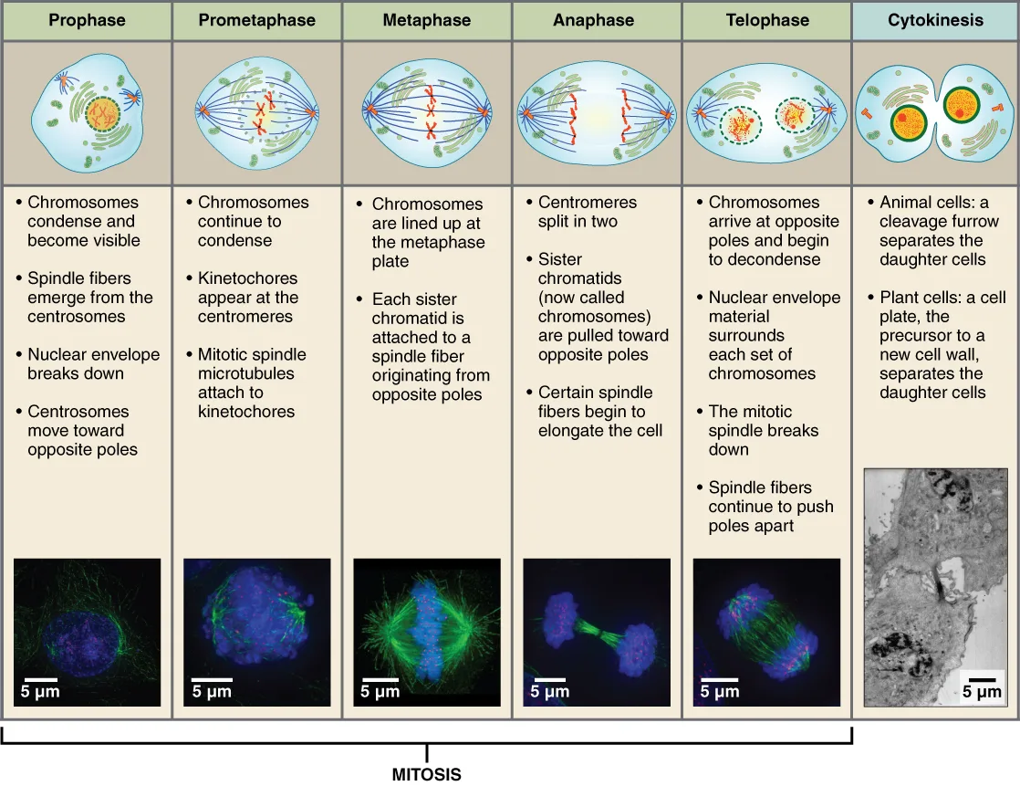
前期は準備の時期である。コンデンシンが染色体に結合して巻き上げ、染色体は太く短い形に凝縮する。このとき、Cdk1がヒストンH1を、Aurora BがヒストンH3をリン酸化する。複製でできた2本の姉妹染色分体は、コヒーシンの輪で束ねられたままでいる。
続いて核膜が崩れて小さな小胞になり、核小体が消える。転写も止まり、小胞体とGolgi装置も細かく断片化する。2つの中心体は細胞の両極へ移動し、そこから紡錘体が組み立てられ始める。この過程はPlkとAurora Aが仕切る。
前中期には、紡錘体の微小管が染色体の動原体に結合する。染色体はセントロメアを先頭にして、細胞の中央の赤道面(中期板)へ向かって動き始める。
なぜ核膜を崩さなければならないのか。紡錘体の微小管は細胞質の中心体から伸びてくる。核膜が残っていると、微小管は核の中の染色体に届かない。そこでCdk1が核膜の裏打ちであるラミンをリン酸化して網目をばらばらにし、核膜を小胞に分解する。核膜が消えるので、転写の装置も染色体から離れ、遺伝子の読み取りは止まる。
なぜ染色体を凝縮するのか。間期のクロマチンは非常に長く、からみ合っている。そのまま引っぱれば切れたり絡まったりする。コンデンシンで短く固めることで、姉妹染色分体どうしをほどき分け、紡錘体で安全に運べる「荷物」の形にしている。
English version
Core
Prophase is the preparation phase. Condensin binds chromosomes and coils them up, and they condense into short, thick forms. At this time Cdk1 phosphorylates histone H1 and Aurora B phosphorylates histone H3. The two sister chromatids made by replication are still held together by rings of cohesin.
Next, the nuclear envelope breaks into small vesicles and the nucleolus disappears. Transcription stops, and the ER and Golgi break into small pieces. The two centrosomes move to opposite poles, and the spindle starts to form from them. Plk and Aurora A control this.
In prometaphase, spindle microtubules attach to the kinetochores of chromosomes. Led by their centromeres, chromosomes start moving toward the equator of the cell (the metaphase plate).
Going further
Why must the nuclear envelope break down? Spindle microtubules grow from centrosomes in the cytoplasm. If the envelope stayed, they could not reach the chromosomes inside the nucleus. So Cdk1 phosphorylates the lamins lining the envelope, takes the network apart, and the envelope breaks into vesicles. With the envelope gone, transcription machinery also leaves the chromosomes, and gene reading stops.
Why condense chromosomes? Interphase chromatin is extremely long and tangled. Pulling it as it is would break or tangle it. Packing it short and firm with condensin lets sister chromatids be untangled from each other and turns them into “parcels” the spindle can move safely.
中期・後期・終期 — 全員がそろうまで待ち、そろったら一斉に引く
中期には、すべての染色体が中期板の上に一列に並ぶ。ここに分裂期の重要な関門、紡錘体チェックポイントがある。紡錘体の組み立てに誤りがあると、ここで進行が止められる。この関門はAPC/CとサイクリンA・Bを介して働く。
後期は、姉妹染色分体が離れることで始まる。束ねていたコヒーシンが酵素で切られ、紡錘体の線維が綱のように引いて、分かれた染色分体(ここから娘染色体と呼ぶ)を両極へ運ぶ。微小管は3種類が役割を分ける。動原体微小管は短くなって染色体を極へ引き寄せ、極間微小管は赤道をまたいで互いに滑り合い極を押し広げ、星状体微小管は細胞の膜側に向かって紡錘体の位置と向きを決め、後期には極をさらに引き離す。
終期には染色体が両極に着き、紡錘体はばらばらになる。ホスファターゼがラミンからリン酸を外し、核膜のかけらを含む小胞が2組の染色体の周りに集まって、新しい核膜ができる。
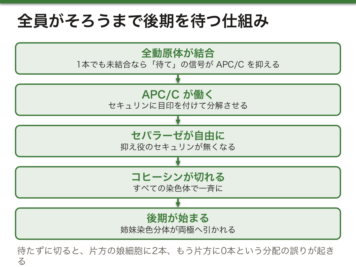
なぜ全員がそろうまで待つ必要があるのか。コヒーシンを切るのはセパラーゼという酵素で、ふだんはセキュリンという抑え役と結合して働けない。APC/Cがセキュリンに目印を付けて分解させると、セパラーゼが自由になり、すべての染色体のコヒーシンが一斉に切られる。1本でも動原体が紡錘体に結合していなければ、その動原体から「待て」の信号が出てAPC/Cを抑える。もし待たずに切ってしまえば、結合していない染色体は行き先が決まらず、片方の娘細胞に2本、もう片方に0本という分配の誤りが起きる。
English version
Core
In metaphase, all chromosomes line up on the metaphase plate. Here sits a key checkpoint of M phase, the spindle checkpoint. If spindle assembly has an error, progress is stopped here. The checkpoint acts through APC/C and cyclins A and B.
Anaphase begins when sister chromatids separate. The cohesin holding them is cut by an enzyme, and spindle fibres pull like ropes, carrying the separated chromatids (now called daughter chromosomes) to the poles. Three kinds of microtubules share the work. Kinetochore microtubules shorten and pull chromosomes to the poles. Interpolar microtubules cross the equator and slide past each other, pushing the poles apart. Astral microtubules reach toward the cell membrane, set the spindle’s position and direction, and in anaphase pull the poles further apart.
In telophase, chromosomes reach the poles and the spindle comes apart. Phosphatases remove phosphates from the lamins, vesicles carrying pieces of the old envelope gather around the two chromosome sets, and new nuclear envelopes form.
Going further
Why wait until every chromosome is ready? Cohesin is cut by an enzyme called separase, which is normally held inactive by a blocker called securin. When APC/C tags securin for destruction, separase is freed and cohesin on all chromosomes is cut at once. If even one kinetochore is not attached to the spindle, it sends a “wait” signal that holds APC/C back. If the cell cut without waiting, the unattached chromosome would have no fixed destination, and one daughter cell might get two copies and the other none.
細胞質分裂 — 動物はくびれ、植物は内側から壁を築くCytokinesis
細胞質分裂は、もとの細胞を実際に2つに分ける過程である。開始の合図は、それまでCdkにリン酸化されていたタンパク質からリン酸が外れることである。
動物細胞では、赤道の位置で細胞膜が内側にくびれ、分裂溝ができる。くびれをつくるのは、アクチンとミオシンIIが重なり合った収縮環で、巾着の紐を締めるように細胞を絞る。分裂溝の位置は、後期の星状体微小管と極間微小管の位置で決まる。
植物細胞は硬い細胞壁に囲まれていて、くびれることができない。代わりに、細胞の中央にできる隔膜形成体(フラグモプラスト)という微小管の足場にGolgi装置由来の小胞が運ばれて融合し、細胞板をつくる。細胞板は中央から外へ広がり、やがて新しい細胞壁になる。
なぜ溝の位置は紡錘体が決めるのか。細胞質分裂は、染色体が分かれたその間で切らなければ意味が無い。紡錘体の中央部(極間微小管が重なる場所)と星状体微小管が、膜の赤道部分に収縮環を組み立てる合図を送るので、溝は自動的に2組の染色体の間にできる。紡錘体が細胞の片側に寄っていれば溝も片側に寄り、大きさのちがう2つの細胞ができる。卵子ができるときの不等な分裂はこの仕組みを利用している。
小器官も分けなければならない。ミトコンドリアは分裂期に小さく断片化する。網の目のようにつながったままでは片方に偏りかねないが、小さな粒に分かれていれば、2つの娘細胞にほぼ均等に配られる。
English version
Core
Cytokinesis is the process that actually splits the cell in two. The start signal is removal of phosphates from proteins that Cdks had phosphorylated.
In animal cells, the membrane pinches inward at the equator, forming the cleavage furrow. The pinching is done by a contractile ring of overlapping actin and myosin II, which squeezes the cell like a drawstring. The furrow’s position is set by the positions of astral and interpolar microtubules in anaphase.
Plant cells are surrounded by a rigid wall and cannot pinch. Instead, vesicles from the Golgi apparatus are carried to a microtubule scaffold in the middle of the cell, the phragmoplast, and fuse to form a cell plate. The plate spreads from the centre outward and becomes a new cell wall.
Going further
Why does the spindle decide where the furrow goes? Cytokinesis is useless unless the cut is made between the separated chromosomes. The middle of the spindle (where interpolar microtubules overlap) and the astral microtubules signal the membrane at the equator to build the contractile ring, so the furrow forms between the two chromosome sets automatically. If the spindle sits to one side, the furrow does too, and two cells of different sizes result. The unequal division that forms the egg uses this mechanism.
Organelles must also be shared. Mitochondria break into small pieces in M phase. A connected network might end up mostly on one side, but small particles can be dealt out almost evenly to the two daughter cells.
減数分裂 — 複製1回・分裂2回で、数を半分にしつつ混ぜるMeiosis
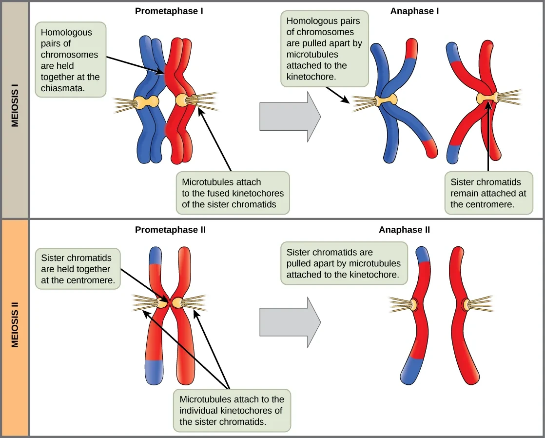
減数分裂は、二倍体の生殖細胞から一倍体の配偶子をつくる、生殖細胞だけの分裂である。DNAの複製は1回、分裂は2回(第一分裂と第二分裂)。生殖巣の中の生殖細胞は、まず有糸分裂で増え、適切な合図を受けると減数分裂に入る。
最も複雑なのは第一分裂前期で、5つの段階に分かれる。細糸期:染色体が凝縮し始める。接合期:相同染色体が全長にわたって並び、シナプトネマ複合体でぴったり貼り合わされる(対合)。太糸期:対合が完成して二価染色体になり、交叉が起きる。複糸期:シナプトネマ複合体がほどけ、相同染色体は離れ始めるが、交叉の場所(キアズマ)でつながったままでいる。移動期:さらに凝縮し、末端のキアズマだけでつながる。
中期Iで二価染色体が赤道面に並び、後期Iで相同染色体どうしが反対の極へ分かれる(姉妹染色分体はまだ離れない)。短い分裂間期(中間期)にはDNAの複製は無く、第二分裂は染色体数nのまま有糸分裂と同じように姉妹染色分体を分ける。
交叉は相同組み換えで起きる。トポイソメラーゼに似た酵素(Spo11)がDNAに計画的に二本鎖切断を入れ、相同染色体の配列を手本に修復するときに、鎖がつなぎ換えられる。なぜ交叉が無いと分配に失敗するのか。交叉とコヒーシンでできたキアズマが相同染色体をつなぎ、両極から引かれたときに張力が生まれる。この張力が「正しく両極につながった」という合図になる。交叉の無い相同染色体はばらばらに動いてしまい、不分離が起きやすい。組み換えの不全は不妊の原因にもなる。
後期Iでは父由来と母由来の染色体がそれぞれランダムに分かれるので、23対の組み合わせは223=約840万通りになる。交叉がこれにさらに新しい組み合わせを加える。
| 有糸分裂 | 減数分裂 | |
|---|---|---|
| 行う細胞 | 体細胞 | 生殖細胞 |
| 分裂の回数 | 1回 | 2回(複製は1回) |
| 相同染色体の対合・交叉 | 無い | 第一分裂前期にある |
| 結果 | 同じ2n細胞が2つ | 異なるn細胞が最大4つ |
English version
Core
Meiosis is a division found only in germ cells. It makes haploid gametes from diploid cells, with one DNA replication and two divisions (meiosis I and II). Germ cells in the gonads first multiply by mitosis, then enter meiosis when they receive the right signals.
The most complex part is prophase I, which has five stages. Leptotene: chromosomes begin to condense. Zygotene: homologous chromosomes line up along their whole length and are glued together by the synaptonemal complex (synapsis). Pachytene: synapsis is complete, each pair is a bivalent, and crossing over occurs. Diplotene: the synaptonemal complex breaks down and homologues begin to separate, but stay joined at the crossover points (chiasmata). Diakinesis: chromosomes condense further and are held only by terminal chiasmata.
In metaphase I, bivalents line up at the equator; in anaphase I, homologous chromosomes go to opposite poles (sister chromatids stay together). There is no DNA replication in the short interphase between (interkinesis), and meiosis II separates sister chromatids like a mitosis, but with n chromosomes.
Going further
Crossing over happens by homologous recombination. A topoisomerase-like enzyme (Spo11) makes planned double-strand breaks, and when they are repaired using the homologous chromosome as a template, the strands are rejoined in new ways. Why does segregation fail without crossovers? Chiasmata, made by crossovers plus cohesin, link the homologues, so tension builds when they are pulled from both poles. This tension is the signal that they are correctly attached to both poles. Homologues without a crossover move independently, and nondisjunction becomes likely. Failed recombination can also cause infertility.
In anaphase I, paternal and maternal chromosomes separate at random, so 23 pairs give 223 = about 8.4 million combinations. Crossing over adds even more new combinations.
| Mitosis | Meiosis | |
|---|---|---|
| Cells | Somatic cells | Germ cells |
| Divisions | One | Two (one replication) |
| Synapsis and crossing over | None | In prophase I |
| Result | Two identical 2n cells | Up to four different n cells |
分裂の誤りと病気 — トリソミー・モザイク・片親性ダイソミー・癌
ヒトで最も多い倍数性は三倍体(3n)で、1つの卵に2つの精子が入る二精子受精や、減数分裂の失敗でできた二倍体の配偶子から生じる。異数性の最も多い原因は、減数分裂での不分離である。
Down症候群(21トリソミー)の約95%は不分離によるもので、残りの多くは転座による。余分な21番は90〜95%が母親由来である。出生児の約2〜4%はモザイク、つまり21トリソミーの細胞と正常な細胞が混ざった状態である。
モザイクとは、受精後に起きた変化によって、1人の中に遺伝的にちがう細胞の集団が2つ以上ある状態をいう。体の細胞にあれば体細胞モザイク、生殖細胞の組織にあれば生殖細胞系列モザイクである。
片親性ダイソミー(UPD)は、1対の染色体の2本をどちらも片方の親から受け継いだ状態である。なぜそんなことが起きるのか。トリソミーの受精卵が、発生のごく早い時期に余分な1本を失って「救済」されることがある。3本のうちどれを失うかは偶然なので、3回に1回ほどは、残った2本が同じ親のものになる。数は正しく戻っても、刷り込み遺伝子の働きが片方に偏る。母親由来の14番を2本もつ例では、低身長・肥満・知的障害・思春期早発などが起き、血液や唾液にはトリソミーの細胞が、皮膚にはほとんど無い、というモザイクの名残が見つかることもある。
ミトコンドリアにもモザイクがある。mtDNAは変化が起きやすく、細胞の中に正常なものと変化したものが混ざる。分裂のときミトコンドリアは染色体ほど厳密には分けられず、ランダムに配られるので、娘細胞ごとに割合が少しずつちがってくる。
有糸分裂の誤りの後、細胞の行き先はいくつかある。染色体が1本足りない(モノソミー)または多い(トリソミー)娘細胞ができる、分裂そのものが失敗して四倍体(4N)になる、アポトーシスで死ぬ、あるいはG0期で止まる。この見張りが壊れた細胞は、誤りを抱えたまま増え続ける。腫瘍細胞の多くは、染色体の切断や異数性が広がったゲノム不安定性を示し、異数性によって癌遺伝子のコピーが増えたり、癌抑制遺伝子が失われたりして、腫瘍の成長に有利になる。
English version
Core
The most common polyploidy in humans is triploidy (3n). It comes from dispermy, when two sperm enter one egg, or from a diploid gamete made by failed meiosis. The most common cause of aneuploidy is nondisjunction in meiosis.
About 95% of Down syndrome (trisomy 21) comes from nondisjunction; most of the rest comes from translocations. The extra chromosome 21 comes from the mother in 90–95% of cases. About 2–4% of affected births are mosaic, with a mix of trisomy 21 cells and normal cells.
Mosaicism means having two or more genetically different cell groups in one person, caused by a change after fertilisation. In body cells it is somatic mosaicism; in germ cell tissue it is germline mosaicism.
Going further
Uniparental disomy (UPD) means both chromosomes of a pair come from the same parent. How can that happen? A trisomic embryo may lose the extra chromosome very early and be “rescued”. Which of the three is lost is random, so in about one case in three, the two remaining copies are from the same parent. The number is correct again, but imprinted genes now work in a one-sided way. In one example with two maternal chromosome 14s, there was short stature, obesity, intellectual disability and early puberty; trisomy cells were found in blood and saliva but hardly in skin, a trace of mosaicism.
Mitochondria can also be mosaic. mtDNA changes easily, so normal and changed copies are mixed in a cell. At division, mitochondria are not shared as strictly as chromosomes; they are dealt out at random, so the proportion differs a little in each daughter cell.
In the clinic
After an error in mitosis, a cell has several possible fates. Daughter cells may have one chromosome too few (monosomy) or too many (trisomy); division may fail and give a tetraploid (4N) cell; the cell may die by apoptosis; or it may stop in G0. Cells whose guards are broken keep multiplying with the error. Most tumour cells show genomic instability, with widespread chromosome breaks and aneuploidy. Aneuploidy can add copies of oncogenes or remove tumour suppressor genes, helping the tumour grow.
まとめ
| 細胞骨格の3種類 — 太さと材料で役割が分かれる | 細胞骨格は、細胞の形を保ち、動くときには形を変え、小器官の位置を決め、変形に耐え、荷物を運ぶ。 |
|---|---|
| モータータンパク質 — 線路の上を、決まった向きに歩く | モータータンパク質は、ATPを分解して得たエネルギーで、細胞骨格の線維の上を動き、力を生む。 |
| 有糸分裂の調節 — CDK・Plk・Aurora・APC/Cのリレー | 体細胞の分裂は、遺伝的に同じ2つの娘細胞をつくり、組織の成長・再生・置き換えを支える。 |
| 前期と前中期 — 染色体を固め、核膜をほどき、紡錘体で捕まえる | 前期は準備の時期である。コンデンシンが染色体に結合して巻き上げ、染色体は太く短い形に凝縮する。 |
| 中期・後期・終期 — 全員がそろうまで待ち、そろったら一斉に引く | 中期には、すべての染色体が中期板の上に一列に並ぶ。 |
| 細胞質分裂 — 動物はくびれ、植物は内側から壁を築く | 細胞質分裂は、もとの細胞を実際に2つに分ける過程である。 |
| 減数分裂 — 複製1回・分裂2回で、数を半分にしつつ混ぜる | 減数分裂は、二倍体の生殖細胞から一倍体の配偶子をつくる、生殖細胞だけの分裂である。 |
| 分裂の誤りと病気 — トリソミー・モザイク・片親性ダイソミー・癌 | ヒトで最も多い倍数性は三倍体(3n)で、1つの卵に2つの精子が入る二精子受精や、減数分裂の失敗でできた二倍体の配偶子から生じる。 |
このノートは医学生が授業の内容を自分の言葉でまとめたもので、診療の判断に使うものではありません。誤りに気づいたらお知らせください。