遺伝子発現とは、遺伝子に書かれた情報を、働くタンパク質またはRNAに変えることである。段階は2つ。DNAの情報を相補的なRNAに写しとる転写と、mRNAの情報を読んでアミノ酸を並べる翻訳である。ただし、rRNA・tRNA・調節性のRNAの遺伝子のように、転写されたRNAそのものが完成品で翻訳されない遺伝子もある。
真核細胞では、転写は核で、翻訳は細胞質で起きる。その間に、帽子をつけ、尾をつけ、イントロンを切り取る加工が入り、合格したmRNAだけが核膜孔を通って外へ出る。翻訳でできた鎖も、そのままでは働かない。正しく折りたたまれ、切られ、糖や脂質をつけられてはじめてタンパク質になる。この章は、この長い工程を順にたどり、最後に「折りたたみに失敗すると何が起きるか」を病気から見る。
- 転写の開始 — プロモーターに装置を組み立て、泡をつくる
- 転写の伸長と終結 — 鋳型は3′→5′に読み、RNAは5′→3′に伸びる
- RNAプロセシング — 帽子・尾・スプライシング、そして出荷検査
- 選択的スプライシング — 1つの遺伝子から、何種類ものタンパク質
- リボソームとtRNA — 暗号を読む機械と、翻訳する辞書
- 翻訳の開始・伸長・終結 — P部位に開始tRNA、A部位に次のtRNA
- タンパク質の構造と成熟 — 配列が形を決め、形が働きを決める
- 折りたたみの病気 — 出られない、溶けない、固まる
転写の開始 — プロモーターに装置を組み立て、泡をつくる
転写は、遺伝子の上流端(5′側)のプロモーターに多くのタンパク質が集まり、転写開始複合体を組み立てるところから始まる。TATAボックスなどに基本転写因子が結合し、そこへRNAポリメラーゼが呼びこまれる。ポリメラーゼは約14塩基対のDNAをほどいて転写バブルをつくり、開始点からリボヌクレオチド(rNTP)をつなぎ始める。最初の2つのrNTPがホスホジエステル結合でつながった時点で、開始は完了とみなされる。
| 真核細胞のRNAポリメラーゼ | 主に転写するもの |
|---|---|
| RNAポリメラーゼI | rRNAの前駆体(28S・18S・5.8S rRNAのもと) |
| RNAポリメラーゼII | タンパク質をコードする遺伝子のほとんど(mRNA) |
| RNAポリメラーゼIII | tRNA、5S rRNA、そのほかの小さなRNA |
原核細胞のRNAポリメラーゼは1種類だけで、構造もより単純である。
なぜ真核細胞は3種類もポリメラーゼを使い分けるのか。作るRNAによって、必要な量と調節のされ方がまったく違うからである。rRNAは細胞のRNAの大半を占め、増殖する細胞では休みなく大量に作り続ける必要がある。tRNAや5S rRNAも量は多いが短い。これに対しmRNAは、遺伝子ごとに、時期と組織に合わせて細かく上げ下げしなければならない。専用の装置を分けておけば、大量生産のラインと、注文生産のラインを互いに干渉させずに回せる。真核のポリメラーゼIIが多数の転写因子と組み合わさって働くのは、この「注文生産」に必要な細かな調節のためである。
猛毒キノコ(タマゴテングタケなど)のα-アマニチンは、8個のアミノ酸からなる環状ペプチドで、RNAポリメラーゼIIに結合してmRNAの合成を極端に遅くする。知られているなかで最も強いポリメラーゼIIの阻害物質である。腸から吸収された毒は肝臓に集まるため、食べてしばらくは消化器症状だけで、数日たってから急性肝不全が現れる。いったん軽くなったように見える時期があることが、診断を遅らせる。
English version
Core
Transcription begins when many proteins gather on the promoter at the upstream (5′) end of a gene and build a transcription initiation complex. General transcription factors bind elements such as the TATA box and recruit RNA polymerase. The polymerase opens about 14 base pairs of DNA to form a transcription bubble and starts joining ribonucleotides (rNTPs) at the start site. Initiation is considered complete when the first two rNTPs are linked by a phosphodiester bond.
| Eukaryotic RNA polymerase | Mainly transcribes |
|---|---|
| RNA polymerase I | The rRNA precursor (source of 28S, 18S and 5.8S rRNA) |
| RNA polymerase II | Almost all protein-coding genes (mRNA) |
| RNA polymerase III | tRNA, 5S rRNA and other small RNAs |
Prokaryotes have only one RNA polymerase, with a simpler structure.
Going further
Why do eukaryotes use three different polymerases? Because the RNAs they make need very different amounts and kinds of control. rRNA is most of the cell’s RNA, and a growing cell must make huge amounts of it nonstop. tRNA and 5S rRNA are also plentiful but short. mRNA, by contrast, must be raised and lowered gene by gene, to suit the time and the tissue. With separate machines, a mass-production line and a made-to-order line can run without getting in each other’s way. Polymerase II works with many transcription factors because made-to-order production needs this fine control.
In the clinic
α-Amanitin, from deadly mushrooms such as the death cap, is a ring-shaped peptide of eight amino acids. It binds RNA polymerase II and slows mRNA synthesis drastically. It is the strongest known inhibitor of polymerase II. The toxin absorbed from the gut collects in the liver, so at first there are only digestive symptoms, and acute liver failure appears days later. A period when the patient seems to improve can delay the diagnosis.
転写の伸長と終結 — 鋳型は3′→5′に読み、RNAは5′→3′に伸びる
伸長の段階では、RNAポリメラーゼが鋳型のDNAの上を1塩基ずつ進み、前方の二本鎖をほどき、後ろで再び閉じていく。新しいリボヌクレオチドは伸びているRNAの3′端につながるので、RNAは5′→3′に伸び、鋳型の鎖は3′→5′の向きに読まれる。バブルの中では、RNAの3′端の約8塩基が鋳型DNAと対をつくったままになっている。
できるRNAは鋳型鎖(アンチセンス鎖)に相補的で、センス鎖(コード鎖)と同じ配列(TがUに替わる)になる。転写はエクソンだけでなくイントロンも含めて端から端まで行われ、伸長因子がそれを助ける。終わりには、AAUAAAというポリA付加シグナルがRNAに写され、それを合図にRNAが切り離され、ポリメラーゼもDNAから離れる。こうして放たれたものを一次転写産物という。
ポリメラーゼ、鋳型DNA、伸びつつあるRNAからなる伸長複合体はきわめて安定で、途中で外れない。外れてしまうと、そこから再開する仕組みが無く、最初からやり直すしかないからである。これが長い遺伝子で効いてくる。転写の速さは毎分千〜数千塩基ほどなので、ヒト最大級の遺伝子の1つであるジストロフィン遺伝子(約2.2 Mb)を最後まで写すには約16時間かかる。その間ずっと、複合体は鋳型の上に留まっていなければならない。なぜイントロンまで写すのかと疑問に思うかもしれないが、ポリメラーゼには「エクソンだけを選んで読む」能力が無い。切り分けは、後で別の装置(スプライソソーム)が受けもつ。
English version
Core
During elongation, RNA polymerase moves along the template DNA one base at a time, opening the double strand in front and closing it behind. New ribonucleotides join the 3′ end of the growing RNA, so RNA grows 5′→3′, and the template strand is read 3′→5′. Inside the bubble, about 8 bases at the 3′ end of the RNA stay paired with the template DNA.
The RNA is complementary to the template (antisense) strand and has the same sequence as the sense (coding) strand (with U in place of T). Transcription runs from end to end, through introns as well as exons, helped by elongation factors. At the end, the polyadenylation signal AAUAAA is copied into the RNA. This signal leads to the RNA being cut free, and the polymerase leaves the DNA. The released RNA is the primary transcript.
Going further
The elongation complex of polymerase, template DNA and growing RNA is very stable and does not fall off along the way. If it did, there would be no way to restart from that point; it would have to begin again. This matters for long genes. Transcription runs at roughly one to several thousand bases per minute, so copying the dystrophin gene (about 2.2 Mb), one of the largest human genes, takes about 16 hours. The complex must stay on the template all that time. You may ask why introns are copied at all. The polymerase simply cannot pick out only the exons. Cutting is left to another machine, the spliceosome, later.
RNAプロセシング — 帽子・尾・スプライシング、そして出荷検査
タンパク質をコードする遺伝子の一次転写産物はmRNA前駆体(pre-mRNA)で、そのままでは使えない。働くmRNAにするための加工をRNAプロセシングという。
①5′キャップ:RNAの5′端がポリメラーゼIIから顔を出すとすぐ、いくつかの酵素が7-メチルグアノシンの帽子をつける(キャッピング)。キャップは、mRNAを分解酵素から守り、核からの輸送を助け、細胞質で翻訳を始める目印になる。②3′端の加工:エンドヌクレアーゼがAAUAAAの少し下流でRNAを切って3′-OHを露出させ、そこにポリA ポリメラーゼがアデニンを長くつなげてポリA尾部をつくる。③スプライシング:スプライソソームというRNAとタンパク質の複合体がイントロンを切り出し、エクソンどうしをつなぐ。イントロンの両端はふつう5′側がGU、3′側がAGで、これが切断の目印になる。
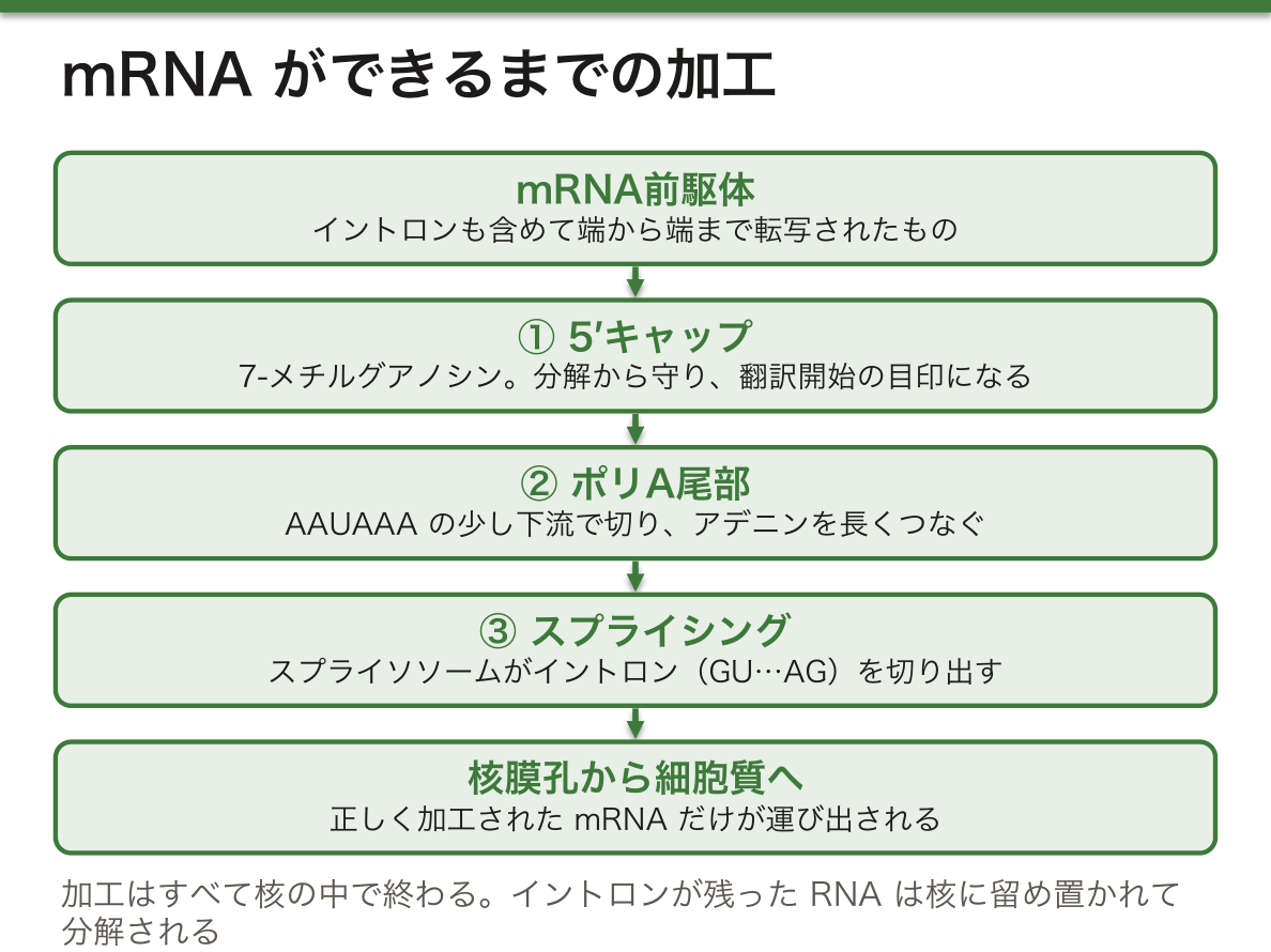
これらの加工はすべて核の中で終わり、完成したmRNAだけが核膜孔複合体を通って細胞質へ運ばれる。この輸送は選択的で、正しく加工されたかどうかと強く結びついている。スプライシングが済むと、つなぎ目の近くに目印のタンパク質が置かれ、それが輸送を助ける。逆に、イントロンが残ったRNAは核に留め置かれて分解される。
なぜこれほど厳しい検査が要るのか。不完全なmRNAが細胞質に出ると、途中で終わった短いタンパク質や、余分なアミノ酸を含んだタンパク質が作られる。そうした断片は働かないだけでなく、正常なタンパク質の邪魔をすることがある。細胞質でも、つなぎ目の目印より手前に終止コドンがあるmRNAは壊される(ナンセンス変異依存mRNA分解)。出荷前と出荷後の二重の検査で、異常な製品を減らしている。
スプライソソームの部品(SF3B1など)の遺伝子の変化は、骨髄異形成症候群などの血液の腫瘍でよく見つかる。また、ナンセンス変異依存mRNA分解は、終止コドンを作る変異(ナンセンス変異)が「短いタンパク質」ではなく「タンパク質が無い」状態になりやすい理由で、遺伝性疾患の重さを左右する。
English version
Core
The primary transcript of a protein-coding gene is pre-mRNA, which cannot be used as it is. The processing that turns it into working mRNA is RNA processing.
(1) 5′ cap: as soon as the 5′ end of the RNA comes out of polymerase II, several enzymes add a 7-methylguanosine cap (capping). The cap protects the mRNA from breakdown enzymes, helps its export from the nucleus, and marks the start point for translation in the cytoplasm. (2) 3′ end processing: an endonuclease cuts the RNA a little downstream of AAUAAA, leaving a free 3′-OH, and poly(A) polymerase adds a long run of adenines, the poly(A) tail. (3) Splicing: the spliceosome, a complex of RNA and protein, cuts out introns and joins exons. Introns usually begin with GU at the 5′ end and end with AG at the 3′ end; these mark the cut sites.
Going further
All of this processing finishes inside the nucleus, and only finished mRNA is carried through the nuclear pore complex to the cytoplasm. This transport is selective and closely tied to correct processing. After splicing, marker proteins are left near each junction, and they help export. RNA that still has an intron is held in the nucleus and broken down.
Why is such a strict check needed? If incomplete mRNA reached the cytoplasm, it would make proteins that stop early or contain extra amino acids. Such pieces not only fail to work but can get in the way of normal proteins. In the cytoplasm, too, an mRNA with a stop codon before a junction marker is destroyed (nonsense-mediated mRNA decay). Checks before and after “shipping” reduce faulty products.
In the clinic
Changes in genes for spliceosome parts (such as SF3B1) are often found in blood cancers such as myelodysplastic syndrome. Nonsense-mediated decay also explains why a variant that creates a stop codon (a nonsense variant) often leads to “no protein” rather than “a short protein”. This affects how severe an inherited disease is.
選択的スプライシング — 1つの遺伝子から、何種類ものタンパク質
選択的スプライシングとは、pre-mRNAの中のエクソンをつなぐか飛ばすかを変えることで、1つの遺伝子から複数のタンパク質(アイソフォーム)を作り分ける仕組みである。エクソンを丸ごと飛ばす、エクソンの端の切る位置をずらす、イントロンを残す、2つのエクソンのどちらか一方だけを使う、といった形がある。ヒトで複数のエクソンをもつ遺伝子のほとんどが、何らかの選択的スプライシングを受ける。
これにより、組織ごと、発生の時期ごと、病気の状態ごとに違うアイソフォームが作られる。たとえば同じ1つの遺伝子から、甲状腺ではカルシウムを下げるホルモンのカルシトニンが、神経では血管を広げるCGRPが作られる。
ヒトのタンパク質をコードする遺伝子は約2万個しかないのに、細胞で働くタンパク質の種類はそれよりはるかに多い。なぜ遺伝子の数が少なくても足りるのか——その大きな理由が選択的スプライシングである。エクソンが機能の部品(膜に刺さる部分、結合する部分など)に対応していることが多いので、部品を組み替えるだけで、局在や結合相手の違うタンパク質が作れる。遺伝子の数ではなく組み合わせの数で多様性を出しているわけで、核膜で転写と翻訳が分けられ、加工の時間がとれる真核細胞ならではの戦略である。
スプライシングの合図そのものが壊れると病気になる。βサラセミアの原因の1つは、β-グロビン遺伝子(HBB)の第1イントロンの先頭のGUがAUに変わるような、5′スプライス部位の1塩基の置換である。目印が消えるとイントロンは切り出されず(または近くの偽の部位で誤って切られ)、イントロンの配列がmRNAに残る。イントロンはアミノ酸を指定するために書かれていないので、読み進めるとすぐに終止コドンに行き当たり、β-グロビンは作られない。αとβのグロビンの量がつり合わなくなり、赤血球が小さく色の薄い貧血になる。エクソンの配列は1文字も変わっていなくても病気になる、という点が大切である。
English version
Core
Alternative splicing lets one gene make several proteins (isoforms) by changing which exons are joined or skipped in the pre-mRNA. Forms include skipping a whole exon, shifting the cut point at an exon edge, keeping an intron, and using only one of two exons. Almost all human genes with several exons undergo some alternative splicing.
This gives different isoforms in different tissues, stages of development and disease states. For example, one gene makes calcitonin, a hormone that lowers calcium, in the thyroid, and CGRP, which widens blood vessels, in nerves.
Going further
Humans have only about 20,000 protein-coding genes, yet cells use far more kinds of protein. Why are so few genes enough? A major reason is alternative splicing. Exons often match functional parts (a part that crosses the membrane, a part that binds something), so rearranging parts makes proteins with different locations or partners. Diversity comes from the number of combinations, not the number of genes. This strategy works in eukaryotes because the nuclear envelope separates transcription from translation and leaves time for processing.
In the clinic
Disease results when the splicing signal itself breaks. One cause of β-thalassaemia is a single base change at the 5′ splice site, such as GU at the start of intron 1 of the β-globin gene (HBB) becoming AU. Without the mark, the intron is not removed (or is cut wrongly at a nearby false site), and intron sequence stays in the mRNA. Introns were not written to code for amino acids, so reading soon hits a stop codon, and no β-globin is made. The balance between α- and β-globin is lost, and the result is anaemia with small, pale red cells. The key point: disease can occur even when not a single letter of the exons has changed.
リボソームとtRNA — 暗号を読む機械と、翻訳する辞書
リボソームはRNAとタンパク質の複合体で、mRNAの上を動きながらアミノ酸を鎖につないでいく。真核細胞では1秒に3〜5個のアミノ酸を加える速さである。どの細胞でもリボソームは大サブユニットと小サブユニットからなり、それぞれ長さの違うrRNAと別のタンパク質の組をもつ。リボソームはtRNAや多くの補助タンパク質も結合させる。
| 原核細胞 | 真核細胞 | |
|---|---|---|
| 全体 | 70S | 80S |
| 大サブユニット | 50S(23S・5S rRNA) | 60S(28S・5.8S・5S rRNA) |
| 小サブユニット | 30S(16S rRNA) | 40S(18S rRNA) |
tRNAは暗号を解く鍵である。各tRNAは3塩基のアンチコドンをもち、mRNAのコドンと相補的に対をつくる。tRNAはアミノアシルtRNA合成酵素によって特定のアミノ酸と高エネルギーの結合でつながり、アミノアシルtRNAになる。この結合のエネルギーが、のちにペプチド結合をつくる力になる。
なぜアミノアシルtRNA合成酵素の正確さが決定的なのか。リボソームが確かめるのはコドンとアンチコドンが対をつくるかどうかだけで、tRNAにどのアミノ酸がついているかは見ていない。もし合成酵素がまちがったアミノ酸をtRNAにつければ、リボソームはそのまま誤ったアミノ酸を組みこんでしまう。つまり遺伝暗号の「辞書」を実際に引いているのは、リボソームではなく20種類の合成酵素である。そのため合成酵素は、形の似たアミノ酸を取りちがえたときに自分で外して付け直す校正の仕組みをもっている。コドンとアミノ酸の対応は、この酵素の識別能力の上に成り立っている。
English version
Core
The ribosome is a complex of RNA and protein that moves along mRNA and joins amino acids into a chain. In eukaryotes it adds 3–5 amino acids per second. In every cell, a ribosome has a large and a small subunit, each with rRNAs of different lengths and its own set of proteins. Ribosomes also bind tRNAs and many helper proteins.
| Prokaryotic cell | Eukaryotic cell | |
|---|---|---|
| Whole | 70S | 80S |
| Large subunit | 50S (23S, 5S rRNA) | 60S (28S, 5.8S, 5S rRNA) |
| Small subunit | 30S (16S rRNA) | 40S (18S rRNA) |
tRNA is the key to the code. Each tRNA has a three-base anticodon that pairs with a complementary codon in the mRNA. The enzyme aminoacyl-tRNA synthetase links a tRNA to a specific amino acid by a high-energy bond, forming an aminoacyl-tRNA. The energy of this bond later drives peptide bond formation.
Going further
Why is the accuracy of aminoacyl-tRNA synthetases so critical? The ribosome only checks whether codon and anticodon pair. It does not check which amino acid is on the tRNA. If a synthetase put the wrong amino acid on a tRNA, the ribosome would build it into the protein without noticing. So the one who really “looks up” the genetic code dictionary is not the ribosome but the 20 synthetases. For this reason, synthetases have a proofreading step that removes and replaces an amino acid when they pick up a similar-looking wrong one. The match between codons and amino acids rests on this enzyme’s ability to tell them apart.
翻訳の開始・伸長・終結 — P部位に開始tRNA、A部位に次のtRNA
開始:まず小サブユニットがmRNAに結合し、開始コドンAUGの位置を決める。そこへ開始用のtRNA(メチオニンをつけたもの)が来て、リボソームのP部位でAUGと対をつくる。以降のtRNAはすべてA部位に入る。最後に大サブユニットが加わって開始複合体ができる。この過程は開始因子が仲立ちする。最初のメチオニンは、多くのタンパク質であとで切り落とされる。
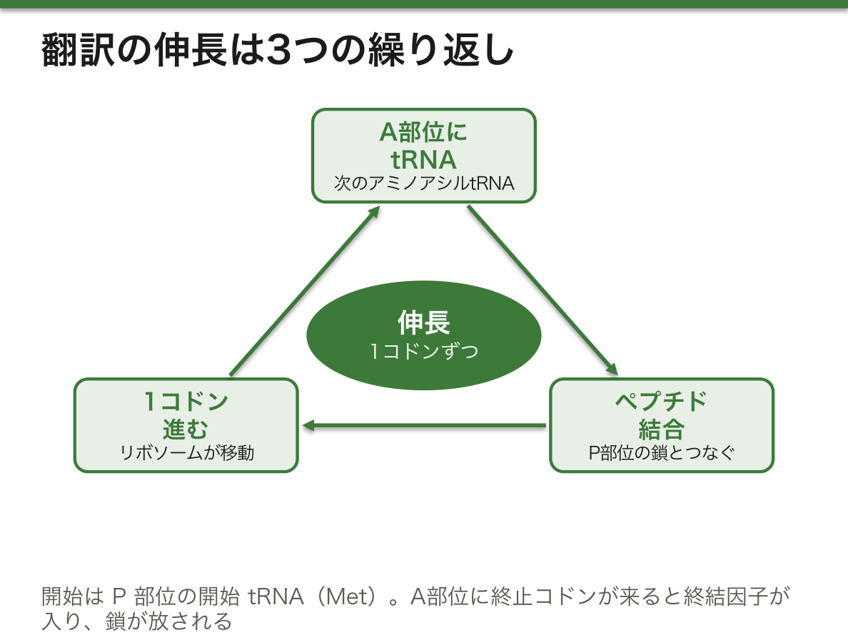
伸長:伸長因子の助けで、次のアミノアシルtRNAがA部位に入り、P部位の鎖とのあいだにペプチド結合ができ、リボソームが1コドンずつmRNAの上を進む。この3つの繰り返しで鎖が伸びる。終結:A部位に終止コドンが来ると、tRNAの代わりに終結因子が入り、完成した鎖が放される。DNAの塩基配列が、ここでようやくアミノ酸の配列に置き換わる。
| 原核細胞 | 真核細胞 | |
|---|---|---|
| 場所 | 細胞質(転写とほぼ同時) | 細胞質(転写と分かれる) |
| 開始位置の決め方 | AUGの少し上流の配列(Shine-Dalgarno配列)がrRNAと対をつくる | 5′キャップから下流へ進み、最初のAUGを探す |
| 最初のアミノ酸 | ホルミル化されたメチオニン(fMet) | メチオニン |
| 開始因子 | 3種 | 多数(9種以上) |
| 終結因子 | 3種 | 2種 |
| mRNAの寿命 | 数秒〜数分 | 数時間〜数日と安定 |
なぜ細菌のmRNAは数分で壊れてよいのか。細菌は環境の変化に数分で応じなければならず、不要になったタンパク質の設計図を残しておくと、作り続けて資源を浪費する。寿命が短ければ、転写を止めれば翻訳もすぐ止まる。真核細胞は、時間をかけて加工し検査したmRNAを長く使い回すほうが得になる。
翻訳の装置は毒素の標的にもなる。ジフテリア毒素は真核細胞の伸長因子eEF2を化学的に修飾して働けなくし、リボソームが前に進めなくなった細胞は死ぬ。腸管出血性大腸菌(O157など)の志賀毒素は、28S rRNAから塩基を1つ取り除いてリボソームを壊し、腎臓の血管の細胞を傷めて溶血性尿毒症症候群を起こす。
English version
Core
Initiation: first the small subunit binds the mRNA and finds the start codon AUG. The initiator tRNA (carrying methionine) comes and pairs with AUG at the ribosome’s P site. All later tRNAs enter the A site. Finally the large subunit joins to form the initiation complex. Initiation factors help this process. In many proteins, the first methionine is cut off later.
Elongation: helped by elongation factors, the next aminoacyl-tRNA enters the A site, a peptide bond forms with the chain at the P site, and the ribosome moves one codon along the mRNA. Repeating these three steps lengthens the chain. Termination: when a stop codon reaches the A site, a release factor enters instead of a tRNA, and the finished chain is released. Here, at last, the DNA base sequence has become an amino acid sequence.
Going further
| Prokaryotic cell | Eukaryotic cell | |
|---|---|---|
| Place | Cytoplasm (almost at the same time as transcription) | Cytoplasm (separate from transcription) |
| Finding the start | A sequence just upstream of AUG (Shine-Dalgarno) pairs with rRNA | Moves down from the 5′ cap to the first AUG |
| First amino acid | Formylmethionine (fMet) | Methionine |
| Initiation factors | 3 | Many (9 or more) |
| Release factors | 3 | 2 |
| mRNA lifetime | Seconds to minutes | Hours to days, stable |
Why can bacterial mRNA break down within minutes? Bacteria must respond to changes in minutes. Keeping blueprints for proteins no longer needed would waste resources on making them. With a short lifetime, stopping transcription quickly stops translation too. Eukaryotic cells, which spend time processing and checking mRNA, do better by using it for a long time.
In the clinic
The translation machinery is also a target of toxins. Diphtheria toxin chemically modifies the eukaryotic elongation factor eEF2 so it cannot work; cells whose ribosomes cannot move forward die. Shiga toxin from enterohaemorrhagic E. coli (such as O157) removes one base from 28S rRNA and wrecks the ribosome. It damages cells of kidney blood vessels and causes haemolytic uraemic syndrome.
タンパク質の構造と成熟 — 配列が形を決め、形が働きを決める
タンパク質の働きは立体構造から生まれ、立体構造はアミノ酸配列で決まる。一次構造はmRNAの配列どおりにペプチド結合でつながったアミノ酸の並び、二次構造は鎖の一部が局所的にとるαヘリックスやβシートなどの形で、主鎖どうしの水素結合で安定する。三次構造は1本の鎖全体の立体配置で、疎水性の側鎖どうしの相互作用(内側に集まる)、極性の側鎖どうしの水素結合、イオン結合、システイン間のジスルフィド結合で支えられる。
正しく折りたたむには助けが要る。シャペロンは伸びつつある鎖に結合して、合成が終わるまで勝手に折りたたまれないよう保護する。タンパク質ジスルフィドイソメラーゼはジスルフィド結合を組み替え、ペプチジルプロリルイソメラーゼはプロリンの前のペプチド結合の向きを変えて、折りたたみを速める。
折りたたんだあとも、多くのタンパク質は加工されて完成する。切断(タンパク質分解による成熟)では、膜を越えて運ばれるタンパク質の先頭の目印(シグナルペプチド)が切られ、大きな前駆体が切られて酵素やホルモンが活性型になる。インスリンはその典型で、プレプロインスリン→プロインスリン→インスリン(とC-ペプチド)と2回切られる。糖鎖の付加は翻訳が終わる前に小胞体で始まり、ゴルジ装置で仕上げられる。脂質の付加は、タンパク質を膜につなぎとめる。正しく折りたたまれなかったり加工に失敗したりしたタンパク質は、プロテアソームやオートファジーで分解される。
なぜシャペロンは「合成が終わるまで待たせる」のか。鎖の前半だけでは、後半の相手が無いまま疎水性の部分がむき出しになり、まちがった相手とくっついて固まりやすい。全体がそろってから折りたたむほうが、正しい形にたどり着きやすいのである。
インスリンとC-ペプチドは1分子のプロインスリンから同じ数だけ切り出されるので、血中のC-ペプチドは自分の膵臓が出しているインスリンの量を表す。注射したインスリンにはC-ペプチドが含まれないため、低血糖の原因が自分で注射したインスリンなのか、インスリンを出す腫瘍なのかを見分けるのに使われる。また、細胞表面のタンパク質を膜につなぐ脂質の錨(GPIアンカー)を作れない血液細胞が増えると、補体から身を守るタンパク質が表面に出せず、赤血球が壊される発作性夜間ヘモグロビン尿症になる。
English version
Core
A protein’s function comes from its 3D structure, and the 3D structure is set by its amino acid sequence. The primary structure is the order of amino acids joined by peptide bonds, following the mRNA. The secondary structure is local shapes such as the α-helix and β-sheet, held by hydrogen bonds between backbone groups. The tertiary structure is the 3D arrangement of the whole chain, held by interactions between hydrophobic side chains (which gather inside), hydrogen bonds between polar side chains, ionic bonds, and disulfide bonds between cysteines.
Correct folding needs help. Chaperones bind the growing chain and keep it from folding on its own until synthesis is finished. Protein disulfide isomerase rearranges disulfide bonds, and peptidyl-prolyl isomerase changes the direction of peptide bonds before proline. Both speed up folding.
Going further
Even after folding, many proteins need processing to be finished. In cleavage (proteolytic maturation), the leading tag (signal peptide) of proteins that cross membranes is cut off, and large precursors are cut to make active enzymes or hormones. Insulin is the classic case: preproinsulin → proinsulin → insulin (plus C-peptide), cut twice. Adding sugars (glycosylation) begins in the ER before translation ends and is finished in the Golgi. Adding lipids anchors proteins to membranes. Proteins that misfold or fail processing are broken down by the proteasome or autophagy.
Why do chaperones make the chain wait until synthesis ends? With only the first half of the chain, hydrophobic parts are exposed without their proper partners from the second half. They easily stick to the wrong partners and clump. Folding after the whole chain is ready makes reaching the right shape more likely.
In the clinic
Insulin and C-peptide are cut from proinsulin in equal numbers, so blood C-peptide shows how much insulin a person’s own pancreas is releasing. Injected insulin contains no C-peptide. This helps tell whether low blood sugar is caused by self-injected insulin or by an insulin-producing tumour. Also, when blood cells that cannot make the lipid anchor (GPI anchor) for surface proteins expand, they cannot display the proteins that protect them from complement. Red cells are destroyed: paroxysmal nocturnal haemoglobinuria.
折りたたみの病気 — 出られない、溶けない、固まる
折りたたみの失敗は、大きく2通りの害を生む。働くべき場所でタンパク質が足りなくなる害と、異常なタンパク質がたまる害である。
α1-アンチトリプシン欠乏症は、その両方が同時に起きる例である。α1-アンチトリプシンは肝細胞で作られて血中に出るプロテアーゼ阻害物質で、肺では好中球のエラスターゼが組織を壊しすぎないよう抑えている。Z型と呼ばれる変異型は正しく折りたためず、肝細胞の小胞体に留め置かれ、分泌されない。小胞体の中で重合してたまったものが肝細胞を傷つけ(肝障害・肝硬変)、一方で肺には届かないため、抑えのきかないエラスターゼが肺の結合組織を壊す(若年の肺気腫)。小胞体にたまった異常タンパク質のうち、溶ける単量体はプロテアソームで、溶けない凝集体はオートファジーで処理される。
アミロイドは、本来は溶けているタンパク質が、βシートを主体とする溶けない線維に組み上がって臓器や組織の細胞の外に沈着したものである。分解されにくく、病気ごとに沈着するタンパク質が決まっている。
Huntington病は、HTT遺伝子のコード領域にあるCAGの繰り返しが不安定に伸びて起きる神経変性疾患である。CAGはグルタミンを指定するので、タンパク質(ハンチンチン)のN末端近くに長いグルタミンの連なりができる。なぜ繰り返しが長いほど早く発症するのか——グルタミンの連なりが長いほど断片が凝集しやすく、神経細胞に害をなすまでの時間が短くなるからである。繰り返しは世代を経て伸びやすく、とくに父親から伝わるときに伸びて、子の世代ほど早く重く発症する(表現促進)。
α1-アンチトリプシン欠乏症の人にとって、喫煙は肺気腫を大きく早める。たばこの煙は肺に炎症を起こして好中球を呼び寄せるうえ、残っている少量のα1-アンチトリプシンを酸化して働けなくするからである。日本ではまれな病気だが、若くして下葉に強い肺気腫が見つかったときや、原因のはっきりしない肝障害では思い出したい。
English version
Core
Folding failure causes two main kinds of harm: too little protein where it should work, and build-up of abnormal protein.
α1-Antitrypsin deficiency shows both at once. α1-Antitrypsin is a protease inhibitor made in liver cells and released into the blood. In the lung, it keeps neutrophil elastase from destroying too much tissue. A variant called Z cannot fold correctly, so it is held in the ER of liver cells and not secreted. It polymerises and builds up in the ER, damaging the liver cells (liver injury, cirrhosis). Meanwhile it does not reach the lung, so unchecked elastase destroys the lung’s connective tissue (emphysema at a young age). Of the abnormal protein in the ER, soluble single molecules are removed by the proteasome, and insoluble clumps by autophagy.
Going further
Amyloid forms when normally soluble proteins build up into insoluble fibres, mainly of β-sheet, that deposit outside cells in organs and tissues. They resist breakdown, and each disease has its own depositing protein.
Huntington’s disease is a neurodegenerative disease caused by unstable expansion of a CAG repeat in the coding region of the HTT gene. CAG codes for glutamine, so the protein (huntingtin) gets a long run of glutamines near its N-terminus. Why does a longer repeat mean earlier onset? A longer glutamine run makes fragments clump more easily, so they harm nerve cells sooner. The repeat tends to grow from one generation to the next, especially when passed from the father, so children are affected earlier and more severely (anticipation).
In the clinic
For people with α1-antitrypsin deficiency, smoking greatly speeds up emphysema. Smoke causes lung inflammation that attracts neutrophils, and it oxidises the small amount of remaining α1-antitrypsin so it cannot work. The disease is rare in Japan, but think of it when severe emphysema of the lower lobes is found in a young person, or when liver injury has no clear cause.
まとめ
| 転写の開始 — プロモーターに装置を組み立て、泡をつくる | 転写は、遺伝子の上流端(5′側)のプロモーターに多くのタンパク質が集まり、転写開始複合体を組み立てるところから始まる。 |
|---|---|
| 転写の伸長と終結 — 鋳型は3′→5′に読み、RNAは5′→3′に伸びる | 伸長の段階では、RNAポリメラーゼが鋳型のDNAの上を1塩基ずつ進み、前方の二本鎖をほどき、後ろで再び閉じていく。 |
| RNAプロセシング — 帽子・尾・スプライシング、そして出荷検査 | タンパク質をコードする遺伝子の一次転写産物はmRNA前駆体(pre-mRNA)で、そのままでは使えない。 |
| 選択的スプライシング — 1つの遺伝子から、何種類ものタンパク質 | 選択的スプライシングとは、pre-mRNAの中のエクソンをつなぐか飛ばすかを変えることで、1つの遺伝子から複数のタンパク質(アイソフォーム)を作り分ける仕組みである。 |
| リボソームとtRNA — 暗号を読む機械と、翻訳する辞書 | リボソームはRNAとタンパク質の複合体で、mRNAの上を動きながらアミノ酸を鎖につないでいく。 |
| 翻訳の開始・伸長・終結 — P部位に開始tRNA、A部位に次のtRNA | 開始:まず小サブユニットがmRNAに結合し、開始コドンAUGの位置を決める。 |
| タンパク質の構造と成熟 — 配列が形を決め、形が働きを決める | タンパク質の働きは立体構造から生まれ、立体構造はアミノ酸配列で決まる。 |
| 折りたたみの病気 — 出られない、溶けない、固まる | 折りたたみの失敗は、大きく2通りの害を生む。 |
このノートは医学生が授業の内容を自分の言葉でまとめたもので、診療の判断に使うものではありません。誤りに気づいたらお知らせください。