細胞は膜で囲まれた箱である。しかし完全に閉じた箱では生きられない。栄養を取りこみ、老廃物を捨て、ホルモンを外に出し、イオンの濃度差を保たなければならない。この章では、細胞の中の膜のネットワーク(内膜系)、細胞膜の構造、そして膜を越えて物を運ぶ方法を、小さな分子と大きな分子(高分子)に分けて扱う。
小さな分子の輸送は、エネルギーを使わずに濃度差に従う(受動輸送)か、エネルギーを使って濃度差に逆らう(能動輸送)かで整理できる。高分子は膜のタンパク質では通れないので、膜そのものを袋にして運ぶ(エンドサイトーシスとエキソサイトーシス)。何が膜を通れて、何が通れないのか、それはなぜかを押さえれば、輸液、利尿薬、糖尿病の薬、遺伝性の高コレステロール血症までが一本の線でつながる。
- 内膜系 — 作る・仕分ける・壊す、をつなぐ膜のネットワーク
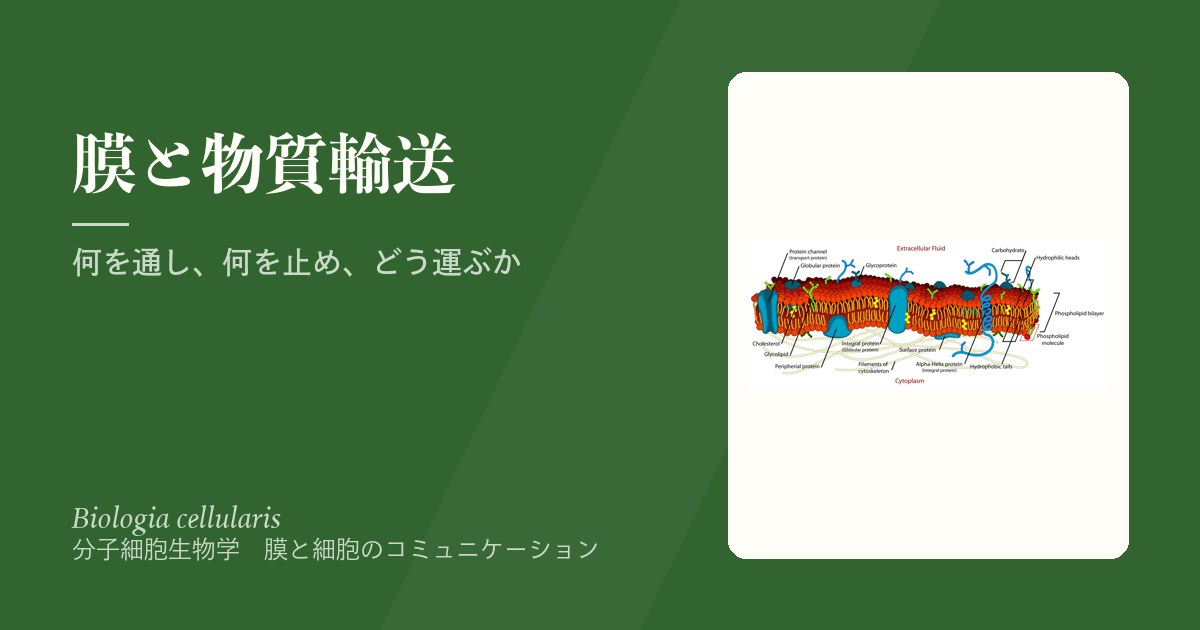
- 細胞膜の構造 — 脂質の二重層に、タンパク質と糖鎖がのる
- 透過性・単純拡散・浸透 — 膜は何を素通りさせるか
- 促進拡散 — 担体は形を変えて運び、チャネルは穴を開ける
- 能動輸送 — ポンプが坂を作り、共輸送がその坂を使う
- チャネルの病気 — 嚢胞性線維症とCFTR
- エンドサイトーシス — 膜を袋にして外のものを取りこむ
- エキソサイトーシスと小胞輸送 — いつでも出すか、合図を待って出すか
内膜系 — 作る・仕分ける・壊す、をつなぐ膜のネットワークReticulum endoplasmicum
内膜系は、核膜、小胞体(ER)、ゴルジ装置、リソソーム、小胞からなる膜の系である。これらは小胞のやりとりでつながり、タンパク質や脂質を作って運ぶ1本の流れをつくる。
| 粗面小胞体 | 滑面小胞体 |
|---|---|
| 膜タンパク質・分泌タンパク質の合成の場 タンパク質の膜への移行、折りたたみ、輸送 糖鎖の付加(グリコシル化) ジスルフィド結合の形成(三次・四次構造の安定化) | 脂質の合成 グリコーゲンや糖質の代謝 ステロイドホルモンの合成(性腺など) 解毒(肝細胞など) Ca2+の貯蔵と放出(筋細胞など) |
ゴルジ装置は、ERから届いたタンパク質の仕分けと出荷(分泌の経路)、糖鎖の加工、脂質と多糖の代謝、そしてリソソームの形成を担う。リソソームは約50種類の加水分解酵素を含む袋で、取りこんだものを分解するほか、古くなった自分の小器官を壊して入れ替えるオートファジーを行う。
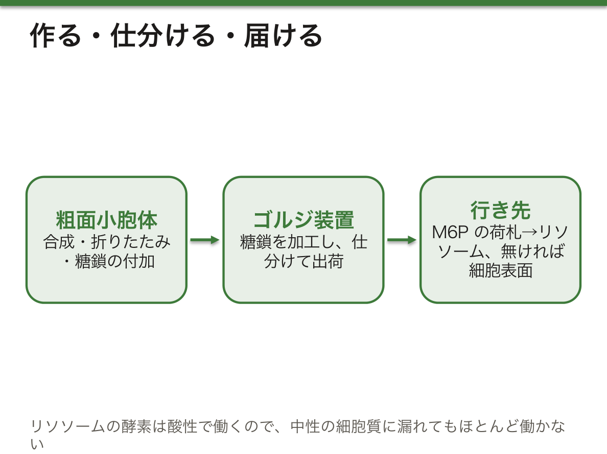
ゴルジ装置は、どうやって「この酵素はリソソーム行き」と見分けるのか。リソソームの酵素には、ゴルジ装置で糖鎖にマンノース6-リン酸という荷札がつけられる。この荷札に結合する受容体が酵素を集めて小胞に詰め、リソソームへ送る。荷札の無いタンパク質は、特別な指示が無いかぎり細胞表面へ向かう。リソソームの内部は酸性に保たれており、酵素は酸性でよく働く。万一酵素が細胞質に漏れても、中性の細胞質ではほとんど働かないので、細胞自体は守られる。
荷札をつける酵素が欠けると、リソソーム酵素がリソソームに届かず細胞の外に分泌されてしまう。これがI細胞病(ムコリピドーシスII)で、血中のリソソーム酵素が高いのに細胞の中には分解できない物質がたまる。個々の酵素が欠けると、Gaucher病やFabry病などのリソソーム病になる。
English version
Core
The endomembrane system is made up of the nuclear envelope, endoplasmic reticulum (ER), Golgi apparatus, lysosomes and vesicles. They are linked by vesicle traffic and form one stream that makes and moves proteins and lipids.
| Rough ER | Smooth ER |
|---|---|
| Site of synthesis of membrane and secreted proteins Protein entry into the membrane, folding and transport Adding sugar chains (glycosylation) Disulfide bond formation (stabilises tertiary and quaternary structure) | Lipid synthesis Glycogen and carbohydrate metabolism Steroid hormone synthesis (gonads) Detoxification (hepatocytes) Storage and release of Ca2+ (muscle cells) |
The Golgi apparatus sorts and ships proteins from the ER (the secretory pathway), modifies sugar chains, handles lipid and polysaccharide metabolism, and forms lysosomes. A lysosome is a sac of about 50 hydrolytic enzymes. It digests material taken in and also performs autophagy, breaking down the cell’s own old organelles so they can be replaced.
Going further
How does the Golgi know that an enzyme belongs in the lysosome? In the Golgi, lysosomal enzymes receive a label on their sugar chain: mannose 6-phosphate. Receptors that bind this label collect the enzymes into vesicles and send them to lysosomes. Proteins without the label go to the cell surface unless there is another signal. The inside of the lysosome is kept acidic, and the enzymes work best in acid. If an enzyme leaks into the cytoplasm, it hardly works at the neutral pH there, so the cell itself is protected.
In the clinic
If the enzyme that adds the label is missing, lysosomal enzymes do not reach the lysosome and are secreted out of the cell instead. This is I-cell disease (mucolipidosis II): lysosomal enzymes are high in the blood, yet undigested material builds up inside cells. Loss of single enzymes causes lysosomal storage diseases such as Gaucher disease and Fabry disease.
細胞膜の構造 — 脂質の二重層に、タンパク質と糖鎖がのるMembrana cellularis
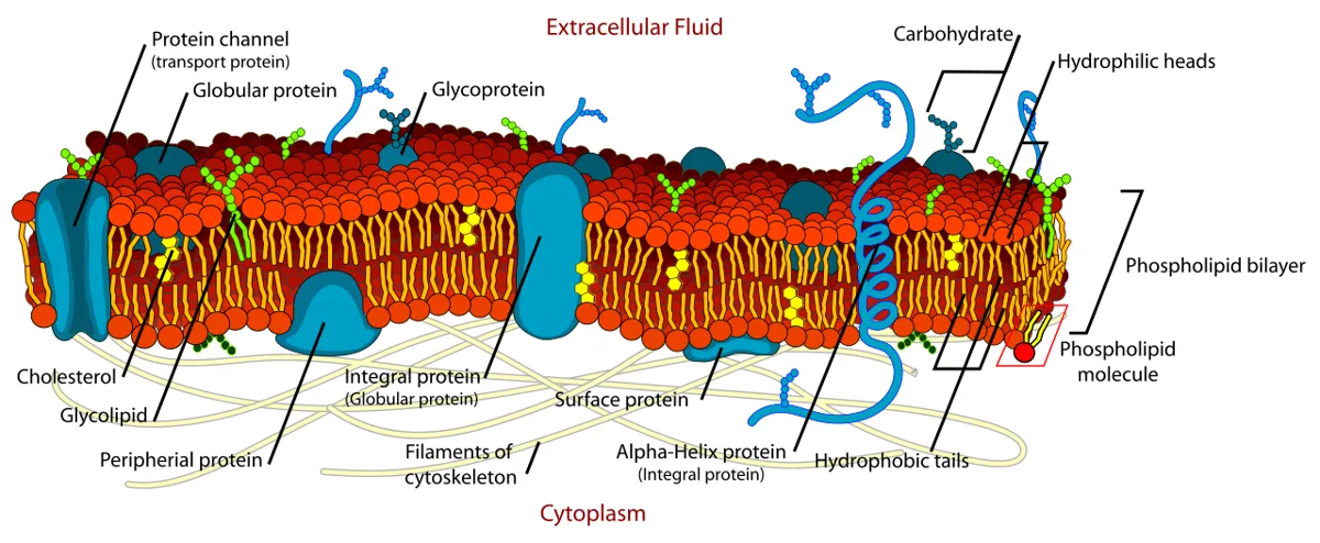
膜の脂質は両親媒性で、水になじむ極性の頭部と、水をはじく尾部をもつ。水の中では疎水効果とファンデルワールス力によって尾部どうしが内側に集まり、頭部が水に向いた脂質二重層が自然にできる。
脂質の主な種類は3つである。グリセロリン脂質は最も多く、グリセロールの骨格にリン酸とアルコール(コリン、セリン、エタノールアミン、イノシトール)がついて、ホスファチジルコリンやホスファチジルセリンなどになる。スフィンゴ脂質はスフィンゴシンからでき、頭部が糖のスフィンゴ糖脂質は細胞膜の脂質の2〜10%を占め、神経組織に特に多い。ステロールは4つの環をもつ構造で、哺乳類の細胞膜にはコレステロールが多い。
膜タンパク質は、二重層を貫く膜貫通(内在性)タンパク質、脂質に共有結合でつなぎ止められた脂質アンカー型タンパク質、膜の片側の表面にだけつく表在性タンパク質に分かれる。膜貫通部分には疎水性のアミノ酸が多い。糖鎖は必ず細胞の外側の面にあり、細胞外基質、増殖因子、抗体などと相互作用する。
細胞膜の働きは、①細胞の中身を囲う(区画化)、②膜に埋めこまれた分子の反応の足場、③選択的な透過性をもつ壁、④物質を運ぶ仕組み、⑤外からの刺激への応答(シグナル伝達)、にまとめられる。
二重層の内側と外側では脂質の組成がちがう(非対称性)。たとえばホスファチジルセリンはふだん内側の層だけにある。なぜこの偏りが大事なのか。アポトーシスが始まると、この偏りが崩れてホスファチジルセリンが外側に出る。マクロファージはこれを「食べてよい」という目印として認識し、死にかけた細胞を回収する。膜の組成の偏り自体が、情報として使われているのである。
English version
Core
Membrane lipids are amphipathic: they have a polar head that likes water and a tail that repels it. In water, the hydrophobic effect and van der Waals forces make the tails gather inside, with the heads facing water, so a lipid bilayer forms by itself.
There are three main kinds of lipids. Phosphoglycerides are the most common: a glycerol backbone with a phosphate and an alcohol (choline, serine, ethanolamine, inositol), giving phosphatidylcholine, phosphatidylserine and so on. Sphingolipids are built from sphingosine; glycosphingolipids, with sugar head groups, make up 2–10% of plasma membrane lipids and are especially common in nervous tissue. Sterols have a four-ring structure; mammalian plasma membranes are rich in cholesterol.
Membrane proteins are integral (transmembrane) proteins that cross the bilayer, lipid-anchored proteins held by covalent bonds to lipids, and peripheral proteins that sit on one surface only. The membrane-crossing parts contain many hydrophobic amino acids. Sugar chains are always on the outer face, where they interact with extracellular matrix, growth factors and antibodies.
Going further
The plasma membrane (1) encloses the cell contents (compartments), (2) is a platform for reactions of embedded molecules, (3) is a barrier with selective permeability, (4) has systems for moving substances, and (5) responds to outside stimuli (signal transduction).
The inner and outer layers have different lipids (asymmetry). For example, phosphatidylserine is normally only in the inner layer. Why does this matter? When apoptosis starts, the asymmetry breaks down and phosphatidylserine appears on the outside. Macrophages read it as an “eat me” signal and remove the dying cell. The uneven make-up of the membrane is itself used as information.
透過性・単純拡散・浸透 — 膜は何を素通りさせるか
水、無機イオン、小さな有機分子(糖、ビタミン、脂肪酸など)は、生物の重さの75〜80%を占める。これらは高分子の材料になり、エネルギーを蓄えて配り、信号としても働く。
細胞膜の透過性は、分子の大きさ・電荷・溶けやすさで決まる。脂質二重層を自力で通れるのは、小さく、比較的疎水性の分子(O2、CO2、ステロイドなど)だけである。こうした分子は膜に溶けこみ、横切り、反対側の水に再び溶け出す。これが単純拡散で、向きは膜の内外の濃度で決まる。濃いほうから薄いほうへの移動はエントロピーを増やす(ΔS>0)ので自由エネルギーが下がり(ΔG<0)、自然に進む。
水が膜を通って、溶質の濃い側へ移動する現象が浸透である。細胞の外の液が細胞内と同じ浸透圧なら等張、高ければ高張、低ければ低張と呼ぶ。高張液では水が出て細胞が縮み(植物細胞では細胞膜が細胞壁からはがれる原形質分離)、低張液では水が入って細胞がふくらむ。
なぜイオンは小さいのに膜を通れないのか。イオンの周りには水分子が強く結びついて殻をつくっている。疎水性の膜の中心に入るには、この水の殻をはがさなければならず、それには大きなエネルギーが要る。だから小さなNa+やK+でも、脂質だけの膜はほとんど通れず、専用のタンパク質(チャネルや担体)を必要とする。水自体は少しは二重層を通れるが、速く大量に動かすには水チャネル(アクアポリン)が使われる。
静脈に入れる輸液が等張(生理食塩水0.9%など)なのは、赤血球や血管の細胞が縮んだり壊れたりしないためである。マラソンのような長時間の運動のあと、汗で失った塩分を補わずに真水だけを大量に飲むと、血液が薄まって低ナトリウム血症になる。血液が脳より低張になると、水が浸透で脳の細胞に入って脳がむくむ。頭蓋骨は広がれないので頭蓋内圧が上がり、頭痛、嘔気、意識の低下、けいれんが起きる。
English version
Core
Water, inorganic ions and small organic molecules (sugars, vitamins, fatty acids) make up 75–80% of living matter by weight. They are building blocks for macromolecules, store and distribute energy, and act as signals.
Membrane permeability depends on a molecule’s size, charge and solubility. Only small, fairly hydrophobic molecules (O2, CO2, steroids) can cross the lipid bilayer on their own. They dissolve into the membrane, cross it, and dissolve again in the water on the other side. This is simple diffusion, and its direction depends on the concentrations on each side. Moving from high to low concentration increases entropy (ΔS>0), so free energy falls (ΔG<0) and it happens on its own.
Osmosis is the movement of water across a membrane towards the side with more solute. A solution with the same osmotic pressure as the cell is isotonic; higher is hypertonic; lower is hypotonic. In a hypertonic solution water leaves and the cell shrinks (in plant cells the membrane pulls away from the wall: plasmolysis); in a hypotonic solution water enters and the cell swells.
Going further
Why can ions not cross the membrane even though they are small? Water molecules bind tightly around an ion and form a shell. To enter the hydrophobic centre of the membrane, the ion must lose this shell, which needs a lot of energy. So even small Na+ and K+ can hardly cross a pure lipid membrane and need special proteins (channels or carriers). Water itself can cross the bilayer a little, but water channels (aquaporins) are used to move it quickly and in large amounts.
In the clinic
IV fluids are isotonic (such as 0.9% saline) so that red blood cells and vessel cells do not shrink or burst. After long exercise such as a marathon, drinking large amounts of plain water without replacing the salt lost in sweat dilutes the blood and causes hyponatraemia. When the blood becomes hypotonic compared with the brain, water moves into brain cells by osmosis and the brain swells. The skull cannot expand, so intracranial pressure rises, with headache, nausea, reduced consciousness and seizures.
促進拡散 — 担体は形を変えて運び、チャネルは穴を開ける
促進拡散は、膜のタンパク質の助けで、極性や電荷をもつ分子(糖、アミノ酸、ヌクレオシド、イオン)を通す輸送である。エネルギーは使わず、常に電気化学的な勾配に従って「下り坂」に進む。助けるタンパク質は2種類ある。
担体タンパク質(輸送体、パーミアーゼ)は、溶質と結合し、形を順に変えながら膜の反対側へ運ぶ。どれも膜を何度も貫く膜貫通タンパク質で、膜を貫く部分はふつうαヘリックスである。1種類の分子だけを濃度勾配に従って運ぶものをユニポーターといい、ブドウ糖輸送体(GLUT)やアミノ酸の輸送体がその例である。
チャネルタンパク質は膜に水を通す通路(孔)を開け、大きさと電荷の合う小さな分子やイオンを通す。ほとんど常に開いている非開閉型(リーク)チャネルと、合図があるときだけ開く開閉型チャネルがある。開閉型は、信号分子の結合で開くリガンド依存性(段階的な電位変化を生む)、膜電位の変化で開く電位依存性(活動電位を生む)、振動や圧力で開く機械刺激依存性に分かれる。水を速く通すアクアポリンもチャネルの一種である。
なぜチャネルは担体よりはるかに速いのか。担体は溶質を1つ(または数個)つかむたびに形を変え、元に戻るという手順をくり返すので、1回ごとに時間がかかる。チャネルは開いている間ずっと通路になっていて、イオンは列をなして流れこむ。そのため1秒あたりに通す数は、チャネルのほうが桁ちがいに多い。一方で、チャネルの孔は狭く、大きさと電荷でイオンを厳密に選ぶので、速さと選択性を両立している。担体は遅いが、形の変化を他のエネルギーと結びつけられるので、次の項目の能動輸送の土台になる。
腎臓の集合管では、抗利尿ホルモン(バソプレシン)の合図でアクアポリン2が細胞膜に運ばれ、水の再吸収が増える。この受容体やアクアポリン2の遺伝子に異常があると、尿を濃縮できない腎性尿崩症になる。
English version
Core
Facilitated diffusion moves polar or charged molecules (sugars, amino acids, nucleosides, ions) across the membrane with the help of membrane proteins. It uses no energy and always goes “downhill” along the electrochemical gradient. Two kinds of proteins help.
Carrier proteins (transporters, permeases) bind a solute and carry it to the other side by changing shape step by step. All are transmembrane proteins that cross the membrane several times, usually as α-helices. A carrier that moves only one kind of molecule down its gradient is a uniporter; glucose transporters (GLUT) and amino acid transporters are examples.
Channel proteins open a water-filled passage (pore) through the membrane and let small molecules or ions of the right size and charge through. Non-gated (leak) channels are open most of the time; gated channels open only when signalled. Gated channels are ligand-gated (open when a signal molecule binds; make graded potentials), voltage-gated (open when membrane potential changes; make action potentials) and mechanically gated (open with vibration or pressure). Aquaporins, which let water through quickly, are also channels.
Going further
Why are channels much faster than carriers? A carrier must change shape and return each time it moves one (or a few) solute molecules, so each cycle takes time. A channel stays a passage while open, and ions flow through in a line. So per second, a channel moves far more. Yet the pore is narrow and selects ions strictly by size and charge, so channels combine speed and selectivity. Carriers are slow, but their shape change can be linked to other energy sources, which is the basis of active transport in the next item.
In the clinic
In the collecting duct of the kidney, the signal of antidiuretic hormone (vasopressin) moves aquaporin-2 to the cell membrane and increases water reabsorption. Defects in the receptor or the aquaporin-2 gene cause nephrogenic diabetes insipidus, in which urine cannot be concentrated.
能動輸送 — ポンプが坂を作り、共輸送がその坂を使う
能動輸送は、別の反応のエネルギーを使って、分子を濃度や電位の勾配に逆らって「上り坂」に運ぶ輸送である。
一次性能動輸送では、ATPの加水分解のエネルギーを直接使う。このポンプはATPアーゼで、代表がNa+/K+-ATPアーゼである。ATP 1分子あたりNa+を3つ細胞の外へ、K+を2つ中へ運び、細胞の外はNa+が多く、中はK+が多い状態を保つ。
二次性能動輸送(共輸送)では、ポンプが作ったイオンの電気化学的勾配に蓄えられたエネルギーを使う。Na+が勾配に従って細胞に流れこむ力に乗せて、別の分子を勾配に逆らって運ぶ。同じ向きに運ぶものを共輸送体(シンポーター)、逆向きに運ぶものを交換輸送体(アンチポーター)という。腸や腎臓でNa+とブドウ糖を一緒に取りこむSGLTは前者、Na+を入れてCa2+やH+を出すNa+/Ca2+交換輸送体やNa+/H+交換輸送体は後者である。
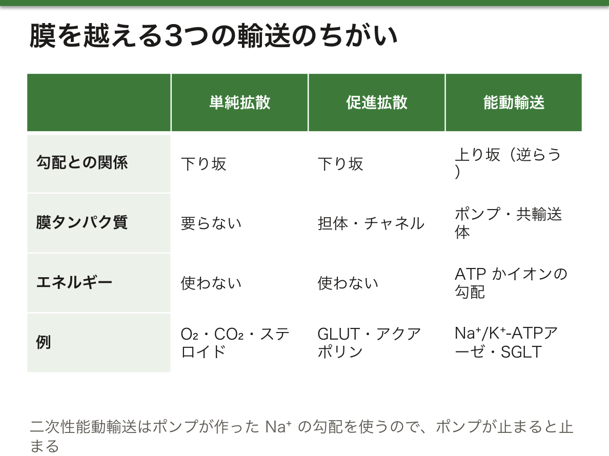
なぜ二次性能動輸送はポンプが止まると止まるのか。共輸送体自身はATPを使わない。使っているのは、Na+/K+-ATPアーゼがATPを消費して作り続けているNa+の濃度差である。ダムにたとえるなら、ポンプが水を汲み上げ、共輸送体はその水が流れ落ちる力で水車を回している。ポンプが止まれば濃度差はやがて消え、共輸送も止まる。細胞が使うATPのかなりの部分がこのポンプに費やされるのは、細胞のたくさんの輸送がこの「坂」に頼っているからである。
強心薬のジゴキシンは心筋のNa+/K+-ATPアーゼを阻害する。細胞内のNa+が増えると、Na+/Ca2+交換輸送体がCa2+を外に出しにくくなり、細胞内のCa2+が増えて収縮力が強まる。SGLT2阻害薬は腎臓の近位尿細管でのブドウ糖の再吸収を止めて、糖を尿に出させる糖尿病の薬で、心不全や慢性腎臓病にも使われている。
English version
Core
Active transport uses energy from another reaction to move molecules against a concentration or electrical gradient, “uphill”.
Primary active transport uses the energy of ATP hydrolysis directly. These pumps are ATPases; the classic one is the Na+/K+-ATPase. For each ATP it moves 3 Na+ out of the cell and 2 K+ in, keeping Na+ high outside and K+ high inside.
Secondary active transport (cotransport) uses the energy stored in the ion gradient made by the pump. The force of Na+ flowing into the cell down its gradient is used to carry another molecule against its gradient. A carrier moving both in the same direction is a symporter; one moving them in opposite directions is an antiporter. SGLT, which takes in Na+ and glucose together in the gut and kidney, is a symporter. The Na+/Ca2+ exchanger and Na+/H+ exchanger, which bring Na+ in and push Ca2+ or H+ out, are antiporters.
Going further
Why does secondary active transport stop when the pump stops? The cotransporter itself uses no ATP. It uses the Na+ concentration difference that the Na+/K+-ATPase keeps making by spending ATP. Think of a dam: the pump lifts the water, and the cotransporter turns a water wheel with the falling water. If the pump stops, the difference slowly disappears and cotransport stops too. A large share of a cell’s ATP goes to this pump because so much transport depends on this “slope”.
In the clinic
The heart drug digoxin blocks the Na+/K+-ATPase in heart muscle. When Na+ inside the cell rises, the Na+/Ca2+ exchanger removes less Ca2+, intracellular Ca2+ rises, and contraction becomes stronger. SGLT2 inhibitors stop glucose reabsorption in the proximal tubule and make glucose leave in the urine. They are diabetes drugs and are also used for heart failure and chronic kidney disease.
チャネルの病気 — 嚢胞性線維症とCFTRMucoviscidosis
嚢胞性線維症(CF)は、ヨーロッパ系の人々で最も多い単一遺伝子病の1つで、常染色体劣性の形式で遺伝する。原因はCFTR(嚢胞性線維症膜貫通コンダクタンス制御因子)の遺伝子の変異である。
CFTRは上皮細胞の膜にあるCl–チャネルで、cAMPとプロテインキナーゼAによるリン酸化で調節され、ATPが結合すると開き、ATPが加水分解されると閉じる。ATPを使うが、それは輸送のエネルギーではなく開閉の合図である。
CFTRが働かないと、気道や膵管の分泌物が異常に粘っこくなり、管をふさぐ。その結果、炎症と組織の破壊が起き、肺と膵臓の両方が傷む。
なぜCl–のチャネル1つの異常で、分泌物がねばねばになるのか。気道の表面では、Cl–が上皮から管の内腔に出ると、それに引かれてNa+が、さらに浸透で水がついていく。これで粘液がちょうどよい水分を保ち、線毛で運ばれる。CFTRが働かないとCl–が出ず、水も出ないので、粘液が乾いて濃くなり、線毛で運べなくなる。膵管でも同じことが起き、消化酵素が腸に届かずに膵臓自体を傷める。水は自分では動けず、イオンの後を浸透でついていくしかないという原則が、病気の形をそのまま決めている。
診断には汗のCl–濃度の検査が使われる。汗腺の管ではCFTRが逆にCl–を回収する役なので、CFでは汗が塩辛くなる。繰り返す肺感染、膵外分泌不全による脂肪便と栄養障害、男性不妊(精管の欠損)が典型である。日本人ではきわめてまれで、慢性の呼吸器感染と膵炎が続く患者で思い浮かべる必要がある。変異の種類に応じた治療薬(CFTRモジュレーター)も実用化されている。
English version
Core
Cystic fibrosis (CF) is one of the most common single-gene disorders in people of European origin and is inherited in an autosomal recessive way. It is caused by variants in the gene for CFTR (cystic fibrosis transmembrane conductance regulator).
CFTR is a Cl– channel in the membrane of epithelial cells. It is regulated by phosphorylation through cAMP and protein kinase A; it opens when ATP binds and closes when ATP is hydrolysed. It uses ATP, but as a signal for opening and closing, not as energy for transport.
When CFTR does not work, secretions in the airways and pancreatic ducts become abnormally thick and block the ducts. This leads to inflammation and tissue destruction in both the lungs and the pancreas.
Going further
Why does a defect in one Cl– channel make secretions sticky? On the airway surface, when Cl– leaves the epithelium into the lumen, Na+ follows, and water follows by osmosis. This keeps mucus at the right water content so cilia can move it. Without CFTR, Cl– does not leave, water does not follow, and mucus dries, thickens and cannot be moved by cilia. The same happens in the pancreatic duct: digestive enzymes do not reach the gut and damage the pancreas itself. The rule that water cannot move by itself and can only follow ions by osmosis decides the shape of the disease.
In the clinic
Diagnosis uses the sweat chloride test. In sweat gland ducts, CFTR works the other way and takes back Cl–, so in CF sweat is salty. Typical features are repeated lung infections, fatty stools and poor nutrition from pancreatic insufficiency, and male infertility (missing vas deferens). CF is extremely rare in Japanese people, so it must be kept in mind in patients with ongoing respiratory infections and pancreatitis. Drugs matched to the type of variant (CFTR modulators) are now in use.
エンドサイトーシス — 膜を袋にして外のものを取りこむEndocytosis
タンパク質、核酸、多糖、脂質などの高分子は、チャネルや担体では運べない。そこで真核細胞は、細胞膜をくぼませて小胞にし、外のものを包んで取りこむ。これがエンドサイトーシスで、3つの形がある。
食作用(ファゴサイトーシス)は、微生物や死んだ細胞などの大きな粒子を取りこむ。粒子が表面の受容体に結合すると、アクチンの働きで偽足が伸び、それが閉じてファゴソームができる。ファゴソームはリソソームと融合してファゴリソソームになり、中身が分解される。プロの食細胞はマクロファージ、好中球、樹状細胞である。
飲作用(ピノサイトーシス)は、溶けている物質を選ばずに液ごと取りこむ。
受容体介在性エンドサイトーシスは、細胞表面の受容体に結合した高分子を選んで取りこむ。受容体は、細胞質側をクラスリンというタンパク質で覆われた被覆ピットに集まり、そこがくびれてクラスリン被覆小胞になる。
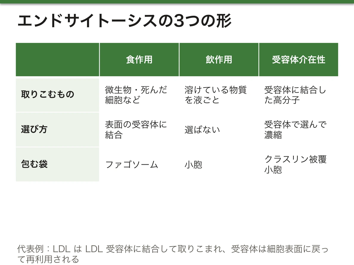
代表例がコレステロールの取りこみである。コレステロールは血液中をLDL(低密度リポタンパク質)という粒子として運ばれる。LDLは細胞表面のLDL受容体に結合し、被覆ピットから取りこまれる。小胞の中が酸性になるとLDLは受容体から離れ、LDLはリソソームで分解されてコレステロールが放出される。受容体のほうは細胞表面に戻されて再利用される。
なぜわざわざ受容体を使うのか。飲作用のように液ごと取りこむと、血中に薄く存在するものは少ししか入らない。受容体が狙った分子を被覆ピットに集めて濃縮するので、効率が数百倍以上に上がる。しかも受容体の数を変えれば取りこみ量を調節できる。
家族性高コレステロール血症は、主にLDL受容体の遺伝子の変異で起き、LDLが細胞に取りこまれずに血中にたまる。若いうちから心筋梗塞を起こし、アキレス腱の肥厚や黄色腫がみられる。スタチンは細胞のコレステロール合成を抑え、その結果LDL受容体を増やして血中のLDLを下げる。PCSK9阻害薬は、LDL受容体を分解へ導くPCSK9を止めて、受容体が再利用される回数を増やす。
English version
Core
Macromolecules such as proteins, nucleic acids, polysaccharides and lipids cannot be moved by channels or carriers. So eukaryotic cells fold in the plasma membrane to form a vesicle, wrap up outside material and take it in. This is endocytosis, and it has three forms.
Phagocytosis takes in large particles such as microorganisms and dead cells. When a particle binds receptors on the surface, actin pushes out pseudopodia, which close around it to form a phagosome. The phagosome fuses with a lysosome to form a phagolysosome, where the contents are digested. The professional phagocytes are macrophages, neutrophils and dendritic cells.
Pinocytosis takes in dissolved material together with fluid, without selection.
Receptor-mediated endocytosis selectively takes in macromolecules bound to surface receptors. The receptors gather in coated pits, whose cytoplasmic side is covered with the protein clathrin; the pits pinch off as clathrin-coated vesicles.
Going further
The classic example is cholesterol uptake. Cholesterol travels in the blood in particles called LDL (low-density lipoprotein). LDL binds the LDL receptor on the cell surface and is taken in through coated pits. When the vesicle becomes acidic, LDL lets go of the receptor; LDL is broken down in lysosomes and cholesterol is released. The receptor is returned to the surface and reused.
Why use receptors at all? Taking in fluid as in pinocytosis brings in only a little of something present at low levels in the blood. Receptors collect the target molecules in coated pits and concentrate them, raising efficiency by hundreds of times or more. Also, the cell can control uptake by changing the number of receptors.
In the clinic
Familial hypercholesterolaemia is caused mainly by variants in the LDL receptor gene. LDL is not taken into cells and builds up in the blood. Patients have heart attacks at a young age and show thickened Achilles tendons and xanthomas. Statins block cholesterol synthesis in cells, which increases LDL receptors and lowers blood LDL. PCSK9 inhibitors block PCSK9, which sends LDL receptors to breakdown, so each receptor is reused more times.
エキソサイトーシスと小胞輸送 — いつでも出すか、合図を待って出すかExocytosis
エキソサイトーシスは、細胞が作った分子を小胞に詰めて細胞の外に放出する過程である。分泌小胞が細胞膜と融合すると、中身が外に出て、小胞の膜は細胞膜の一部になる。2つの形がある。
| 構成性エキソサイトーシス | 調節性エキソサイトーシス | |
|---|---|---|
| 行う細胞 | すべての細胞 | 分泌の専門の細胞 |
| 合図 | 不要(作られたら順次出す) | 特定の合図(多くは細胞内Ca2+の上昇) |
| 運ぶもの | 多くの膜タンパク質や細胞外基質の成分 | ホルモン、神経伝達物質、サイトカインなど |
小胞輸送には、小胞の被覆(コート)の種類による分担がある。クラスリン被覆小胞は細胞膜とトランスゴルジ網からの輸送、COPII被覆小胞はERからゴルジ装置へ、COPI被覆小胞はゴルジ装置からERへの逆向きの輸送やゴルジ槽どうしの輸送を担う。被覆は積み荷を集めて小胞の形を作り、目的の膜と融合する前に脱ぎ捨てられる。こうして2つの膜の細胞質側の面が直接触れ合い、融合できる。
調節性エキソサイトーシスの代表は、膵島のβ細胞からのインスリン分泌である。①血糖が上がるとブドウ糖がβ細胞に入り、代謝されてATP/ADP比が上がる。②ATPが細胞膜のATP感受性K+チャネルを閉じる。③K+が出られなくなって膜が脱分極する。④電位依存性Ca2+チャネルが開いてCa2+が流れこむ。⑤Ca2+の上昇を合図にインスリンの小胞が細胞膜と融合する。
なぜ代謝がそのまま分泌の合図になるのか。β細胞はブドウ糖を「測る」専用の受容体をもつ代わりに、ブドウ糖を燃やした結果のATPの量をチャネルの開閉に結びつけている。代謝の速さが血糖をそのまま反映するので、血糖センサーとして働く。なお、エキソサイトーシスで足された膜は、ほぼ同じ速さでエンドサイトーシスによって回収され、細胞の表面積が保たれる。
糖尿病の薬のスルホニル尿素薬は、ATP感受性K+チャネルを直接閉じて、血糖に関係なくインスリンを出させる。そのため効きすぎると低血糖を起こしやすい。このチャネルの遺伝子が活性化する変異は新生児糖尿病の原因になり、スルホニル尿素薬がよく効く。
English version
Core
Exocytosis is how a cell packs molecules it has made into vesicles and releases them outside. When a secretory vesicle fuses with the plasma membrane, its contents go out and its membrane becomes part of the plasma membrane. There are two forms.
| Constitutive exocytosis | Regulated exocytosis | |
|---|---|---|
| Which cells | All cells | Specialised secretory cells |
| Signal | None needed (released as made) | A specific signal (often a rise in intracellular Ca2+) |
| Cargo | Many membrane proteins and extracellular matrix components | Hormones, neurotransmitters, cytokines |
Vesicle traffic is divided by the type of coat. Clathrin-coated vesicles carry cargo from the plasma membrane and the trans-Golgi network; COPII-coated vesicles go from the ER to the Golgi; COPI-coated vesicles go back from the Golgi to the ER and between Golgi cisternae. The coat collects cargo and shapes the vesicle, and it is thrown off before fusion with the target membrane, so the cytosolic faces of the two membranes can touch directly and fuse.
Going further
The classic example of regulated exocytosis is insulin secretion from pancreatic β cells. (1) When blood glucose rises, glucose enters the β cell and is metabolised, so the ATP/ADP ratio rises. (2) ATP closes ATP-sensitive K+ channels in the membrane. (3) K+ cannot leave, and the membrane depolarises. (4) Voltage-gated Ca2+ channels open and Ca2+ flows in. (5) The rise in Ca2+ signals insulin vesicles to fuse with the membrane.
Why does metabolism itself become the signal for secretion? Instead of having a receptor that “measures” glucose, the β cell links the amount of ATP made by burning glucose to the opening and closing of a channel. Because the rate of metabolism reflects blood glucose directly, the cell works as a glucose sensor. Membrane added by exocytosis is taken back by endocytosis at nearly the same rate, so the cell’s surface area stays the same.
In the clinic
The diabetes drugs sulfonylureas close ATP-sensitive K+ channels directly and release insulin regardless of blood glucose, so too strong an effect easily causes hypoglycaemia. Activating variants in the genes for this channel cause neonatal diabetes, which responds well to sulfonylureas.
まとめ
| 内膜系 — 作る・仕分ける・壊す、をつなぐ膜のネットワーク | 内膜系は、核膜、小胞体(ER)、ゴルジ装置、リソソーム、小胞からなる膜の系である。 |
|---|---|
| 細胞膜の構造 — 脂質の二重層に、タンパク質と糖鎖がのる | 膜の脂質は両親媒性で、水になじむ極性の頭部と、水をはじく尾部をもつ。 |
| 透過性・単純拡散・浸透 — 膜は何を素通りさせるか | 水、無機イオン、小さな有機分子(糖、ビタミン、脂肪酸など)は、生物の重さの75〜80%を占める。 |
| 促進拡散 — 担体は形を変えて運び、チャネルは穴を開ける | 促進拡散は、膜のタンパク質の助けで、極性や電荷をもつ分子(糖、アミノ酸、ヌクレオシド、イオン)を通す輸送である。 |
| 能動輸送 — ポンプが坂を作り、共輸送がその坂を使う | 能動輸送は、別の反応のエネルギーを使って、分子を濃度や電位の勾配に逆らって「上り坂」に運ぶ輸送である。 |
| チャネルの病気 — 嚢胞性線維症とCFTR | 嚢胞性線維症(CF)は、ヨーロッパ系の人々で最も多い単一遺伝子病の1つで、常染色体劣性の形式で遺伝する。 |
| エンドサイトーシス — 膜を袋にして外のものを取りこむ | タンパク質、核酸、多糖、脂質などの高分子は、チャネルや担体では運べない。 |
| エキソサイトーシスと小胞輸送 — いつでも出すか、合図を待って出すか | エキソサイトーシスは、細胞が作った分子を小胞に詰めて細胞の外に放出する過程である。 |
このノートは医学生が授業の内容を自分の言葉でまとめたもので、診療の判断に使うものではありません。誤りに気づいたらお知らせください。