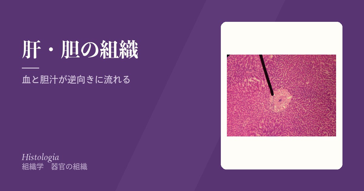
肝臓の組織学は「血液と胆汁が反対向きに流れている」という一点を掴めば、あとは全部そこから導ける。
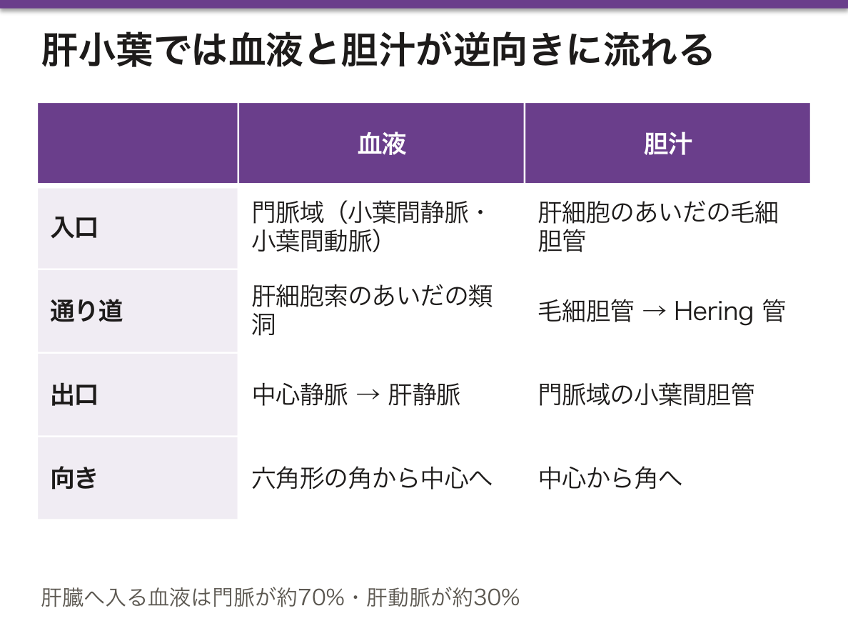
- 血液——門脈域(外側)から 類洞 を通って 中心静脈(内側)へ。外から内へ
- 胆汁——肝細胞のあいだの 毛細胆管 から門脈域の胆管へ。内から外へ
同じ肝細胞が、片側の面で血液とやりとりし、反対側の面で胆汁を出す。1個の細胞が二方向を向いている——これが肝臓の作りの本質である。
この流れの向きから、どこが最初に傷むかが決まる。血液は外側から入るので、酸素も栄養も毒物も、まず門脈域の近くの細胞に届く。中心静脈のまわりはいちばん最後に酸素が届く場所——だからうっ血も虚血も中心静脈周囲から始まる。
膵臓は同じ器官の中に外分泌部(腺房)と内分泌部(膵島)が同居する。内分泌部は内分泌腺の章にある。ここでは外分泌部を扱う。
- 肝小葉と門脈域
- 類洞と Disse 腔
- 胆道 — 毛細胆管から十二指腸まで
- 肝細胞 — 1つの細胞が3方向を向いている
- 胆嚢 — 濃縮するためだけの袋
- 肝の再生 — 切っても戻る、唯一の大きな実質臓器
- 肝星細胞 — ビタミン A を溜め、線維化の主役に転じる細胞
肝小葉と門脈域Lobulus hepatis
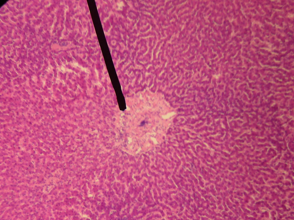
肝臓の構造上の単位。直径1〜2mm の六角形。
- 中心静脈——六角形の中心。ここから肝静脈へ集まる
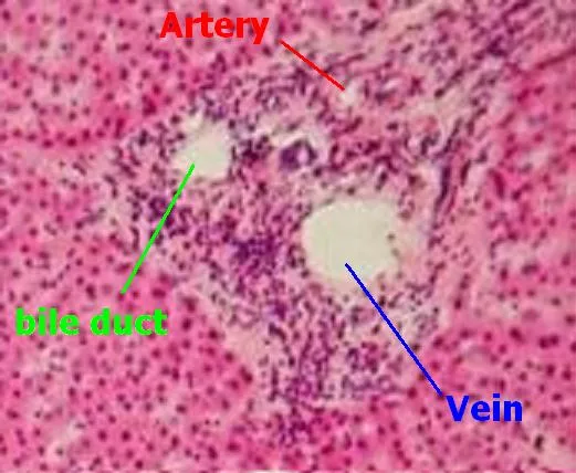
- 門脈域(グリソン鞘)——六角形の角。小葉間動脈・小葉間静脈・小葉間胆管の3つが束になる(三つ組)
- 肝細胞索——中心静脈から放射状に並ぶ肝細胞の列
血液は角から中心へ、胆汁は中心から角へ流れる。
肝臓へ入る血液は門脈が約70%・肝動脈が約30%。栄養は門脈、酸素は主に肝動脈が運ぶ。
同じ組織を3通りの区切り方で見ることができ、目的によって使い分ける。
- 肝小葉——中心静脈が真ん中。形(組織像)を説明するのに便利
- 門脈小葉——門脈域が真ん中。胆汁の流れ(外分泌腺として)を説明する区切り
- 肝腺房——2つの門脈域を結ぶ軸を中心に、血流の順に帯1・2・3と分ける。病変の分布を説明するのに最も役に立つ
帯1(門脈域寄り)は酸素が豊富、帯3(中心静脈寄り)は最も乏しい。ここから病変の場所が予測できる。
- 虚血・うっ血性肝障害——酸素の届きにくい帯3(小葉中心性)から壊死する。右心不全でできるにくずく肝がこれ
- アセトアミノフェン中毒——毒性代謝物を作るチトクロムP450 が帯3に多いので、やはり中心静脈周囲が壊死する
- ウイルス性肝炎——炎症細胞は門脈域に集まる。だから線維化も門脈域から始まる
「どの帯が傷んでいるか」を見れば原因の系統が絞れる——酸素と代謝の地図が、そのまま病理の地図になっている。
肝小葉のどこが傷むかで、原因が推定できる。血流は門脈域から中心静脈へ流れるので、中心静脈周囲(3帯)が最も酸素が薄い。
中心静脈周囲(3帯)の壊死 — 虚血(ショック肝)・うっ血(右心不全)・アセトアミノフェン中毒。薬物代謝酵素が最も多い帯でもあるため、中毒性肝障害はここから起こる。うっ血肝の割面が「にくずく肝」に見えるのはこの分布による。
門脈域周囲(1帯) — ウイルス性肝炎の界面肝炎・胆汁うっ滞。
肝硬変では小葉構造が壊れ、再生結節を線維が取り囲む。門脈血が類洞を通れなくなる(門脈圧亢進)と同時に、肝細胞が血液と接する面積が減る(肝不全)——2つの結果が同じ構造の破壊から出ている。
English version
Core
The structural unit of the liver: a hexagon 1–2 mm across.
- Central vein — at the centre of the hexagon, draining to the hepatic veins.
- Portal tract (Glisson’s capsule) — at the corners. Interlobular artery, vein and bile duct bound together (the portal triad).
- Hepatic cords — rows of hepatocytes radiating from the central vein.
Blood flows from corner to centre; bile from centre to corner.
The blood entering the liver is about 70 per cent portal, 30 per cent arterial. The portal vein carries the nutrients, the hepatic artery chiefly the oxygen.
Going further
The same tissue can be divided three ways, and each is used for a different purpose.
- Hepatic lobule — the central vein in the middle. Convenient for describing the histological appearance.
- Portal lobule — the portal tract in the middle. The division that describes the flow of bile (the liver as an exocrine gland).
- Hepatic acinus — centred on the axis joining two portal tracts, divided into zones 1, 2 and 3 in the order of blood flow. The most useful for describing the distribution of lesions.
Zone 1 (near the portal tract) is rich in oxygen, zone 3 (near the central vein) the poorest. From this the site of a lesion can be predicted.
- Ischaemic and congestive injury — necrosis begins in zone 3, where oxygen arrives least (centrilobular). The nutmeg liver of right heart failure is this.
- Paracetamol poisoning — the cytochrome P450 that makes the toxic metabolite is concentrated in zone 3, so again it is the perivenular region that necroses.
- Viral hepatitis — the inflammatory cells gather in the portal tracts, so fibrosis also begins there.
Seeing which zone is damaged narrows the class of cause — the map of oxygen and metabolism is directly the map of pathology.
In the clinic
Where in the lobule the damage lies allows the cause to be inferred. Blood flows from portal tract to central vein, so the perivenular region (zone 3) has the thinnest oxygen.
Perivenular (zone 3) necrosis — ischaemia (shock liver), congestion (right heart failure), paracetamol poisoning. It is also the zone richest in drug-metabolising enzymes, so toxic liver injury starts here. The cut surface of a congested liver looks like nutmeg because of this distribution.
Periportal (zone 1) — interface hepatitis of viral hepatitis, cholestasis.
In cirrhosis the lobular architecture is destroyed and regenerative nodules are surrounded by fibrous tissue. Portal blood can no longer pass through the sinusoids (portal hypertension) and at the same time the area over which hepatocytes meet blood falls (liver failure) — two results issuing from the destruction of the same structure.
類洞と Disse 腔Sinusoides hepatis
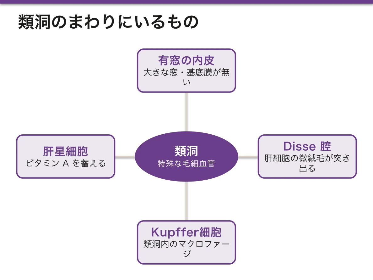
肝細胞索のあいだを流れる、特殊な毛細血管。
- 内皮に大きな窓があり、基底膜が無い——ふつうの毛細血管と決定的に違う
- Disse 腔——内皮と肝細胞のあいだの隙間。ここに肝細胞の微絨毛が突き出る
- Kupffer 細胞——類洞内のマクロファージ。門脈から来た細菌・老化赤血球を処理する
- 肝星細胞(伊東細胞)——Disse 腔にあり、ビタミンAを脂肪滴に蓄える
基底膜が無く内皮に窓があることは、肝臓が「血液の成分と直接やりとりする臓器」であるために必須である。アルブミンも凝固因子もリポ蛋白も、関門を挟まずに血漿と行き来する。
この開けっ放しの構造が、病気になると閉じる。慢性の障害では肝星細胞が活性化して筋線維芽細胞に変わり、コラーゲンを作ってDisse 腔に基底膜を作ってしまう(類洞の毛細血管化)。
結果、2つのことが同時に起こる。
- 物質のやりとりができなくなる——肝細胞が生きていても機能が落ちる
- 類洞が硬く狭くなる——門脈圧が上がる
肝硬変の門脈圧亢進は「肝臓が硬いから」ではなく、類洞が塞がるからである。だから食道静脈瘤も腹水も脾腫も、この一つの変化から出てくる。
星細胞がビタミンAを貯めているという事実にも意味がある。活性化すると脂肪滴(ビタミンA)を失って線維を作る細胞に変わる——貯蔵係が破壊者に転職する。線維化を止める薬の標的が星細胞なのはこのためである。
肝生検で見るのは細胞の死に方より線維の伸び方である。門脈域から始まった線維が隣の門脈域や中心静脈へ橋を架け(橋渡し線維化)、やがて肝細胞を取り囲んで偽小葉を作った状態が肝硬変である。
ここから先が非可逆とされてきたが、原因(ウイルス・アルコール・脂肪)を除けば線維化は戻りうることが分かってきた。C型肝炎の直接作用型抗ウイルス薬で線維化が改善するのは、星細胞の活性化が止まるためである。
肝硬変では肝細胞癌のリスクが線維化の段階に比例して上がる。だから病期分類が、そのまま画像による定期監視の間隔を決める。
English version
Core
A specialised capillary running between the hepatic cords.
- The endothelium has large fenestrations and there is no basal lamina — the decisive difference from an ordinary capillary.
- Space of Disse — the gap between endothelium and hepatocyte, into which the microvilli of the hepatocyte project.
- Kupffer cell — the macrophage within the sinusoid, dealing with bacteria arriving by the portal vein and with aged red cells.
- Hepatic stellate (Ito) cell — in the space of Disse, storing vitamin A in lipid droplets.
Going further
The absence of a basal lamina and the fenestrated endothelium are essential if the liver is to be an organ that exchanges directly with the constituents of the blood. Albumin, clotting factors and lipoproteins all pass to and from the plasma with no barrier between.
This wide-open structure closes when disease occurs. In chronic injury the stellate cells are activated, become myofibroblasts, make collagen and lay down a basal lamina in the space of Disse (capillarisation of the sinusoid). Two things then happen at once.
- Exchange of substances fails — function falls although the hepatocytes are alive.
- The sinusoids stiffen and narrow — the portal pressure rises.
Portal hypertension in cirrhosis is not because the liver is hard but because the sinusoids are obstructed. So oesophageal varices, ascites and splenomegaly all issue from this one change.
That the stellate cell stores vitamin A also has meaning. On activation it loses its lipid droplets (the vitamin A) and becomes a cell that makes fibrous tissue — the storekeeper changes trade and becomes the destroyer. This is why the stellate cell is the target of drugs intended to stop fibrosis.
In the clinic
What is looked at in a liver biopsy is less how the cells die than how the fibrous tissue extends. Fibrosis starting in a portal tract bridges to the next portal tract or to a central vein (bridging fibrosis), and when it finally surrounds the hepatocytes to form pseudolobules, that is cirrhosis.
Beyond this point was long held to be irreversible, but it is now known that if the cause is removed (virus, alcohol, fat) the fibrosis can regress. Fibrosis improves after direct-acting antivirals for hepatitis C because the activation of the stellate cells stops.
In cirrhosis the risk of hepatocellular carcinoma rises in proportion to the stage of fibrosis, so the staging itself sets the interval of imaging surveillance.
胆道 — 毛細胆管から十二指腸までVesica biliaris
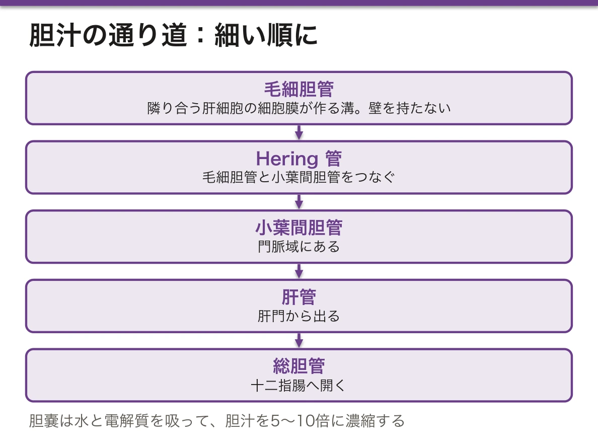
胆汁の通り道を、細い順に。
- 毛細胆管——隣り合う肝細胞の細胞膜が作る溝。壁を持たない。密着結合で血液側と隔てられる
- Hering 管 → 小葉間胆管(門脈域)→ 肝管 → 総胆管
- 胆嚢——単層円柱上皮。粘膜下層を持たない。粘膜ヒダが深く落ち込む(Rokitansky-Aschoff 洞)
胆嚢の仕事は濃縮で、水と電解質を吸って胆汁を5〜10倍にする。
毛細胆管に壁が無いことが、肝障害の検査値を説明する。血液と胆汁を隔てているのは肝細胞どうしの密着結合だけなので、細胞が壊れるとその境が破れ、胆汁が直接血中へ漏れる。
だから検査値は2系統に分かれ、どちらが上がるかで障害の型が読める。
- AST・ALT——肝細胞の中身。細胞が壊れたことを示す(肝細胞障害型)
- ALP・γ-GT——胆管側の細胞膜にある酵素。胆汁がうっ滞すると誘導されて増える(胆汁うっ滞型)
ALP が上がるのは「胆管が詰まったから漏れる」のではなく、うっ滞そのものが酵素の産生を増やすからである。だから閉塞では ALP と γ-GT が並んで上がる。ALP だけ上がって γ-GT が正常なら、出所は肝臓ではなく骨(骨芽細胞)である——同じ酵素が2か所から出るという事実が、そのまま鑑別になる。
胆嚢に粘膜下層が無いことも重要で、胆嚢癌は早期から筋層を越えて肝へ直接広がる。壁の層が少ない分、進行が速い。
Rokitansky-Aschoff 洞は粘膜が筋層へ落ち込んだ憩室様の構造で、慢性胆嚢炎で目立つ。ここに胆汁が滞ると炎症が繰り返される。
閉塞性黄疸では直接(抱合型)ビリルビンが上がり、尿が濃くなり、便が白くなる。抱合されたビリルビンは水溶性なので尿に出られるが、腸へ届かないためウロビリノーゲンが作られない。色の変化3つが、すべて抱合という一段階から説明できる。
痛みの出方にも構造が効く。胆嚢は内臓痛として心窩部に感じられるが、炎症が壁を越えて壁側腹膜に及ぶと右季肋部の限局した痛みに変わる(Murphy 徴候)。痛みの場所が移ったら、炎症が層を越えた合図である。
English version
Core
The path of bile, from the finest upwards.
- Bile canaliculus — a groove formed by the membranes of adjacent hepatocytes. It has no wall of its own, and is sealed off from the blood side by tight junctions.
- Canals of Hering → interlobular bile duct (in the portal tract) → hepatic duct → common bile duct.
- Gallbladder — simple columnar epithelium. No submucosa. The mucosal folds sink deeply in (Rokitansky–Aschoff sinuses).
The gallbladder’s work is concentration, absorbing water and electrolytes to concentrate the bile 5–10 fold.
Going further
That the canaliculus has no wall explains the biochemical tests of liver injury. All that separates blood from bile is the tight junction between hepatocytes, so when cells are destroyed that boundary breaks and bile leaks directly into the blood. Hence the tests fall into two groups, and which of them rises tells you the type of injury.
- AST, ALT — the contents of the hepatocyte. They show that cells have been destroyed (hepatocellular pattern).
- ALP, γ-GT — enzymes on the membrane facing the bile duct. Cholestasis induces them (cholestatic pattern).
ALP rises not because a blocked duct leaks but because the cholestasis itself increases production of the enzyme. So in obstruction ALP and γ-GT rise together. If ALP alone is raised with a normal γ-GT, the source is not liver but bone (osteoblasts) — the fact that one enzyme comes from two places is itself the differential.
That the gallbladder has no submucosa also matters: carcinoma of the gallbladder crosses the muscle early and spreads directly into the liver. With fewer layers in the wall, it advances faster.
In the clinic
Rokitansky–Aschoff sinuses are diverticulum-like invaginations of mucosa into the muscle, prominent in chronic cholecystitis. Bile stagnating in them makes the inflammation recur.
In obstructive jaundice the conjugated (direct) bilirubin rises, the urine darkens and the stool becomes pale. Conjugated bilirubin is water-soluble and so can appear in the urine, but as it does not reach the gut no urobilinogen is made. All three changes in colour are explained by the single step of conjugation.
Structure tells in the pain too. The gallbladder is felt as visceral pain in the epigastrium, but when inflammation crosses the wall to the parietal peritoneum it becomes localised right hypochondrial pain (Murphy’s sign). A shift in the site of the pain is the signal that inflammation has crossed a layer.
肝細胞 — 1つの細胞が3方向を向いているHepatocytus
肝細胞は多面体で、面ごとに違う仕事をするのが最大の特徴。
| 面 | 向く先 | 働き |
|---|---|---|
| 血管側(基底面) | Disse 腔=類洞 | 微絨毛を出して物質を取り込み、蛋白を血中へ分泌 |
| 胆管側(頂端面) | 毛細胆管——隣の肝細胞との間の溝 | 胆汁を分泌。トランスポーター(BSEP・MRP2)が並ぶ |
| 側面 | 隣の肝細胞 | 密着結合・ギャップ結合 |
毛細胆管には固有の壁が無い——隣り合う肝細胞の細胞膜が作る溝で、周囲を密着結合が封じている。「管」なのに管を作る細胞がいないという珍しい構造。
細胞内には滑面小胞体が豊富(薬物代謝・グリコーゲン代謝)、ペルオキシソーム(長鎖脂肪酸のβ酸化・カタラーゼ)、多数のミトコンドリア。核は多倍体・二核のものが多い。
「血と胆汁が逆向きに流れる」という肝の設計が、この細胞の極性から生まれる。血液は門脈域 → 類洞 → 中心静脈へ、胆汁は毛細胆管 → 門脈域の胆管へ——同じ肝細胞索の中を、正反対の向きに流れる。
密着結合が壊れると胆汁が血中へ漏れる——これが胆汁うっ滞の細胞レベルの姿。血液検査で直接ビリルビンと ALP・γ-GTP が上がるのは、この漏出と、胆管上皮の酵素誘導による。
肝小葉の場所によって肝細胞の性格が違う(代謝の帯状分布)——これが病変の分布を決める。
| 帯 | 位置 | 性格 | 侵されやすいもの |
|---|---|---|---|
| ゾーン1 | 門脈域の周り | 酸素が豊富。糖新生・尿素合成・β酸化 | ウイルス性肝炎、鉄の沈着 |
| ゾーン3 | 中心静脈の周り | 酸素が乏しい。解糖・脂質合成・薬物代謝(CYP2E1) | 虚血(うっ血肝の中心性壊死)、アセトアミノフェン中毒、アルコール性肝障害 |
アセトアミノフェンがゾーン3を壊すのは、そこに CYP2E1 が集中していて毒性代謝物 NAPQI が作られるから。アルコールは CYP2E1 を誘導するので、常習飲酒者では通常量でも肝障害が起こりうる。解毒にはN-アセチルシステイン(グルタチオンを補う)。
面ごとに仕事が違うので、どの面が壊れたかで検査値の型が変わる。
肝細胞の障害(細胞質・血管側) — AST・ALT が上がる。ウイルス性肝炎・薬物性・虚血。
胆管側(毛細胆管)の障害 — ALP・γ-GTP が上がる。胆汁うっ滞。「どちらが優位か」で肝細胞性か胆汁うっ滞性かを分けるのが肝障害を見たときの最初の作業になる。
AST/ALT 比も手がかりになる。ALT 優位=ウイルス性・脂肪肝、AST 優位(比 2 以上)=アルコール性(AST はミトコンドリアにも多く、アルコールでビタミン B6 が不足して ALT の合成が落ちる)。
肝細胞は合成もしているので、アルブミンと PT(プロトロンビン時間)が肝の予備能を映す。Child–Pugh 分類はこれらを組み合わせている。
English version
Core
The hepatocyte is polyhedral, and its chief feature is that each face does a different job.
| Face | Faces onto | Work |
|---|---|---|
| Vascular (basal) | Space of Disse = sinusoid | Microvilli take up substances; proteins are secreted into the blood |
| Biliary (apical) | Bile canaliculus — the groove between neighbouring hepatocytes | Secretes bile; transporters (BSEP, MRP2) are lined up here |
| Lateral | The neighbouring hepatocyte | Tight junctions, gap junctions |
The canaliculus has no wall of its own — a groove formed by the membranes of adjacent hepatocytes and sealed around by tight junctions. An unusual structure: a duct with no cell to make it.
The cytoplasm is rich in smooth endoplasmic reticulum (drug and glycogen metabolism), peroxisomes (β-oxidation of long-chain fatty acids, catalase) and numerous mitochondria. Many nuclei are polyploid or the cells binucleate.
Going further
The liver’s design — blood and bile flowing in opposite directions — arises from the polarity of this cell. Blood runs portal tract → sinusoid → central vein, bile runs canaliculus → duct in the portal tract: opposite directions within the same hepatic cord.
When the tight junctions break, bile leaks into the blood — this is cholestasis at the cellular level. Direct bilirubin, ALP and γ-GT rise in the blood from that leakage together with induction of the enzymes of the biliary epithelium.
Hepatocytes differ in character according to where they lie in the lobule (metabolic zonation) — and this decides the distribution of lesions.
| Zone | Position | Character | Susceptible to |
|---|---|---|---|
| Zone 1 | Around the portal tract | Oxygen-rich. Gluconeogenesis, urea synthesis, β-oxidation | Viral hepatitis, iron deposition |
| Zone 3 | Around the central vein | Oxygen-poor. Glycolysis, lipogenesis, drug metabolism (CYP2E1) | Ischaemia (centrilobular necrosis of the congested liver), paracetamol poisoning, alcoholic injury |
Paracetamol destroys zone 3 because CYP2E1 is concentrated there and makes the toxic metabolite NAPQI. Alcohol induces CYP2E1, so in a habitual drinker liver injury can occur at an ordinary dose. The antidote is N-acetylcysteine (replenishing glutathione).
In the clinic
Because each face does a different job, which face is damaged changes the pattern of the tests.
- Injury to the hepatocyte (cytoplasm, vascular face) — AST and ALT rise. Viral, drug-induced, ischaemic.
- Injury on the biliary side (canaliculus) — ALP and γ-GT rise. Cholestasis.
Deciding which predominates — hepatocellular or cholestatic — is the first task on seeing abnormal liver tests.
The AST/ALT ratio is also a clue. ALT predominant = viral or fatty liver; AST predominant (ratio above 2) = alcoholic (AST is also abundant in mitochondria, and alcohol causes vitamin B6 deficiency which reduces ALT synthesis).
The hepatocyte also synthesises, so albumin and the prothrombin time mirror hepatic reserve. The Child–Pugh classification combines them.
胆嚢 — 濃縮するためだけの袋Vesica biliaris
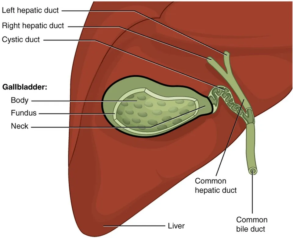
胆嚢の壁は消化管の4層構造から「粘膜筋板」と「粘膜下層」が欠けている——これが組織学上の最大の特徴で、臨床に直結する。
| 層 | 内容 |
|---|---|
| 粘膜 | 単層円柱上皮(微絨毛を持ち、吸収に適する)+粘膜固有層。高いヒダを作る |
| 筋層 | 不規則に走る平滑筋(層をなさない) |
| 外膜/漿膜 | 肝に接する面は外膜、遊離面は漿膜 |
粘膜のヒダが深く落ち込んで筋層を貫くことがあり、Rokitansky-Aschoff 洞と呼ばれる(正常でも見られるが、腺筋腫症で著明)。
胆嚢の仕事は濃縮——Na⁺ を能動的に吸収し、水がそれに従うことで、肝から来た胆汁を5〜10倍に濃縮して蓄える。CCK(脂肪が十二指腸へ来ると分泌)で収縮し、Oddi 括約筋は弛緩する。
粘膜下層が無いことの帰結が2つある。
①胆嚢癌の予後が悪い——粘膜下層という「緩衝地帯」が無く、筋層の外はすぐ漿膜下層、その先は肝実質。癌は薄い壁を短時間で貫き、肝へ直接浸潤する。だから早期に見つけないと切除しても再発する。
②T分類が他臓器と対応しない——胃や大腸の「粘膜下層まで=早期癌」という枠組みがそのまま使えない。
胆石の成り立ちも濃縮機能から説明できる。胆汁はコレステロール・胆汁酸・リン脂質の絶妙な比でミセルを作り、コレステロールを溶かしている。濃縮の過程でこの比が崩れると析出する。
| 石の種類 | 成分 | 背景 |
|---|---|---|
| コレステロール石 | コレステロール | 4F(Female・Forty・Fatty・Fertile)、急激な減量、経口避妊薬 |
| 黒色石 | ビリルビンカルシウム | 溶血、肝硬変 |
| ビリルビンカルシウム石(褐色石) | — | 胆道感染——細菌のβ-グルクロニダーゼが抱合ビリルビンを遊離させる |
急性胆嚢炎は胆嚢管の閉塞から始まる——出口が詰まり、濃縮された胆汁が粘膜を化学的に傷害し、そこに感染が加わる。Murphy 徴候(右季肋部を圧迫しながら吸気させると痛みで息が止まる)が古典的所見。
粘膜下層と粘膜筋板が無いことが、胆嚢癌の深達度分類と予後を特殊にしている。層が少ないぶん、早く漿膜に達し、早く肝へ直接浸潤する。
胆嚢摘出標本で偶然に癌が見つかることがあり、深達度によっては追加の肝切除とリンパ節郭清が必要になる。
Rokitansky–Aschoff 洞 — 粘膜上皮が筋層内へ落ち込んだ憩室様の構造。慢性胆嚢炎で目立つようになり、胆嚢腺筋腫症の本体でもある。
胆嚢は濃縮するだけの袋である。だから摘出しても消化は保たれる(胆汁は肝から直接流れる)。術後に脂っこい食事で下痢しやすいのは濃縮と貯留の機能を失ったからである。
English version
Core
The wall of the gallbladder lacks the muscularis mucosae and the submucosa of the four-layered gut wall — its greatest histological feature, and one with direct clinical consequences.
| Layer | Contents |
|---|---|
| Mucosa | Simple columnar epithelium (microvilli, suited to absorption) + lamina propria. Forms tall folds |
| Muscle | Smooth muscle running irregularly (not in layers) |
| Adventitia / serosa | Adventitia where it meets the liver, serosa on the free surface |
The mucosal folds may sink deeply and penetrate the muscle as Rokitansky–Aschoff sinuses (present normally, but prominent in adenomyomatosis).
The gallbladder’s work is concentration — Na⁺ is absorbed actively and water follows, concentrating the bile from the liver 5–10 fold for storage. CCK (released when fat reaches the duodenum) contracts it while the sphincter of Oddi relaxes.
Going further
The absence of a submucosa has two consequences.
- ① The prognosis of gallbladder cancer is poor — there is no submucosal buffer zone; outside the muscle is the subserosa and immediately beyond it the liver parenchyma. The tumour crosses the thin wall quickly and invades the liver directly, so unless found early it recurs after resection.
- ② The T classification does not correspond to that of other organs — the framework of "into the submucosa = early cancer" used in stomach and colon cannot be applied.
The formation of gallstones is also explained by the concentrating function. Bile holds cholesterol in solution in micelles at an exact ratio of cholesterol, bile acids and phospholipid. If that ratio breaks down during concentration, it precipitates.
| Stone | Composition | Background |
|---|---|---|
| Cholesterol stone | Cholesterol | The four Fs (female, forty, fatty, fertile), rapid weight loss, oral contraceptives |
| Black pigment stone | Calcium bilirubinate | Haemolysis, cirrhosis |
| Brown pigment stone | Calcium bilirubinate | Biliary infection — bacterial β-glucuronidase frees conjugated bilirubin |
Acute cholecystitis begins with obstruction of the cystic duct — the exit blocks, the concentrated bile injures the mucosa chemically, and infection is added. Murphy’s sign (pressure in the right hypochondrium arrests inspiration through pain) is the classic finding.
In the clinic
The absence of submucosa and muscularis mucosae makes the staging and prognosis of gallbladder cancer peculiar. With fewer layers it reaches the serosa early and invades the liver directly early. Cancer is sometimes found incidentally in a cholecystectomy specimen, and depending on the depth, additional liver resection and lymph node dissection are required.
Rokitansky–Aschoff sinuses — diverticulum-like invaginations of mucosal epithelium into the muscle. They become prominent in chronic cholecystitis and are the substance of adenomyomatosis.
The gallbladder is only a bag for concentrating, so digestion is preserved after removal (bile flows straight from the liver). Diarrhoea after fatty meals postoperatively is the loss of the functions of concentration and storage.
肝の再生 — 切っても戻る、唯一の大きな実質臓器Regeneratio hepatis
肝細胞は普段はほとんど分裂していない(G0 期)が、失われると一斉に細胞周期へ入る。
7割を切除しても、数週間でほぼ元の重量に戻る——ただし葉の形は戻らない。残った葉が肥大する(代償性肥大)。
関わる因子——肝細胞増殖因子(HGF)・IL-6・TNF-α・上皮成長因子。止める側は TGF-β。
大量に失われて肝細胞が分裂できないときは、肝前駆細胞(Hering 管の卵円細胞)が動員される。
再生できるのは、線維の骨組み(細網線維)が残っている場合になる。
「骨組みが残れば元に戻り、壊れれば結節になる」という一線が、肝疾患の帰結を決めている。肝硬変は、再生が起きないのではなく、間違った形で起きている状態になる(修復と創傷治癒の章)——「再生結節と線維の隔壁が、血流の道を壊す」。
だから肝硬変では、再生しているのに機能が戻らない(肝の各論の章)——「細胞は増えても、類洞と胆管との位置関係が失われている」。
この再生能があるから、生体肝移植が成り立つ(肝の外科解剖の章)——「提供者も受容者も、数か月で必要な大きさまで戻る」。
切除の限界は「残る肝の量」で決まる——「術前に残肝容積を計算し、足りなければ門脈塞栓術で残す側を先に肥大させる」。
再生と発癌は、同じ土台の上にある(発癌の分子機構の章)——「慢性肝炎で壊れては再生を繰り返す肝から、肝細胞癌が出る」。
肝は再生するが、線維化は元に戻らない。この非対称が予後を決める。
肝切除は残肝の量と機能で決まる。正常肝なら70%まで切除できるが、肝硬変があると再生能が落ちて肝不全になる。ICG 検査・Child–Pugh 分類で術前に評価する。
肝硬変では再生が「結節」として起こる。秩序を失った再生であり、肝細胞癌の温床になる。肝硬変患者には定期的な画像と AFP による監視が要る。
肝星細胞(Ito 細胞)が線維化の主役である。ふだんはビタミン A を蓄えているが、障害を受けると筋線維芽細胞様に変化してコラーゲンを出す。原因を取り除けば線維化がある程度戻ることがC 型肝炎の抗ウイルス療法後に確認されている。
English version
Core
Hepatocytes normally scarcely divide (they are in G0), but when they are lost they enter the cell cycle together. Even after 70 per cent is resected, the weight returns almost to normal within some weeks — though the shape of the lobes does not: the remaining lobes hypertrophy (compensatory hypertrophy).
Factors involved — hepatocyte growth factor (HGF), IL-6, TNF-α, epidermal growth factor. TGF-β is on the side that stops it.
When the loss is massive and the hepatocytes cannot divide, hepatic progenitor cells (the oval cells of the canals of Hering) are recruited.
Regeneration is possible when the fibrous scaffold (the reticulin framework) survives.
Going further
The single line — if the scaffold survives it returns to normal, if it is destroyed it becomes nodular — decides the outcome of liver disease. Cirrhosis is not the absence of regeneration but regeneration taking the wrong form — regenerative nodules and fibrous septa destroy the paths of blood flow.
So in cirrhosis function does not return although regeneration is occurring — the cells multiply, but the relation of sinusoid to bile duct is lost.
Because of this capacity, living donor liver transplantation is possible — both donor and recipient return to the size they need within some months.
The limit of resection is set by the volume of liver that remains — the residual volume is calculated beforehand, and if it is insufficient, portal vein embolisation hypertrophies the side to be kept first.
Regeneration and carcinogenesis rest on the same ground — hepatocellular carcinoma arises from a liver that has repeatedly been destroyed and regenerated in chronic hepatitis.
In the clinic
The liver regenerates, but fibrosis does not reverse. This asymmetry decides the prognosis.
Hepatic resection is limited by the volume and the function of the remnant. A normal liver tolerates removal of 70 per cent, but with cirrhosis the capacity to regenerate falls and liver failure follows. The ICG test and the Child–Pugh classification assess this beforehand.
In cirrhosis regeneration occurs as nodules — regeneration that has lost its order, and a seedbed for hepatocellular carcinoma. Patients with cirrhosis need regular imaging and AFP surveillance.
The hepatic stellate (Ito) cell is the protagonist of fibrosis. Normally it stores vitamin A, but on injury it becomes myofibroblast-like and secretes collagen. That fibrosis can partly regress once the cause is removed has been confirmed after antiviral therapy for hepatitis C.
肝星細胞 — ビタミン A を溜め、線維化の主役に転じる細胞
Disse 腔にいる細胞で、ふだんは目立たない。2 つの顔を持つ。
| 静止期 | ビタミン A(レチノイド)を脂肪滴として貯蔵 | 体内のビタミン A の 80% がここにある。HE では小さな空胞として見える程度 |
| 活性化 | 筋線維芽細胞に変わる | 脂肪滴を失い、コラーゲンを大量に作り、収縮する(平滑筋アクチンが陽性になる) |
肝硬変の線維は、この細胞が作っている。肝細胞が壊れて Kupffer 細胞と血小板が TGF-β を出すと、星細胞が活性化する。原因が何であれ(ウイルス・アルコール・脂肪肝・自己免疫)、最終的な線維化の経路は共通している。
収縮するという性質が、門脈圧亢進の理解を変えた。
門脈圧が上がるのは線維で物理的に狭くなるからだけではない。活性化した星細胞が類洞を締めている——つまり可逆な成分がある。一酸化窒素が不足すると締まりが強くなるので、血管作動性の薬が効く余地がここにある。
線維化は可逆でありうるという理解も、この細胞から出てきた。原因を取り除くと(C 型肝炎の治癒・禁酒・減量)、活性化した星細胞がアポトーシスや静止期への復帰を起こし、線維が減ることが実際に示されている。「肝硬変は不可逆」という古い理解は、少なくとも一部で修正された。
⚠️ ビタミン A の過剰摂取は、星細胞を過形成させて肝障害を起こす。サプリメントや、ホッキョクグマやイヌイットの伝承にあるような動物の肝の摂取で報告がある。
同じ「静止している細胞が、傷を機に筋線維芽細胞へ変わる」構図は、腎の間質細胞・肺の線維芽細胞・膵の星細胞にも共通する。線維化という現象は、臓器を越えて同じ設計で起きている。
C 型肝炎を治癒させた患者でも、線維化が進んでいれば肝細胞癌の監視を続ける。線維は減りうるが、それまでに蓄積した変異は消えない。
English version
Core
A cell in the space of Disse, inconspicuous in health. It has two faces.
| State | What it does | |
|---|---|---|
| Quiescent | Stores vitamin A (retinoid) as lipid droplets | 80 per cent of the body’s vitamin A is here. On H&E, no more than small vacuoles |
| Activated | Becomes a myofibroblast | Loses its lipid droplets, makes collagen in quantity, and contracts (becomes positive for smooth muscle actin) |
The fibrous tissue of cirrhosis is made by this cell. When hepatocytes are destroyed and Kupffer cells and platelets release TGF-β, the stellate cell is activated. Whatever the cause — virus, alcohol, fatty liver, autoimmunity — the final pathway of fibrosis is shared.
Going further
That it contracts changed the understanding of portal hypertension. The pressure rises not only because fibrous tissue narrows the vessels physically: activated stellate cells squeeze the sinusoids — that is, there is a reversible component. A shortage of nitric oxide tightens the grip, which is where vasoactive drugs find room to work.
The understanding that fibrosis can be reversible also came from this cell. Remove the cause (cure of hepatitis C, abstinence, weight loss) and the activated stellate cells undergo apoptosis or return to quiescence, and the fibrous tissue has actually been shown to diminish. The old teaching that cirrhosis is irreversible has been revised, at least in part.
⚠️ Excess vitamin A causes hyperplasia of the stellate cells and liver injury, reported from supplements and from eating animal liver of the kind described in the traditions of polar bear and Inuit hunting.
The same pattern — a quiescent cell turning into a myofibroblast on injury — is shared by the interstitial cells of the kidney, the fibroblasts of the lung and the stellate cells of the pancreas. Fibrosis is a phenomenon that occurs to the same design across organs.
In the clinic
Even in a patient cured of hepatitis C, surveillance for hepatocellular carcinoma continues if fibrosis has advanced. The fibrous tissue may diminish, but the mutations accumulated up to then do not disappear.
まとめ
| 肝小葉と門脈域 | 肝臓の構造上の単位。直径1〜2mm の六角形。 |
|---|---|
| 類洞と Disse 腔 | 肝細胞索のあいだを流れる、特殊な毛細血管。 |
| 胆道 — 毛細胆管から十二指腸まで | 胆汁の通り道を、細い順に。毛細胆管——隣り合う肝細胞の細胞膜が作る溝。 |
| 肝細胞 — 1つの細胞が3方向を向いている | 肝細胞は多面体で、面ごとに違う仕事をするのが最大の特徴。 |
| 胆嚢 — 濃縮するためだけの袋 | 胆嚢の壁は消化管の4層構造から「粘膜筋板」と「粘膜下層」が欠けている——これが組織学上の最大の特徴で、臨床に直結する。 |
| 肝の再生 — 切っても戻る、唯一の大きな実質臓器 | 肝細胞は普段はほとんど分裂していない(G0 期)が、失われると一斉に細胞周期へ入る。 |
| 肝星細胞 — ビタミン A を溜め、線維化の主役に転じる細胞 | Disse 腔にいる細胞で、ふだんは目立たない。 |
このノートは医学生が授業の内容を自分の言葉でまとめたもので、診療の判断に使うものではありません。誤りに気づいたらお知らせください。