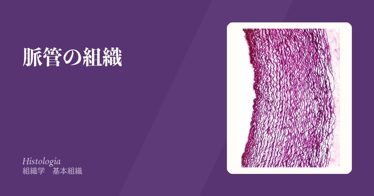
血管の壁はどこでも3層である。内側から内膜・中膜・外膜。違うのはどの層が厚いかだけで、それはその血管が受け持つ仕事から決まる。
- 圧の変動を吸収する大動脈 → 中膜が弾性線維だらけ
- 血流量を配分する筋型動脈 → 中膜が平滑筋だらけ
- 血液を溜める静脈 → 壁が薄く内腔が広い。弁がある
- 物質を交換する毛細血管 → 内皮1層と基底膜だけ
そして内皮細胞がこの章の主役になる。単なる裏打ちではなく、凝固・血管緊張・炎症・透過性のすべてを制御する巨大な内分泌器官である。動脈硬化も、敗血症も、DICも、糖尿病合併症も、出発点は内皮の機能不全にある。
- 内膜
- 中膜
- 粥状硬化の組織 — 内膜で何が起きているか
- 毛細血管
- 奇網と臓器特有の毛細血管 — 毛細血管が2回続く場所
- 静脈
- 動静脈吻合 — 毛細血管を通さずに、血を短絡させる
- 血管を養う血管と、血管を支配する神経
- 血管内皮 — たった1層が、これだけの仕事をしている
- 血管新生 — 新しい血管が要るときと、要らないのに作られるとき
- 血管の加齢 — 硬くなる、という変化
内膜Tunica intima
最内層。内皮(単層扁平上皮)+基底膜+わずかな結合組織。動脈では外側の境に内弾性板がある。
内皮の働きは受け身ではない。
- 抗血栓——プロスタサイクリン(PGI₂)・NO・トロンボモジュリン・ヘパラン硫酸
- 血管緊張の調節——NO(拡張)とエンドセリン(収縮)
- 白血球の通行管理——セレクチン・ICAM/VCAM を出して接着させる
- 透過性の門番
内皮が傷むと、抗血栓から血栓促進へ性質が反転する。これが動脈硬化と血栓症を結ぶ接点である。
NO は内皮型NO合成酵素(eNOS)がアルギニンから作り、平滑筋のグアニル酸シクラーゼ → cGMP を介して弛緩させる。この経路に薬が並ぶ——硝酸薬(NOを供給)、PDE5阻害薬(cGMPの分解を止める。だから硝酸薬との併用が禁忌)、可溶性グアニル酸シクラーゼ刺激薬。
内皮機能不全は動脈硬化の最初期の変化であり、糖尿病・高血圧・喫煙・脂質異常のすべてが共通してここを傷つける。だから危険因子が重なるほど直線的でなく相乗的にリスクが上がる。
内皮が傷むことが、血栓症のすべての出発点である。Virchow の3徴(血管内皮の傷害・血流の停滞・血液凝固能の亢進)の筆頭に内皮が来る。
内皮は「固めない」働きを常に出し続けている——プロスタサイクリン・NO・トロンボモジュリン・ヘパラン硫酸。傷つくとこの抑制が外れ、同時にコラーゲンと組織因子が露出して一気に凝固へ傾く。
NO を作れなくなるのが内皮機能障害で、高血圧・糖尿病・喫煙・脂質異常症に共通する最初の変化である。動脈硬化はここから始まる。
ニトログリセリンは NO を外から与える薬である。内皮が壊れていても効くのはこのためで、PDE5 阻害薬との併用で致命的な低血圧になる。
内皮由来の腫瘍が血管肉腫と Kaposi 肉腫(HHV-8)である。
English version
Core
The innermost layer: endothelium (simple squamous) plus basal lamina and a little connective tissue. In arteries an internal elastic lamina marks its outer boundary.
The endothelium is not passive.
- Antithrombotic — prostacyclin (PGI₂), NO, thrombomodulin, heparan sulphate.
- Regulation of tone — NO (dilates) and endothelin (constricts).
- Traffic control of leukocytes — selectins, ICAM and VCAM.
- Gatekeeper of permeability.
Going further
When the endothelium is injured its character reverses from antithrombotic to prothrombotic. This is the junction between atherosclerosis and thrombosis.
NO is made from arginine by endothelial NO synthase (eNOS) and relaxes smooth muscle through guanylate cyclase and cGMP. The drugs line up along this pathway — nitrates (supplying NO), PDE5 inhibitors (preventing the breakdown of cGMP, hence contraindicated with nitrates), and soluble guanylate cyclase stimulators.
Endothelial dysfunction is the earliest change of atherosclerosis, and diabetes, hypertension, smoking and dyslipidaemia all damage this same point. Which is why risk rises synergistically rather than additively as the factors accumulate.
In the clinic
Injury to the endothelium is the starting point of all thrombosis. Endothelial injury heads Virchow’s triad, with stasis and hypercoagulability.
The endothelium is continuously issuing signals not to clot — prostacyclin, NO, thrombomodulin, heparan sulphate. When it is damaged that restraint is removed and, at the same time, collagen and tissue factor are exposed, tipping everything at once towards coagulation.
Failure to make NO is endothelial dysfunction, the first change common to hypertension, diabetes, smoking and dyslipidaemia. Atherosclerosis begins here.
Nitroglycerin is a drug that supplies NO from outside, which is why it works even when the endothelium is damaged — and why combination with a PDE5 inhibitor causes fatal hypotension.
Tumours of endothelial origin are angiosarcoma and Kaposi sarcoma (HHV-8).
中膜Tunica media
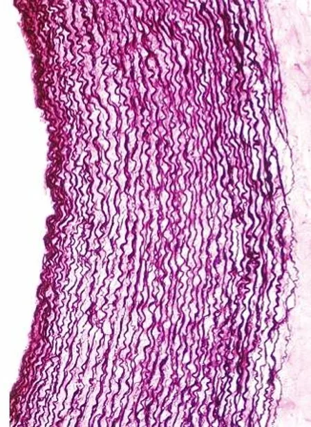
平滑筋と弾性線維の層。ここの構成で動脈が2つに分かれる。
- 弾性型動脈(大動脈・肺動脈・腕頭動脈など太い動脈)——弾性板が何層も重なる。心拍動を平滑化する(Windkessel)
- 筋型動脈(それ以外のほとんど)——平滑筋が主体。収縮して血流の配分を変える
外側の境に外弾性板。静脈では中膜が薄い。
中膜の性質が血管ごとに違う病気を作る。
- 大動脈解離——弾性型動脈の中膜が裂ける。嚢胞性中膜壊死(Marfan・加齢・高血圧)が基盤。内膜が裂けて血液が中膜内へ入り、偽腔を作る
- 細動脈硬化——高血圧では硝子様変性(血漿蛋白の染み込み)、悪性高血圧では類線維素壊死+onion skin 様の増殖
- Mönckeberg 型中膜石灰化——中膜だけが石灰化する。内腔は狭くならないので虚血は起こさないが、血管が硬く圧迫できなくなりABI が偽高値になる(糖尿病・CKD)
粥状硬化(アテローム)は内膜の病変で、中膜の病変とは別物である。「どの層の病気か」で症状も検査所見も変わる——これが組織学を臨床で使うということの実例になる。
中膜の型が、どの大動脈疾患になるかを決める。
大動脈解離 — 内膜が裂けて中膜の層のあいだに血液が入り込む。中膜が弱くなる素因(Marfan 症候群のフィブリリン異常・嚢胞性中膜壊死)と高血圧が重なって起こる。引き裂かれるような激痛・血圧の左右差・縦隔拡大。Stanford A 型は緊急手術。
大動脈の弾性(Windkessel 効果)が失われると脈圧が開く。高齢者の収縮期高血圧は弾性線維の変性で大動脈が硬くなった結果であり、「血管が硬い」ことが数字に現れている。
筋型動脈は交感神経で口径を変える——末梢血管抵抗の主役で、降圧薬(Ca 拮抗薬・α遮断薬)の作用点になる。
巨細胞性動脈炎は中膜の弾性板を破壊し、側頭動脈の圧痛・顎跛行・視力障害を起こす。失明を防ぐため、生検を待たずステロイドを開始する。
English version
Core
The layer of smooth muscle and elastic fibres. Its composition divides the arteries into two.
- Elastic arteries (aorta, pulmonary trunk, brachiocephalic) — many layers of elastic laminae. They smooth the pulsation of the heart (Windkessel).
- Muscular arteries (almost all the rest) — chiefly smooth muscle. They contract to redistribute flow.
An external elastic lamina marks the outer boundary. In veins the media is thin.
Going further
The character of the media creates a different disease in each kind of vessel.
- Aortic dissection — the media of an elastic artery splits. Cystic medial necrosis (Marfan, ageing, hypertension) is the substrate: the intima tears, blood enters the media and creates a false lumen.
- Arteriolosclerosis — in hypertension, hyaline change (plasma protein permeating the wall); in malignant hypertension, fibrinoid necrosis with onion-skin proliferation.
- Mönckeberg medial calcification — the media alone calcifies. The lumen is not narrowed so there is no ischaemia, but the vessel becomes rigid and incompressible, giving a falsely high ABI (diabetes, CKD).
Atheroma is a disease of the intima and is a different thing from disease of the media. Which layer is diseased changes both the symptoms and the investigations — a concrete example of what it means to use histology clinically.
In the clinic
The type of media determines which aortic disease occurs.
Aortic dissection — the intima tears and blood enters between the layers of the media. It arises when a predisposition weakening the media (fibrillin abnormality in Marfan syndrome, cystic medial necrosis) coincides with hypertension. Tearing pain, a difference in blood pressure between the arms, mediastinal widening. Stanford type A requires emergency surgery.
When the elasticity of the aorta (the Windkessel effect) is lost the pulse pressure widens. Systolic hypertension in the elderly is the result of degenerate elastic fibres stiffening the aorta — "stiff vessels" appearing as a number.
Muscular arteries change calibre under sympathetic control — the principal determinant of peripheral resistance and the site of action of antihypertensives (calcium channel blockers, α blockers).
Giant cell arteritis destroys the elastic lamina of the media, causing temporal tenderness, jaw claudication and visual loss. Start corticosteroids without waiting for the biopsy, to prevent blindness.
粥状硬化の組織 — 内膜で何が起きているか
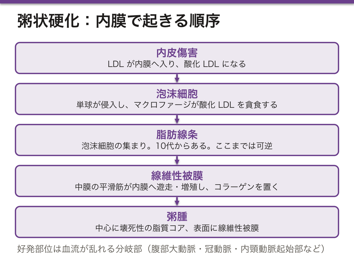
内膜に脂質と細胞が溜まってプラークを作る病変。段階を追うと組織像が繋がる。
- 内皮傷害——LDLが内膜へ入り酸化LDLになる
- 単球が侵入し、マクロファージが酸化LDLを貪食して泡沫細胞になる
- 脂肪線条——泡沫細胞の集まり。10代からすでにある。ここまでは可逆
- 中膜の平滑筋が内膜へ遊走・増殖し、コラーゲンを置いて線維性被膜を作る
- 粥腫——中心に壊死性の脂質コア、表面に線維性被膜
好発部位は血流が乱れる分岐部(腹部大動脈・冠動脈・膝窩動脈・内頸動脈起始部)。
臨床的に決定的なのはプラークの大きさではなく安定性である。
- 安定プラーク——線維性被膜が厚く脂質コアが小さい。内腔を狭めるので労作性狭心症を起こすが、破綻しにくい
- 不安定プラーク——被膜が薄く、脂質コアが大きく、炎症細胞が多い。狭窄は軽度でも破綻して急性冠症候群を起こす
だから心筋梗塞の多くは、直前まで狭窄が軽かった部位で起きる。狭窄率だけを見ていては予測できない——この事実がスタチンの意義を説明する。スタチンはLDLを下げるだけでなくプラークを安定化させる(被膜を厚くし炎症を抑える)ため、狭窄の改善以上にイベントを減らす。
マクロファージがコレステロールを取り込むのはスカベンジャー受容体経由で、LDL受容体と違いフィードバック抑制を受けない。だから取り込み続けて泡沫細胞になる。「制御されない受容体」があることが病気を作る。
プラークは「大きさ」ではなく「壊れやすさ」で危険が決まる。
不安定プラーク — 脂質コアが大きく、線維性被膜が薄く、炎症細胞が多い。狭窄が軽くても破綻して急性冠症候群になる。安定狭心症で見つかる高度狭窄のほうが、むしろ破綻しにくい——「狭さ」と「危なさ」は別である。
スタチンの効果は LDL 低下だけでは説明できない。プラークを安定化させる(被膜を厚くし、炎症を抑える)ことがイベント抑制の主要な機序と考えられている。
破綻すると組織因子が露出して血栓ができる。完全閉塞なら ST 上昇型心筋梗塞、不完全なら非 ST 上昇型/不安定狭心症。抗血小板薬が急性期治療の中心になる理由がここにある。
好発部位は血流が乱れる分岐部——頸動脈分岐部・冠動脈近位・腹部大動脈・腸骨動脈。
English version
Core
A lesion in which lipid and cells accumulate in the intima to form a plaque. Follow the stages and the histology connects.
- Endothelial injury — LDL enters the intima and is oxidised.
- Monocytes enter; macrophages ingest oxidised LDL and become foam cells.
- Fatty streak — a collection of foam cells. Present from the teens, and still reversible at this stage.
- Medial smooth muscle migrates into the intima, proliferates and lays down collagen to form the fibrous cap.
- Atheroma — a necrotic lipid core with a fibrous cap over it.
Sites of predilection are bifurcations, where flow is disturbed: abdominal aorta, coronary arteries, popliteal artery, origin of the internal carotid.
Going further
What matters clinically is not the size of the plaque but its stability.
- Stable plaque — thick fibrous cap, small lipid core. It narrows the lumen and causes exertional angina, but does not readily rupture.
- Unstable plaque — thin cap, large lipid core, many inflammatory cells. Even with mild stenosis it ruptures and causes an acute coronary syndrome.
So most myocardial infarctions occur at sites where the stenosis was mild until just before. Looking only at the degree of stenosis cannot predict it — and this fact explains the value of statins: they not only lower LDL but stabilise the plaque (thickening the cap and suppressing inflammation), reducing events beyond any improvement in stenosis.
Macrophages take up cholesterol through the scavenger receptor, which, unlike the LDL receptor, is not subject to feedback inhibition. So they keep taking it up and become foam cells. The existence of an unregulated receptor creates the disease.
In the clinic
The danger of a plaque is decided by how easily it breaks, not by how large it is.
Unstable plaque — large lipid core, thin fibrous cap, many inflammatory cells. Even with mild stenosis it ruptures and causes an acute coronary syndrome. A tight stenosis found in stable angina is in fact less likely to rupture — "narrow" and "dangerous" are different things.
The effect of statins cannot be explained by LDL lowering alone. Stabilisation of the plaque — a thicker cap and suppressed inflammation — is thought to be the principal mechanism of event reduction.
On rupture, tissue factor is exposed and thrombus forms: complete occlusion gives ST-elevation infarction, incomplete gives non-ST-elevation infarction or unstable angina. This is why antiplatelet therapy is central in the acute phase.
毛細血管Vas capillare
内皮1層と基底膜だけ。周囲に周皮細胞(ペリサイト)が乗る。ここでのみ物質交換が起きる。
内皮の隙間の作り方で3型に分かれ、どの臓器にあるかが決まる。
- 連続型——隙間が最も少ない。筋・肺・皮膚・中枢神経(脳では密着結合が加わり血液脳関門)
- 有窓型——内皮に小さな窓がある。腎糸球体・内分泌腺・小腸粘膜・脈絡叢(濾過や分泌が盛んな場所)
- 洞様(不連続型)——大きな隙間があり基底膜も不完全。肝・脾・骨髄(細胞そのものが通る場所)
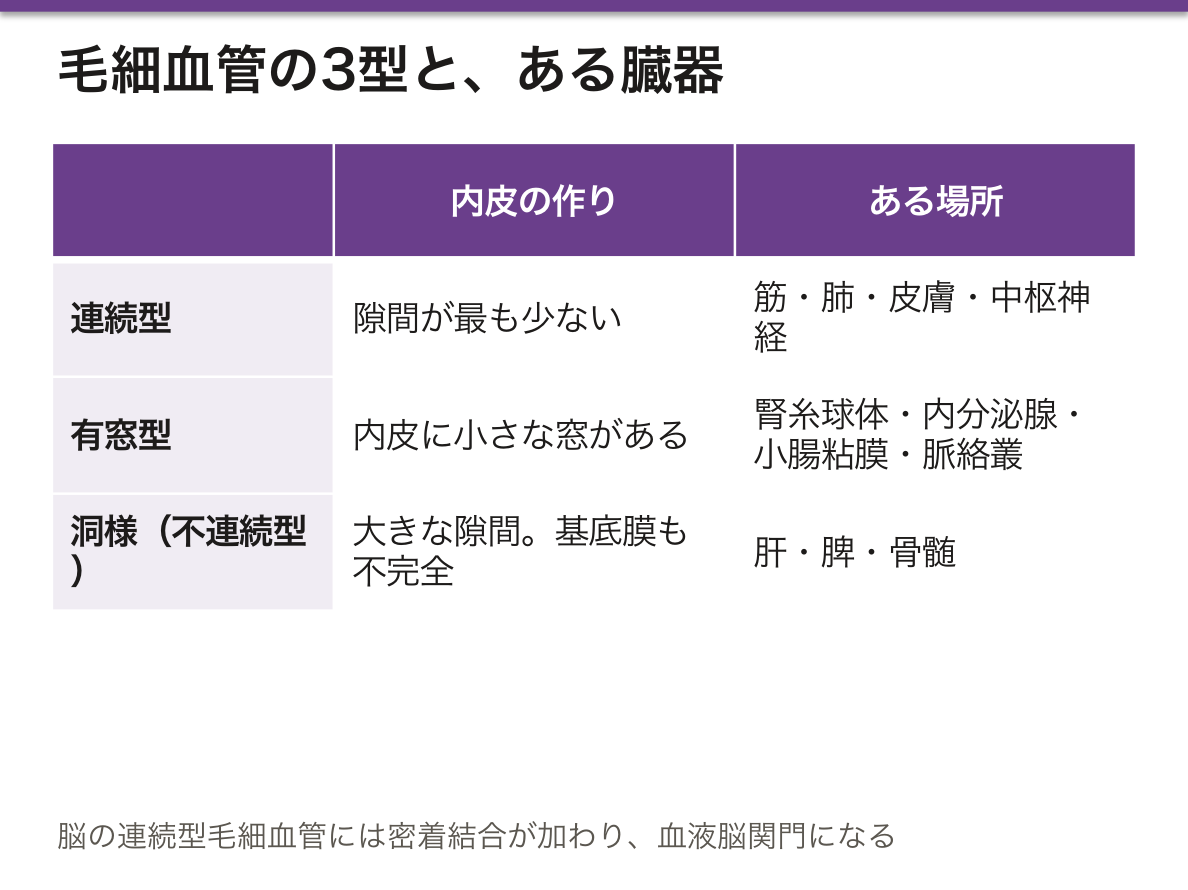
隙間の大きさが、その臓器で何が起きるかを決めている——この対応が要点である。
糸球体では有窓内皮+基底膜+足細胞のスリット膜という3層の関門が働く。物理的な大きさの制限(size barrier)と、ヘパラン硫酸の負電荷による制限(charge barrier)の両方があり、どちらが壊れるかで蛋白尿の性質が変わる。
肝の類洞は基底膜が無く、内皮と肝細胞のあいだのDisse 腔で血漿が直接肝細胞に触れる。だからアルブミンや凝固因子を効率よく血中へ出せる。慢性肝障害ではここに星細胞由来のコラーゲンが沈着して類洞の毛細血管化が起き、物質交換が落ちると同時に門脈圧が上がる。
周皮細胞は毛細血管の安定と血流の微調整を担う。糖尿病網膜症では周皮細胞が最初に脱落し、支えを失った壁が膨らんで毛細血管瘤になる——眼底所見の最初期の変化がこれである。
内皮の型が、その臓器で何が漏れるかを決める。
有窓型(腎糸球体・内分泌腺・腸管)は水と小分子をよく通す。糸球体では、窓+基底膜+足細胞のスリット膜の3層が大きさと電荷で濾過を制御する。この選択性が壊れるのが蛋白尿である。
連続型で密着結合が強いのが血液脳関門である。
洞様(類洞:肝・脾・骨髄)は隙間が大きく、細胞さえ通る。血球が骨髄から血中へ出られるのも、肝で血漿が肝細胞に直接触れられるのもこの型による。
周皮細胞が失われるのが糖尿病網膜症の最初期で、毛細血管瘤ができる。眼底に見える点状出血と毛細血管瘤は、周皮細胞の脱落の帰結である。
English version
Core
One layer of endothelium and a basal lamina, with pericytes on the outside. Exchange of substances occurs only here.
Three types by how the gaps are made, and this decides which organ they are in.
- Continuous — fewest gaps. Muscle, lung, skin, CNS (where tight junctions are added to make the blood–brain barrier).
- Fenestrated — small windows in the endothelium. Renal glomerulus, endocrine glands, intestinal mucosa, choroid plexus — places of active filtration or secretion.
- Sinusoidal (discontinuous) — large gaps and an incomplete basal lamina. Liver, spleen, bone marrow — places where whole cells pass.
Going further
The size of the gap determines what happens in that organ — this correspondence is the point.
In the glomerulus three barriers act in series: fenestrated endothelium, basal lamina and the slit diaphragms of the podocytes. There is a restriction by size and a restriction by the negative charge of heparan sulphate, and which one fails changes the character of the proteinuria.
The hepatic sinusoid has no basal lamina, and plasma touches the hepatocyte directly in the space of Disse. This is how albumin and clotting factors can be exported efficiently. In chronic liver disease, collagen from stellate cells is deposited here and the sinusoid becomes capillarised, so exchange falls at the same time as portal pressure rises.
Pericytes stabilise the capillary and fine-tune flow. In diabetic retinopathy the pericytes are lost first, and the unsupported wall balloons into a microaneurysm — the earliest change on fundoscopy.
In the clinic
The type of endothelium decides what leaks in that organ.
Fenestrated types (glomerulus, endocrine glands, intestine) pass water and small molecules readily. In the glomerulus, windows plus basal lamina plus podocyte slit diaphragms control filtration by size and charge. Loss of that selectivity is proteinuria.
A continuous type with strong tight junctions is the blood–brain barrier.
Sinusoidal types (liver, spleen, marrow) have gaps large enough for cells to pass — which is how blood cells leave the marrow and how plasma touches hepatocytes directly.
Loss of pericytes is the earliest change of diabetic retinopathy, producing microaneurysms. The dot haemorrhages and microaneurysms seen on the fundus are the consequence of pericyte loss.
奇網と臓器特有の毛細血管 — 毛細血管が2回続く場所Rete mirabile
奇網(rete mirabile、「すばらしい網」)は、同じ種類の血管のあいだに挟まった毛細血管網をいう。ふつうは動脈 → 毛細血管 → 静脈だが、奇網では毛細血管の前後が同じ種類になる。
| 種類 | 並び | 例 |
|---|---|---|
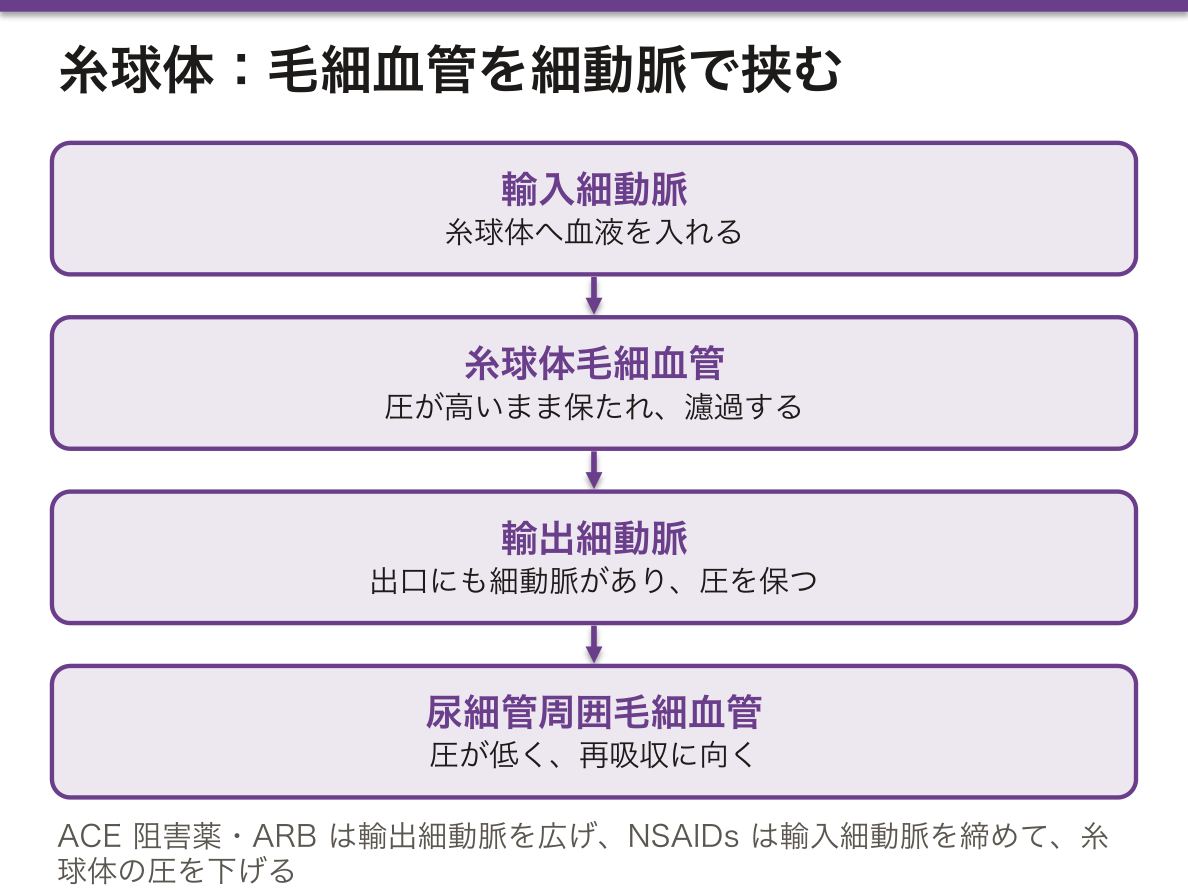
| 動脈性の奇網 | 動脈(細動脈)→ 毛細血管 → 動脈(細動脈) | 腎の糸球体(輸入細動脈 → 糸球体毛細血管 → 輸出細動脈) |
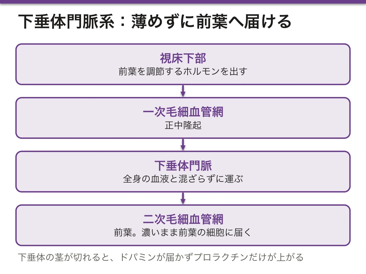
| 静脈性の奇網(門脈系) | 静脈 → 毛細血管 → 静脈 | 肝(腸の毛細血管 → 門脈 → 類洞 → 中心静脈)、下垂体門脈系(正中隆起の一次毛細血管網 → 下垂体門脈 → 前葉の二次毛細血管網) |
臓器ごとの毛細血管も、働きに合わせて形が違う。
| 臓器 | 毛細血管 | 理由 |
|---|---|---|
| 脳 | 連続性。内皮が密着結合でつながる | 血液脳関門 |
| 腎糸球体 | 有窓性で、窓に隔膜が無い | 大量の濾過 |
| 内分泌腺・腸の絨毛 | 有窓性(隔膜あり) | ホルモンや吸収物の出入り |
| 肝・脾・骨髄 | 類洞(不連続性)。肝ではマクロファージ(Kupffer 細胞)が並ぶ | 大きな分子や細胞が出入りする |
なぜ毛細血管を2回続けるのか。理由は臓器ごとに違う。
- 糸球体——出口にも細動脈(輸出細動脈)があるので、糸球体の中の圧が高いまま保たれ、濾過ができる。そのあとの尿細管周囲の毛細血管は圧が低く、今度は再吸収に向く
- 下垂体門脈系——視床下部のホルモンを、全身の血液で薄めずに濃いまま前葉へ直接届ける
- 肝——腸で吸収したものを、全身に出る前にまず肝細胞に通す
この名前は、古代の医師 Galen が動物の頭蓋底で見た、動脈が細かな網に分かれて再び集まる構造に由来する。
ACE 阻害薬・ARBは輸出細動脈を広げるので、糸球体の圧が下がる。両側の腎動脈狭窄のように、輸出細動脈を締めて圧を保っている腎臓では急に GFR が落ちる。NSAIDs は反対に輸入細動脈を締めて GFR を下げる。
下垂体の柄(茎)が圧迫されたり切れたりすると、門脈で運ばれていたドパミンが前葉に届かなくなり、プロラクチンだけが上がる。ほかの前葉ホルモンは、刺激が届かなくなって下がる。
肝の門脈系があるので、飲んだ薬の一部は全身に出る前に肝で代謝される(初回通過効果)。ニトログリセリンを舌下で使うのは、門脈を通らずに効かせるためである。門脈の流れが肝硬変で妨げられると、門脈圧亢進症になり、食道静脈瘤ができる。
English version
Core
A rete mirabile ("wonderful net") is a capillary network placed between two vessels of the same kind. Usually the order is artery → capillary → vein, but in a rete mirabile the vessels before and after the capillaries are of the same type.
| Type | Order | Examples |
|---|---|---|
| Arterial rete | artery (arteriole) → capillaries → artery (arteriole) | renal glomerulus (afferent arteriole → glomerular capillaries → efferent arteriole) |
| Venous rete (portal system) | vein → capillaries → vein | liver (intestinal capillaries → portal vein → sinusoids → central vein); hypophyseal portal system (primary plexus in the median eminence → portal veins → secondary plexus in the anterior lobe) |
Capillaries of each organ also differ in form to suit their job.
| Organ | Capillary | Reason |
|---|---|---|
| Brain | continuous; endothelium joined by tight junctions | blood–brain barrier |
| Glomerulus | fenestrated, without diaphragms | heavy filtration |
| Endocrine glands, intestinal villi | fenestrated (with diaphragms) | passage of hormones and absorbed material |
| Liver, spleen, marrow | sinusoids (discontinuous); in the liver lined by macrophages (Kupffer cells) | large molecules and cells pass in and out |
Going further
Why put two capillary beds in a row? The reason differs by organ.
- Glomerulus — an arteriole at the exit (efferent arteriole) keeps the pressure inside the glomerulus high for filtration. The peritubular capillaries after it have low pressure and suit reabsorption.
- Hypophyseal portal system — delivers hypothalamic hormones to the anterior lobe directly and concentrated, without dilution in the general blood.
- Liver — passes everything absorbed from the gut through hepatocytes first, before it reaches the body.
The name comes from a structure Galen saw at the base of the skull in animals, where an artery splits into a fine net and joins again.
In the clinic
ACE inhibitors and ARBs widen the efferent arteriole and lower glomerular pressure. In kidneys that keep pressure by tightening the efferent arteriole, as in bilateral renal artery stenosis, GFR falls suddenly. NSAIDs do the opposite: they narrow the afferent arteriole and lower GFR.
If the pituitary stalk is compressed or cut, dopamine carried by the portal veins no longer reaches the anterior lobe, and only prolactin rises. Other anterior-lobe hormones fall because their stimulating signals no longer arrive.
Because of the hepatic portal system, part of an oral drug is metabolised by the liver before reaching the body (first-pass effect). Sublingual nitroglycerin works by avoiding the portal route. When cirrhosis blocks portal flow, portal hypertension develops and oesophageal varices form.
静脈Vena
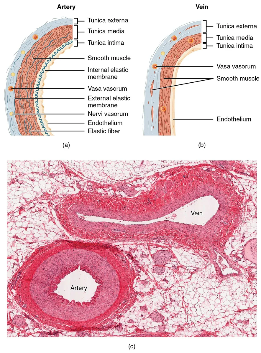
同じ径の動脈と比べて壁が薄く、内腔が広く、断面がつぶれて見える。中膜の平滑筋が少なく、外膜が相対的に厚い。
弁を持つ(四肢の中〜小静脈にとくに多い)。逆流を防ぎ、筋ポンプと組んで血液を心臓へ送る。
全血液量の約60〜70%が静脈系にある。容量血管と呼ばれる所以。
「容量血管」であることが循環動態の理解に効く。静脈の緊張(交感神経支配)が変わるだけで心臓に戻る血液量(前負荷)が大きく動く。
- 敗血症・アナフィラキシー——静脈が拡張して血液が末梢に貯留し、前負荷が落ちて血圧が下がる(分布異常性ショック)
- 硝酸薬——主に静脈を拡張させ前負荷を下げる。だから狭心症で心臓の仕事量が減る
弁が壊れると逆流し、静脈圧が上がって下肢静脈瘤になる。長期化するとうっ滞性皮膚炎・色素沈着(ヘモジデリン)・難治性潰瘍(内果周囲)へ進む。動脈性潰瘍が足趾・踵にでき境界明瞭で疼痛が強いのと対照的で、「どこにできるか」で原因血管が分かる。
血栓の性質も動静脈で違う。静脈血栓は赤色血栓(フィブリンと赤血球が主体)で抗凝固薬が効き、動脈血栓は白色血栓(血小板が主体)で抗血小板薬が効く。血流が遅いか速いかが、血栓の成分と薬の選択を決めている。
弁が壊れると、逆流して圧が上がる。
下肢静脈瘤 — 弁不全で逆流し、表在静脈が拡張・蛇行する。長時間の立位・妊娠・肥満・加齢が危険因子。進行するとうっ滞性皮膚炎・色素沈着・難治性潰瘍(足関節内果の上)。圧迫療法が基本になる。
深部静脈血栓症 — Virchow の3徴。片側性の下腿の腫脹と疼痛。肺血栓塞栓症へ直結するので長期臥床・術後・長距離移動での予防(早期離床・弾性ストッキング・抗凝固)が要る。
壁が薄く内腔が広いので採血と静脈路の確保に使う。静脈は容量血管で、全血液量の約2/3を蓄えている——出血時の代償にも、ニトログリセリンによる前負荷軽減にもこの性質が関わる。
English version
Core
Compared with an artery of the same calibre, the wall is thinner, the lumen wider, and the cross-section appears collapsed. The media has little smooth muscle and the adventitia is relatively thick.
Valves are present, especially in the medium and small veins of the limbs; they prevent reflux and, with the muscle pump, return blood to the heart.
About 60–70 per cent of the blood volume is in the venous system — hence "capacitance vessels".
Going further
Being capacitance vessels is what makes them matter haemodynamically. A change in venous tone (sympathetically controlled) alone moves a large volume of the blood returning to the heart (the preload).
- Sepsis and anaphylaxis — the veins dilate, blood pools peripherally, preload falls and the pressure drops (distributive shock).
- Nitrates — dilate the veins chiefly, reducing preload; this is how they reduce cardiac work in angina.
When the valves fail, reflux raises the venous pressure and produces varicose veins; with time, stasis dermatitis, pigmentation (haemosiderin) and intractable ulceration around the medial malleolus. By contrast an arterial ulcer forms on the toes or heel, is sharply demarcated and is very painful — where the ulcer is tells you which vessel is at fault.
The character of thrombus also differs: venous thrombus is red (fibrin and red cells) and responds to anticoagulants; arterial thrombus is white (platelets) and responds to antiplatelet drugs. Whether the flow is slow or fast determines both the composition of the clot and the choice of drug.
In the clinic
When the valves fail, blood refluxes and the pressure rises.
Varicose veins — valvular incompetence with reflux, dilating and tortuous superficial veins. Prolonged standing, pregnancy, obesity and age are risk factors. Progression brings stasis dermatitis, pigmentation and intractable ulceration above the medial malleolus. Compression is the basis of treatment.
Deep vein thrombosis — Virchow’s triad. Unilateral calf swelling and pain. Because it leads directly to pulmonary embolism, prophylaxis is required with prolonged bed rest, after surgery and on long journeys (early mobilisation, graduated stockings, anticoagulation).
Thin walls and a wide lumen make veins the route for venepuncture and cannulation. They are capacitance vessels holding about two-thirds of the blood volume — which bears both on compensation for haemorrhage and on the reduction of preload by nitroglycerin.
動静脈吻合 — 毛細血管を通さずに、血を短絡させるAnastomosis arteriovenosa
細動脈と細静脈を、毛細血管を経ずに直接つなぐ短絡路。壁に厚い平滑筋があり、交感神経の支配で開閉する。
あるのは体温調節に関わる場所になる——指趾の末端・手掌と足底・鼻尖・耳介・口唇。
| 開くと | 閉じると |
|---|---|
| 血が皮膚表面を大量に流れ、熱を捨てる | 血が深部へ回り、熱を保つ。指先は冷たくなる |
糸球(glomus)——特殊化した動静脈吻合。厚い被膜の中に、上皮様に見える平滑筋細胞(glomus 細胞)と豊富な神経終末を持つ。
肺と肝にも短絡路があり、病的に開くと問題になる。
「血液が毛細血管を通らない」ということが、そのまま病態になる。短絡した血は、酸素を渡さずに静脈へ帰る(呼吸の章)——「酸素を投与しても改善しないシャントの本質が、この構造にある」。
肝肺症候群は、肺の血管が拡張して短絡になる状態になる(肝硬変の合併症の章)——「起坐位で低酸素が悪化する(platypnea・orthodeoxia)という珍しい向きを取る」。
体温調節の面では、この短絡が最も速く働く仕組みになる(体温を保つ章)——「寒いと指先から冷たくなるのは、末梢が犠牲になっているのではなく、中心を守っているからになる」。
glomus 腫瘍は、この構造から生じる(手の疾患の章)——「爪下の激痛・寒冷で増悪・小さくて画像に写りにくい。痛みの割に腫瘤が見えない」。
遺伝性出血性末梢血管拡張症(Osler 病)では、全身に動静脈奇形ができる(出血の型の章)——「反復する鼻出血・口唇と舌の毛細血管拡張・肺と脳の動静脈奇形が並ぶ」。
動静脈吻合は体温調節の弁である。寒いと閉じて熱を逃さず、暑いと開いて放熱する。末梢が冷たくなるのは、循環が悪いからだけではない。
Raynaud 現象 — 寒冷や精神的ストレスで指趾の細動脈が過剰に収縮し、蒼白 → 紫 → 発赤の三相性の色調変化を示す。膠原病(全身性強皮症・SLE)の初発症状のことがあるので、爪郭毛細血管を観察し、自己抗体を調べる。
グロムス腫瘍 — 動静脈吻合(グロムス装置)由来の良性腫瘍。爪床下に好発し、激痛・寒冷過敏・限局した圧痛という3徴が非常に特徴的である。小さくても激烈に痛むので、指の原因不明の痛みで思い出す。
透析用の動静脈シャントは、この短絡を人工的に作ったものである。
English version
Core
A shunt connecting an arteriole directly to a venule without passing through a capillary bed. Its wall has thick smooth muscle and it opens and closes under sympathetic control.
Found where thermoregulation matters — fingertips and toes, palms and soles, tip of the nose, auricle, lips.
- Open — blood flows in quantity through the skin surface and heat is lost.
- Closed — blood is diverted to the core and heat is conserved; the fingers become cold.
The glomus body is a specialised arteriovenous anastomosis: within a thick capsule, epithelioid smooth muscle cells (glomus cells) with abundant nerve endings.
Shunts also exist in lung and liver and cause trouble when they open pathologically.
Going further
That the blood does not pass through a capillary is itself the pathology. Shunted blood returns to the vein without giving up oxygen — the essence of a shunt that does not improve with supplemental oxygen lies in this structure.
Hepatopulmonary syndrome is a state in which the pulmonary vessels dilate into shunts — and takes the unusual form of hypoxia that worsens on sitting up (platypnoea–orthodeoxia).
In thermoregulation this shunt is the fastest-acting mechanism — the fingers go cold in the cold not because the periphery is being neglected but because the core is being protected.
Glomus tumour arises from this structure — severe subungual pain, worse with cold, small and hard to see on imaging: pain out of all proportion to a visible mass.
In hereditary haemorrhagic telangiectasia (Osler disease) arteriovenous malformations form throughout the body — recurrent epistaxis, telangiectasia of the lips and tongue, and arteriovenous malformations of lung and brain.
In the clinic
The arteriovenous anastomosis is the valve of thermoregulation. It closes in the cold to retain heat and opens in the heat to lose it. Cold extremities are not only a matter of poor circulation.
Raynaud phenomenon — cold or emotional stress causes excessive constriction of the digital arterioles, producing the triphasic colour change white → blue → red. It may be the presenting symptom of a connective tissue disease (systemic sclerosis, SLE), so examine the nailfold capillaries and test for autoantibodies.
Glomus tumour — a benign tumour of the glomus apparatus, characteristically subungual, with the highly distinctive triad of severe pain, cold sensitivity and pinpoint tenderness. Remember it for unexplained pain in a digit — it hurts violently even when small.
An arteriovenous fistula for dialysis is this shunt created artificially.
血管を養う血管と、血管を支配する神経Vasa vasorum
太い血管の壁は、内腔の血液からは栄養を受け取れない。拡散で届くのは内側の約1/3(内膜と中膜内層)までで、それより外側は壁の中を走る細い血管=vasa vasorumが養う。
| 分布 | |
|---|---|
| 動脈 | 外膜と中膜の外側1/3まで |
| 静脈 | より内側まで入り込む——静脈血は酸素が乏しいため |
神経は外膜に分布し(nervi vasorum)、大部分が交感神経。α1受容体を介して血管収縮、β2受容体で骨格筋の血管は拡張する。血管平滑筋には副交感神経がほとんど来ない——血圧の調節は「締める側の強弱」で行われる。
vasa vasorum が病気の舞台になる。
| 病態 | vasa vasorum の関与 |
|---|---|
| 梅毒性大動脈炎(第3期梅毒) | 上行大動脈の vasa vasorum に閉塞性内膜炎が起こる → 中膜が虚血で壊死・弾性線維が失われる → 上行大動脈瘤と大動脈弁閉鎖不全。「上行大動脈に限局する」のは、そこに vasa vasorum が最も発達しているから |
| 粥状硬化のプラーク内出血 | プラークが厚くなると拡散では足りず、vasa vasorum が新生してプラーク内へ侵入。この脆い新生血管が破綻してプラーク内出血 → 急激な狭窄・破綻を起こす。急性冠症候群の引き金の一つ |
| 大動脈解離 | 中膜の中を血流が裂き進む。偽腔が vasa vasorum の破綻から始まるという説もある |
「拡散で届く距離には限りがある」という原則は、腫瘍にもそのまま当てはまる。腫瘍が直径1〜2 mm を超えると拡散では養えず、血管新生を起こさなければ成長できない——VEGF がその引き金で、抗VEGF抗体(ベバシズマブ)はこの一点を狙う薬。血管新生を止めることが治療になるという発想は、血管壁の栄養の原理から出ている。
栄養血管が詰まると、血管そのものが壊れる。
梅毒性大動脈炎 — 第三期梅毒でvasa vasorum の閉塞性動脈内膜炎が起こり、中膜が壊死して上行大動脈瘤・大動脈弁閉鎖不全になる。上行大動脈に vasa vasorum が最も多いことが好発部位を説明する。
大動脈解離でもvasa vasorum の破綻が壁内血腫の起点になりうる。
血管の神経支配——交感神経が収縮させる。脊髄損傷で交感神経が絶たれると、末梢血管が拡張して神経原性ショックになる。徐脈を伴うことが出血性ショック(頻脈)との鑑別点である。
大動脈と頸動脈洞の圧受容器は血圧の急変を感知する。頸動脈洞を圧迫すると徐脈・血圧低下——頸動脈洞症候群では失神の原因になる。
English version
Core
The wall of a large vessel cannot be nourished from the blood in its lumen. Diffusion reaches only about the inner third (intima and inner media); beyond that it is supplied by fine vessels running within the wall, the vasa vasorum.
- Artery — adventitia and the outer third of the media.
- Vein — penetrating further inwards, because venous blood is poor in oxygen.
Nerves run in the adventitia (nervi vasorum) and are mostly sympathetic: constriction through α1 receptors, and dilatation of skeletal muscle vessels through β2. Vascular smooth muscle receives virtually no parasympathetic supply — blood pressure is regulated by varying how hard it is squeezed.
Going further
The vasa vasorum become the stage for disease.
| Condition | Role of the vasa vasorum |
|---|---|
| Syphilitic aortitis (tertiary syphilis) | Obliterative endarteritis of the vasa vasorum of the ascending aorta → ischaemic necrosis of the media with loss of elastic fibres → aneurysm and aortic regurgitation. It is confined to the ascending aorta because that is where the vasa vasorum are most developed |
| Intraplaque haemorrhage | As a plaque thickens, diffusion no longer suffices and vasa vasorum grow into it. These fragile new vessels rupture, causing intraplaque haemorrhage with abrupt stenosis or rupture — one trigger of the acute coronary syndrome |
| Aortic dissection | Blood splits through the media; some hold that the false lumen begins with rupture of a vasa vasorum |
The principle that diffusion reaches only so far applies equally to tumours. Beyond 1–2 mm in diameter a tumour cannot be nourished by diffusion and cannot grow without angiogenesis — VEGF is the trigger, and anti-VEGF antibodies (bevacizumab) aim at exactly that point. The idea that stopping angiogenesis is treatment comes out of the principle of how a vessel wall is fed.
In the clinic
When the nutrient vessels are occluded, the vessel itself is destroyed.
Syphilitic aortitis — in tertiary syphilis, obliterative endarteritis of the vasa vasorum causes necrosis of the media, giving an ascending aortic aneurysm and aortic regurgitation. That the vasa vasorum are most numerous in the ascending aorta explains the site.
In aortic dissection too, rupture of a vasa vasorum can be the origin of an intramural haematoma.
Innervation — the sympathetic system constricts. When it is interrupted in spinal cord injury the peripheral vessels dilate and neurogenic shock follows; the accompanying bradycardia distinguishes it from haemorrhagic shock, which is tachycardic.
The baroreceptors of the aorta and carotid sinus detect sudden changes in pressure. Pressure on the carotid sinus produces bradycardia and hypotension — a cause of syncope in carotid sinus syndrome.
血管内皮 — たった1層が、これだけの仕事をしているEndotheliocytus
単層扁平上皮。全身に広げると数百 m² になる、体で最大の「臓器」の一つ。
| 仕事 | 出しているもの |
|---|---|
| 血管を広げる/締める | 一酸化窒素・プロスタサイクリン / エンドセリン |
| 固めない | トロンボモジュリン・ヘパラン硫酸・組織プラスミノゲン活性化因子 |
| 固める(傷ついたとき) | von Willebrand 因子・組織因子・PAI-1 |
| 白血球を通す | セレクチン・ICAM-1・VCAM-1 |
| 関門になる | 密着結合(脳・網膜・精巣) |
Weibel-Palade 小体——内皮に固有の顆粒。von Willebrand 因子と P-セレクチンを貯めている。
形は場所で違う——連続型・有窓型(腎糸球体・内分泌腺・腸)・洞様型(肝・脾・骨髄)。
内皮は「何もしていないときに、何かをしている」細胞になる。平時は固めない・広げるほうへ働き続けており、それをやめることが病気になる——「静かであることが、能動的な状態になっている」。
だから内皮機能障害が、動脈硬化の最初の段階になる。一酸化窒素が減り、接着分子が出て、白血球が壁に入り込む(一酸化窒素の項)——「プラークができる前から、内皮は変わっている」。
敗血症も、内皮の病気として理解できる。組織因子が出て凝固が始まり、透過性が上がって血管内から水が抜ける(敗血症の章)——「輸液しても血圧が上がらず、浮腫だけが増える理由になる」。
播種性血管内凝固は、この裏返しになる(止血の各論の章)——「全身の内皮が同時に凝固側へ傾くと、固めながら出血する」。
有窓型内皮のある場所が、そのまま「濾す・出す・取り込む」臓器と一致する(糸球体の章)——「構造の型を覚えるより、その臓器が何をしているかから導ける」。
内皮は「体で最大の内分泌器官」と言ってよい。1層が全身に広がり、血管の緊張・凝固・炎症・透過性を制御している。
敗血症は内皮の病気である。広範な内皮の活性化と傷害により血管拡張(低血圧)・透過性亢進(浮腫と血管内容量の減少)・微小血栓(DIC)が同時に起こる。3つの症状が別々の病態ではなく、1つの細胞の破綻から出ていると読める。
糖尿病の合併症も内皮の病気として一続きに理解できる。細小血管障害(網膜・腎・神経)と大血管障害(心・脳・末梢動脈)。
血管性浮腫 — ブラジキニンによる透過性亢進。ACE 阻害薬はブラジキニンを分解できなくするため空咳と血管性浮腫を起こす。遺伝性血管性浮腫(C1 インヒビター欠損)では抗ヒスタミン薬もステロイドも効かない——機序が違えば薬が違う。
English version
Core
A simple squamous epithelium. Spread out it covers several hundred square metres — one of the largest "organs" in the body.
| Task | What it releases |
|---|---|
| Dilate / constrict | Nitric oxide, prostacyclin / endothelin |
| Prevent clotting | Thrombomodulin, heparan sulphate, tissue plasminogen activator |
| Promote clotting (when injured) | von Willebrand factor, tissue factor, PAI-1 |
| Admit leukocytes | Selectins, ICAM-1, VCAM-1 |
| Act as a barrier | Tight junctions (brain, retina, testis) |
Weibel–Palade bodies are granules peculiar to endothelium, storing von Willebrand factor and P-selectin.
The form varies with the site — continuous, fenestrated (glomerulus, endocrine glands, gut) and sinusoidal (liver, spleen, marrow).
Going further
The endothelium is a cell that is doing something when it appears to be doing nothing. At rest it works continuously in the direction of not clotting and of dilating, and stopping that is the disease — being quiet is an active state.
So endothelial dysfunction is the first stage of atherosclerosis: nitric oxide falls, adhesion molecules appear and leukocytes enter the wall — the endothelium has already changed before any plaque exists.
Sepsis can also be understood as a disease of the endothelium. Tissue factor appears and coagulation begins; permeability rises and water leaves the vessels — which is why fluid does not raise the blood pressure and only the oedema increases. Disseminated intravascular coagulation is the reverse of the same coin — when the endothelium of the whole body tips towards coagulation at once, the patient clots and bleeds simultaneously.
The places with fenestrated endothelium coincide exactly with organs that filter, secrete or absorb — rather than memorising the types of structure, derive them from what the organ does.
In the clinic
The endothelium may fairly be called the largest endocrine organ in the body: one cell layer spread throughout, controlling vascular tone, coagulation, inflammation and permeability.
Sepsis is a disease of the endothelium. Widespread activation and injury produce vasodilatation (hypotension), increased permeability (oedema and loss of intravascular volume) and microthrombosis (DIC) simultaneously. The three are not separate processes but the failure of one cell.
The complications of diabetes can be understood as a single continuum of endothelial disease — microvascular (retina, kidney, nerve) and macrovascular (heart, brain, peripheral arteries).
Angio-oedema — increased permeability from bradykinin. ACE inhibitors prevent the breakdown of bradykinin and cause dry cough and angio-oedema. In hereditary angio-oedema (C1 inhibitor deficiency) neither antihistamines nor corticosteroids work — a different mechanism means a different drug.
血管新生 — 新しい血管が要るときと、要らないのに作られるときAngiogenesis
2つの作られ方がある——血管新生(既存の血管から芽が伸びる)と、脈管形成(前駆細胞から新しく作る。主に胎生期)。
芽が伸びるときの役割分担。
- 先端細胞(tip cell)——糸状仮足を伸ばして方向を決める。分裂しない
- 茎細胞(stalk cell)——後ろで分裂し、管腔を作る
主な因子——VEGF(低酸素で HIF を介して誘導される)・アンジオポエチン・FGF・PDGF(周皮細胞を呼ぶ)。
抑える側——トロンボスポンジン・アンジオスタチン・エンドスタチン。
正常の成人でこれが起こるのは、創傷治癒・月経周期の子宮内膜・胎盤だけになる。
「低酸素が、血管を呼ぶ」という一本の流れが、腫瘍と網膜症の両方を説明する。足りないから作る、という素直な仕組みが、量の制御を失うと病気になる(HIF の項)——「呼ぶ信号が、止まらない」。
腫瘍は 1〜2 mm を超えると、自前の血管なしには育てない(腫瘍総論の章)——「だから血管新生を止める薬(ベバシズマブ)が治療になる」。
ただし腫瘍血管は未熟で、周皮細胞が乏しく漏れやすい——「だから浮腫が起こり、薬の届き方も不均一になる」。
抗 VEGF 薬の副作用は、正常な血管新生を止めることから来る(分子標的薬の章)——「高血圧・蛋白尿・創傷治癒の遅れ・消化管穿孔。手術の前後は休薬する」。
眼科では、同じ薬を眼内に打つ(網膜と黄斑の章)——「糖尿病網膜症・加齢黄斑変性・網膜静脈閉塞症。全身への曝露を避けて、局所だけに効かせている」。
血管新生は、腫瘍が大きくなるための必須条件である。血管を伴わない腫瘍は 1〜2mm 程度までしか育たない(拡散の限界)。だから血管新生を止めることが治療になる。
抗 VEGF 薬 — ベバシズマブ(大腸癌・肺癌など)。副作用が機序から予測できる——高血圧・蛋白尿・出血・創傷治癒の遅延・消化管穿孔・血栓症。手術の前後は一定期間休薬するのはこのためである。
眼科では抗 VEGF 硝子体注射が加齢黄斑変性・糖尿病黄斑浮腫・網膜静脈閉塞症の標準治療になっている。「要らない血管が作られる」病気の代表である。
糖尿病網膜症の増殖期では虚血網膜が VEGF を出して新生血管を作るが、その血管は脆く、硝子体出血と牽引性網膜剥離を招く。汎網膜光凝固は虚血網膜を減らして VEGF の産生を断つ治療である。
English version
Core
There are two ways vessels are made — angiogenesis (sprouting from an existing vessel) and vasculogenesis (formation from precursors, chiefly in the embryo).
Division of labour in a sprout:
- Tip cell — extends filopodia and decides the direction. Does not divide.
- Stalk cell — divides behind it and forms the lumen.
Principal factors — VEGF (induced by hypoxia through HIF), angiopoietin, FGF, PDGF (which recruits pericytes). Inhibitors — thrombospondin, angiostatin, endostatin.
In the normal adult this occurs only in wound healing, the endometrium during the menstrual cycle, and the placenta.
Going further
The single thread that "hypoxia calls for vessels" explains both tumours and retinopathy. The straightforward mechanism of building because there is a shortage becomes disease when the control of quantity is lost — the signal that calls does not stop.
A tumour beyond 1–2 mm cannot grow without vessels of its own — which is why a drug that stops angiogenesis (bevacizumab) is a treatment. But tumour vessels are immature, poor in pericytes and leaky — hence oedema, and uneven delivery of drugs.
The adverse effects of anti-VEGF drugs follow from stopping normal angiogenesis — hypertension, proteinuria, delayed wound healing, gastrointestinal perforation. They are withheld around the time of surgery.
In ophthalmology the same drug is injected into the eye — diabetic retinopathy, age-related macular degeneration, retinal vein occlusion. Systemic exposure is avoided and the effect confined to the site.
In the clinic
Angiogenesis is a necessary condition for a tumour to grow. Without vessels a tumour reaches only 1–2 mm, the limit of diffusion. So stopping angiogenesis is treatment.
Anti-VEGF drugs — bevacizumab (colorectal and lung cancer). The adverse effects can be predicted from the mechanism: hypertension, proteinuria, haemorrhage, delayed wound healing, gastrointestinal perforation, thrombosis. This is why they are withheld for a period before and after surgery.
In ophthalmology, intravitreal anti-VEGF injection is the standard treatment for age-related macular degeneration, diabetic macular oedema and retinal vein occlusion.
The representative disease of "vessels made where they are not wanted" is proliferative diabetic retinopathy, where ischaemic retina releases VEGF and forms new vessels that are fragile and cause vitreous haemorrhage and tractional detachment. Panretinal photocoagulation reduces the ischaemic retina and cuts off the production of VEGF.
血管の加齢 — 硬くなる、という変化
加齢による変化は動脈硬化(粥状硬化)とは別のもので、誰にでも起こる。中心にあるのは「弾性が失われ、硬くなる」こと。
内膜——びまん性に肥厚する。内皮下に平滑筋細胞と結合組織が増え、内皮の再生能が落ちる。
中膜——弾性板が断裂し、石灰化し、コラーゲンに置き換わる。エラスチンは半減期が数十年で、ほとんど作り直されないタンパクなので、失われた分は戻らない。
外膜——線維化する。
機能への帰結——大動脈が硬くなるとWindkessel(風船)効果が失われる。心臓が押し出した血を大動脈が受け止めて拡張期に送り出す仕組みが働かなくなり、収縮期血圧が上がり、拡張期血圧は下がる=脈圧が開く。高齢者の収縮期高血圧はこれで説明される。
静脈では弁が機能不全に陥り静脈瘤が増える。毛細血管は密度が減り、創傷治癒が遅れる。
English version
Core
The changes of age are distinct from atherosclerosis and happen to everyone. At the centre is the loss of elasticity — becoming stiff.
- Intima — diffuse thickening; smooth muscle and connective tissue increase subendothelially and the regenerative capacity of the endothelium falls.
- Media — the elastic laminae fragment, calcify and are replaced by collagen. Elastin has a half-life of decades and is scarcely remade, so what is lost does not return.
- Adventitia — becomes fibrotic.
Functional consequence — when the aorta stiffens, the Windkessel effect is lost. The mechanism by which the aorta receives the ejected blood and delivers it during diastole no longer works, so the systolic pressure rises and the diastolic falls: the pulse pressure widens. This is the explanation of systolic hypertension in the elderly.
Going further
In veins the valves become incompetent and varicosities increase. Capillary density falls, and wound healing is delayed.
まとめ
| 内膜 | 最内層。内皮(単層扁平上皮)+基底膜+わずかな結合組織。 |
|---|---|
| 中膜 | 平滑筋と弾性線維の層。ここの構成で動脈が2つに分かれる。 |
| 粥状硬化の組織 — 内膜で何が起きているか | 内膜に脂質と細胞が溜まってプラークを作る病変。 |
| 毛細血管 | 内皮1層と基底膜だけ。周囲に周皮細胞(ペリサイト)が乗る。 |
| 奇網と臓器特有の毛細血管 — 毛細血管が2回続く場所 | 奇網(rete mirabile、「すばらしい網」)は、同じ種類の血管のあいだに挟まった毛細血管網をいう。 |
| 静脈 | 同じ径の動脈と比べて壁が薄く、内腔が広く、断面がつぶれて見える。 |
| 動静脈吻合 — 毛細血管を通さずに、血を短絡させる | 細動脈と細静脈を、毛細血管を経ずに直接つなぐ短絡路。 |
| 血管を養う血管と、血管を支配する神経 | 太い血管の壁は、内腔の血液からは栄養を受け取れない。 |
| 血管内皮 — たった1層が、これだけの仕事をしている | 単層扁平上皮。全身に広げると数百 m² になる、体で最大の「臓器」の一つ。 |
| 血管新生 — 新しい血管が要るときと、要らないのに作られるとき | 2つの作られ方がある——血管新生(既存の血管から芽が伸びる)と、脈管形成(前駆細胞から新しく作る。 |
| 血管の加齢 — 硬くなる、という変化 | 加齢による変化は動脈硬化(粥状硬化)とは別のもので、誰にでも起こる。 |
このノートは医学生が授業の内容を自分の言葉でまとめたもので、診療の判断に使うものではありません。誤りに気づいたらお知らせください。