造影CTから肝実質・門脈・肝静脈・腫瘍を分けて取り出す実習。セグメンテーションの理論が「効かない」場面を実際に体験する章である。
肝は閾値法が最も通用しない臓器で、だからこそ練習の題材に選ばれている。
- 肝のセグメンテーションと Couinaud 区域
- Mimics での実際の手順 — 肝の実習
- Couinaud 区域と、なぜ血管を分けるのか
肝のセグメンテーションと Couinaud 区域
造影のタイミングで見えるものが変わる。
| 相 | タイミング | よく見えるもの |
|---|---|---|
| 動脈相 | 約 25〜35 秒 | 肝動脈・多血性腫瘍(HCC) |
| 門脈相 | 約 60〜80 秒 | 門脈・肝実質(最もコントラストが高い) |
| 平衡相 | 約 3 分 | 肝静脈・遅延濃染する腫瘍 |
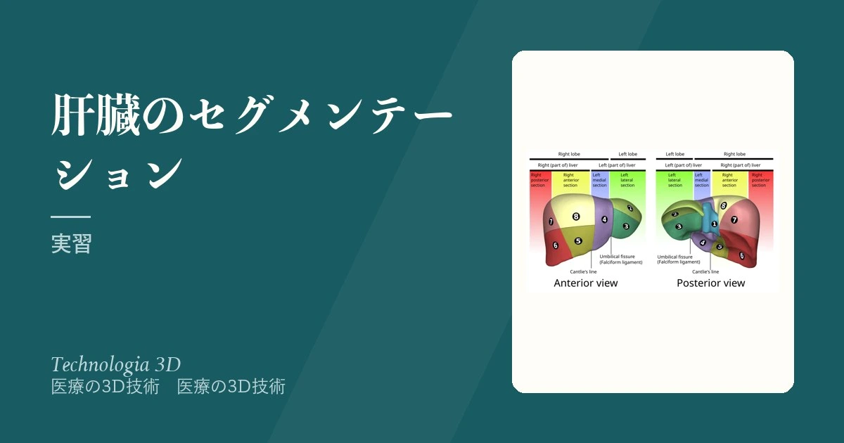
Couinaud 区域:肝を門脈の分岐と3本の肝静脈で8区域に分ける。区域 I は尾状葉。手術の単位は形態的な葉ではなく、この機能的な区域である。
肝の3Dモデルが術前計画で使われる理由は、区域が体表から見えないからである。切除線は血管の走行でしか決まらない——実際の手術では術中エコーで確認するが、事前に立体で把握しておくと安全域の設定が変わる。
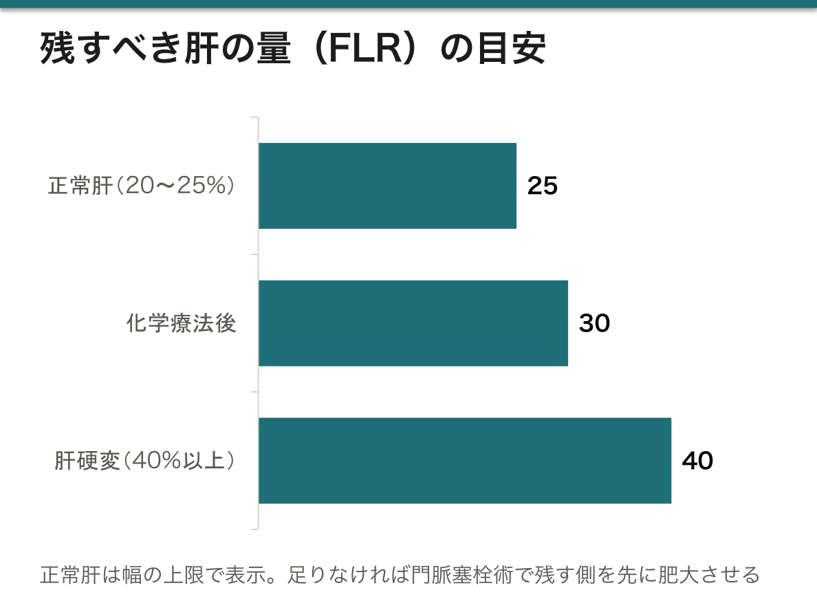
残肝容積(FLR: future liver remnant)の計算が最も実務的な出力である。セグメンテーションで切除予定部分と残る部分の体積を分け、正常肝では 20〜25%、化学療法後は 30%、肝硬変では 40% 以上を残せるかを判定する。足りなければ門脈塞栓術で残す側を先に肥大させる。体積という定量値が、術式そのものを変える。
実習で手こずるのは血管である。門脈と肝静脈は HU 値が近く、相が同じなら区別できない。2つの相のデータを別々にセグメンテーションして重ねるのが基本手順で、細い枝は領域拡張が漏れ出す(leakage)ため手で切る。「自動化できない部分がどこか」を体で知ることが、この実習の目的である。
肝の変異は多い。右後区域枝が左枝から出る、副肝動脈が上腸間膜動脈から出る(replaced right hepatic artery、約15%)など。教科書の図に合わせて修正してはいけない——モデルは患者の実物を写すためにある。
English version
Core
What is visible changes with the timing of contrast.
| Phase | Timing | What is seen well |
|---|---|---|
| Arterial | About 25–35 s | Hepatic artery, hypervascular tumour (HCC) |
| Portal venous | About 60–80 s | Portal vein, liver parenchyma (the highest contrast) |
| Equilibrium | About 3 min | Hepatic veins, tumours with delayed enhancement |
The Couinaud segments: the liver is divided into eight by the branches of the portal vein and the three hepatic veins. Segment I is the caudate lobe. The unit of surgery is not the morphological lobe but this functional segment.
Going further
3D models of the liver are used in planning because the segments cannot be seen from the surface. The line of resection can only be set by the course of the vessels — in the operation itself it is confirmed by intraoperative ultrasound, but grasping the anatomy in three dimensions beforehand changes how the safety margin is set.
Calculating the future liver remnant (FLR) is the most practical output. Segmentation separates the volume to be resected from the volume that will remain, and the judgement is whether 20–25 per cent in a normal liver, 30 per cent after chemotherapy, and 40 per cent or more in cirrhosis can be left. If not, portal vein embolisation is used first to hypertrophy the side that will remain. A quantitative volume changing the operation itself.
What causes trouble in the practical is the vessels. The portal and hepatic veins have close HU values and cannot be distinguished within one phase. Segmenting two phases separately and overlaying them is the basic procedure, and the fine branches leak with region growing and must be cut by hand. Learning in the body where automation cannot reach is the purpose of this practical.
Variation in the liver is common. The right posterior branch arising from the left, an accessory hepatic artery from the superior mesenteric (a replaced right hepatic artery, about 15 per cent). Do not correct the model to match the textbook figure — the model exists to reproduce the actual patient.
Mimics での実際の手順 — 肝の実習
使うのは .mcsx ファイル(Materialise Mimics のプロジェクト)。手順の骨格は次のとおり。
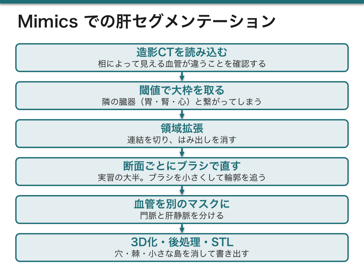
①造影CTを読み込む——肝実質・門脈・肝静脈が造影される相(フェーズ)が違うことを確認する。②閾値で大枠を取る——ただし肝実質と隣の臓器(胃・腎・心)はHUが近く、そのまま繋がってしまう。③領域拡張で連結を切り、はみ出しを消す。④断面を1枚ずつブラシで直す——実習の大半はここ。ブラシの大きさを小さくして、輪郭を追う。⑤血管を別のマスクとして分ける。⑥3D化して、穴・棘・小さな島を後処理で消す。⑦STL で書き出す。
肝が題材に選ばれている理由は「閾値法が最も通用しない臓器」だから。周囲との濃度差が小さく、形の個体差が大きく、境界が造影のタイミングに左右される。セグメンテーションの理論が破れる場面を、手を動かして体験する実習である。
作業の中心はブラシ操作の手順(消す・塗る・輪郭を確定する・領域を広げる)である。「うまくいかなければブラシの大きさを変えて、根気よく繰り返す」——これが実際の作業の質を決める。
English version
Core
What is used is a .mcsx file (a Materialise Mimics project). The skeleton of the procedure is as follows.
① Load the contrast CT — confirm that the phases in which liver parenchyma, portal vein and hepatic veins are enhanced are different. ② Take the outline by thresholding — but the liver parenchyma and the neighbouring organs (stomach, kidney, heart) have close HU and become joined. ③ Cut the connections and remove the overflow with region growing. ④ Correct slice by slice with the brush — most of the practical is here. Reduce the brush size and follow the contour. ⑤ Separate the vessels as another mask. ⑥ Make the 3D and remove holes, spikes and small islands in post-processing. ⑦ Export as STL.
The liver was chosen as the subject because it is the organ on which thresholding works least. The density difference from its surroundings is small, the individual variation of shape is large, and the boundary depends on the timing of contrast. It is the practical in which the theory of segmentation is experienced failing, with one’s own hands.
Going further
The core of the work is the sequence of brush operations (erase, paint, fix the contour, grow the region). "If it does not go well, change the brush size and repeat patiently" — and this is what decides the quality of the actual work.
Couinaud 区域と、なぜ血管を分けるのか
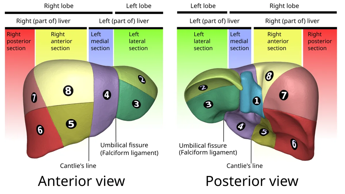
肝は外から見た葉(形態的)と、血管支配による区域(機能的)が一致しない。切除の単位になるのは後者である。
Couinaud 分類——門脈の枝が支配する範囲で8区域に分ける。境界は3本の肝静脈(右・中・左)と門脈の左右分岐の面。各区域は自分の門脈枝・肝動脈枝・胆管枝を持ち、独立して切り出せる。
だから実習で門脈と肝静脈を別々に分ける——血管が分かって初めて、切除線が引ける。腫瘍の位置だけでは切除範囲は決まらない。
数字の並び——尾状葉が I、左葉が II・III・IV、右葉が V〜VIII。時計回りに数える(前面から見て)。
解剖の肝臓と同じ内容を、画像から立体を作る側から見ている。解剖で「肝の H 字」を指すのと、CT から区域を切り出すのは同じ知識である。
English version
Core
In the liver, the lobes seen from outside (morphological) and the segments by vascular supply (functional) do not agree. What becomes the unit of resection is the latter.
The Couinaud classification — division into eight by the territory supplied by a branch of the portal vein. The boundaries are the three hepatic veins (right, middle, left) and the plane of the left–right portal division. Each segment has its own portal branch, hepatic arterial branch and bile duct, and can be removed independently.
Hence the portal and hepatic veins are segmented separately in the practical — only when the vessels are known can a line of resection be drawn. The position of the tumour alone does not settle the extent of resection.
The order of the numbers — the caudate lobe is I, the left lobe II, III and IV, the right lobe V to VIII. They are counted clockwise (seen from the front).
Going further
The same content as the liver in anatomy, seen from the side of building a solid from an image. Pointing out the "H" of the liver in anatomy and cutting the segments out of a CT are the same knowledge.
まとめ
| 肝のセグメンテーションと Couinaud 区域 | 造影のタイミングで見えるものが変わる。相タイミングよく見えるもの動脈相約 25〜35 秒肝動脈・多血性腫瘍(HCC)門脈相約 60〜80 秒門脈・肝実質(最もコントラストが高い)平… |
|---|---|
| Mimics での実際の手順 — 肝の実習 | 使うのは .mcsx ファイル(Materialise Mimics のプロジェクト)。 |
| Couinaud 区域と、なぜ血管を分けるのか | 肝は外から見た葉(形態的)と、血管支配による区域(機能的)が一致しない。 |
このノートは医学生が授業の内容を自分の言葉でまとめたもので、診療の判断に使うものではありません。誤りに気づいたらお知らせください。