脳硬膜は「2枚である」ことが全ての出発点である。2枚が離れれば静脈洞、2枚が落ち込めばひだ、ひだが仕切れば頭蓋腔の区画——ヘルニアの型まで、すべてここから出てくる。
標本で確かめるときは、硬膜を残した標本で、ひだ4つ → 洞を血の流れる順に → 槽と進めると整理しやすい。洞は名前を羅列するのではなく、「どこから来てどこへ抜けるか」を言えると強い(出口は内頸静脈ひとつだけである)。
この範囲は臨床との距離がいちばん近い。硬膜外血腫と硬膜下血腫、くも膜下出血、海綿静脈洞血栓症、水頭症——どれも「どの膜のあいだに何が溜まったか」で名前が付いている。膜の順番を外から数えられれば、病名の意味がそのまま読める。
- 脳硬膜と、その4つのひだ
- 硬膜静脈洞 — 血の流れる順に覚える
- くも膜下槽 — 髄液が溜まる場所
- 脳脊髄液の循環 — 作る・流す・戻す
- 脊髄硬膜
- 大脳鎌
- 小脳鎌
- 鞍隔膜
- 小脳テント
- 上矢状静脈洞
- 下矢状静脈洞
- 直静脈洞
- 静脈洞交会
- 後頭静脈洞
- 横静脈洞
- S状静脈洞
- 上錐体静脈洞
- 下錐体静脈洞
- 海綿静脈洞
- 小脳延髄槽(大槽)
- 橋小脳槽
- 脚間槽
- 交叉槽
- 大大脳静脈槽
- 脳硬膜と、その4つのひだDura mater encephali
- 硬膜静脈洞 — 血の流れる順に覚えるSinus durae matris
- くも膜下槽 — 髄液が溜まる場所Cisterna subarachnoidea
- 脳脊髄液の循環 — 作る・流す・戻すCirculatio liquoris cerebrospinalis
- 脊髄硬膜Dura mater spinalis
- 大脳鎌Falx cerebri
- 小脳鎌Falx cerebelli
- 鞍隔膜Diaphragma sellae
- 小脳テントTentorium cerebelli
- 上矢状静脈洞Sinus sagittalis superior
- 下矢状静脈洞Sinus sagittalis inferior
- 直静脈洞Sinus rectus
- 静脈洞交会Confluens sinuum
- 後頭静脈洞Sinus occipitalis
- 横静脈洞Sinus transversus
- S状静脈洞Sinus sigmoideus
- 上錐体静脈洞Sinus petrosus superior
- 下錐体静脈洞Sinus petrosus inferior
- 海綿静脈洞Sinus cavernosus
- 小脳延髄槽(大槽)Cisterna cerebellomedullaris
- 橋小脳槽Cisterna pontocerebellaris
- 脚間槽Cisterna interpeduncularis
- 交叉槽Cisterna chiasmatica
- 大大脳静脈槽Cisterna venae magnae cerebri
- まとめ
脳硬膜と、その4つのひだDura mater encephali
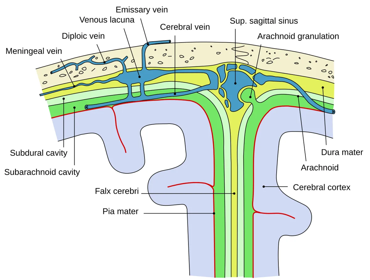
脳硬膜は2葉からなる。外葉=頭蓋骨の内骨膜、内葉=髄膜固有の層。脊髄硬膜が1枚なのに対し、脳硬膜は2枚——ここが両者の最大の違いである。
2葉が離れるところに静脈洞ができ、内葉が2枚重なって内側へ落ち込むところにひだができる。ひだは4つ。
| ひだ | 分けるもの |
|---|---|
| Falx cerebri(大脳鎌) | 左右の大脳半球(正中・矢状) |
| Tentorium cerebelli(小脳テント) | 後頭葉と小脳(水平) |
| Falx cerebelli(小脳鎌) | 左右の小脳半球 |
| Diaphragma sellae(鞍隔膜) | 下垂体窩の上蓋(漏斗が貫く) |
Tentorium cerebelli が脳を上下に仕切るため、頭蓋腔はテント上(supratentorial)とテント下(infratentorial)に分かれる。この境が臨床用語として日常的に使われるのは、腫瘍の好発部位も、ヘルニアの型も、この面を境に変わるからである。
テントの前縁が作る開口が incisura tentorii(テント切痕)で、ここを中脳が通る。テント上の圧が上がると鉤 uncus がこの切痕へ落ち込む(鉤ヘルニア)。大脳鎌の下縁の下を帯状回が越えれば帯状回ヘルニア、大後頭孔へ小脳扁桃が落ちれば大孔ヘルニア——ひだの形がそのままヘルニアの型を決めている。
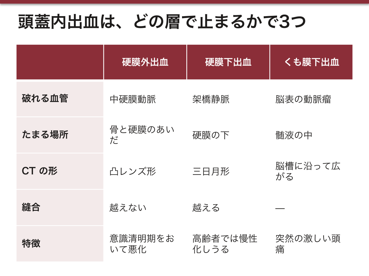
頭蓋内出血は、髄膜のどの層で止まるかで3つに分かれる。硬膜の構造がそのまま血腫の形を決める。
硬膜外出血(EDH)——硬膜動脈(中硬膜動脈)の破綻。骨と硬膜のあいだにたまるので、縫合を越えて広がらず、CT で凸レンズ形になる。側頭骨骨折に伴い、意識清明期(lucid interval)をおいて急速に悪化する。
硬膜下出血(SDH)——脳表から硬膜静脈洞へ渡る架橋静脈が引きちぎれる。三日月形で、縫合を越えて広がる。高齢者・アルコール多飲者では2週以上たって慢性硬膜下血腫として現れる。
くも膜下出血(SAH)——脳表の動脈瘤破裂。髄液の中に出るので血腫を作らず、脳槽に沿って広がる。突然の激しい頭痛。数時間たつとヘモグロビンの分解産物により髄液が黄色くなる(キサントクロミー)。
出典:イラスト解剖学 誌面575頁「髄膜と出血」
English version
Core
The cranial dura is two layers: an outer layer = the inner periosteum of the skull, and an inner layer proper to the meninges. Where the spinal dura is one layer, the cranial dura is two — this is the greatest difference between them.
Where the two layers separate, a venous sinus forms; where two thicknesses of the inner layer fold inwards, a septum forms.
There are four septa.
| Fold | Separates |
|---|---|
| Falx cerebri | The two cerebral hemispheres (median, sagittal) |
| Tentorium cerebelli | Occipital lobe from cerebellum (horizontal) |
| Falx cerebelli | The two cerebellar hemispheres |
| Diaphragma sellae | The roof of the hypophyseal fossa (pierced by the infundibulum) |
Going further
Because the tentorium cerebelli partitions the brain into upper and lower, the cranial cavity divides into supratentorial and infratentorial. This boundary is used as everyday clinical language because both the sites of predilection of tumours and the types of herniation change across it.
The opening formed by the free anterior margin of the tentorium is the incisura tentorii, through which the midbrain passes. When supratentorial pressure rises, the uncus descends into this notch (uncal herniation).
If the cingulate gyrus passes beneath the free edge of the falx it is a subfalcine herniation; if the cerebellar tonsils descend into the foramen magnum it is tonsillar herniation — the shape of the folds decides the type of the herniation.
In the clinic
Intracranial haemorrhage divides into three by the meningeal layer at which it stops. The structure of the dura decides the shape of the haematoma.
Extradural haematoma (EDH) — rupture of a meningeal artery (the middle meningeal). It collects between bone and dura, so it does not cross the sutures and is biconvex (lens-shaped) on CT. It accompanies a temporal bone fracture and deteriorates rapidly after a lucid interval.
Subdural haematoma (SDH) — tearing of the bridging veins running from the surface of the brain to a dural sinus. Crescent-shaped, and it crosses the sutures. In the elderly and in heavy drinkers it appears more than two weeks later as a chronic subdural haematoma.
Subarachnoid haemorrhage (SAH) — rupture of an aneurysm on the surface of the brain. Bleeding into the CSF, it forms no haematoma but spreads along the cisterns. Sudden violent headache. After some hours the breakdown products of haemoglobin turn the CSF yellow (xanthochromia).
硬膜静脈洞 — 血の流れる順に覚えるSinus durae matris
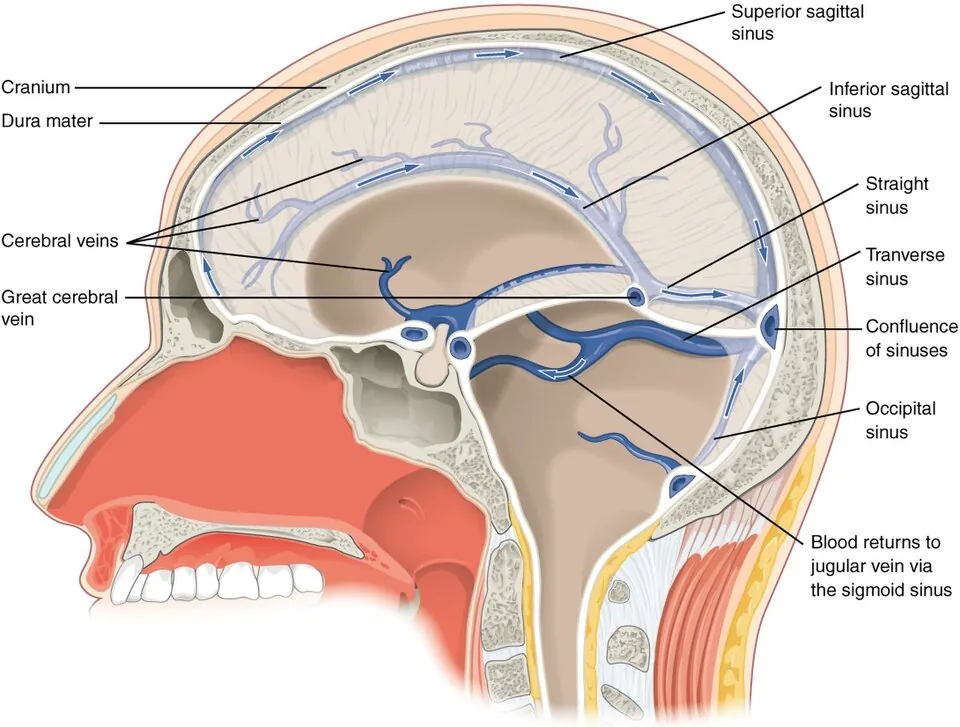
硬膜の2葉のあいだの静脈路。壁に平滑筋も弁も無いので、つぶれず、逆流しうる。
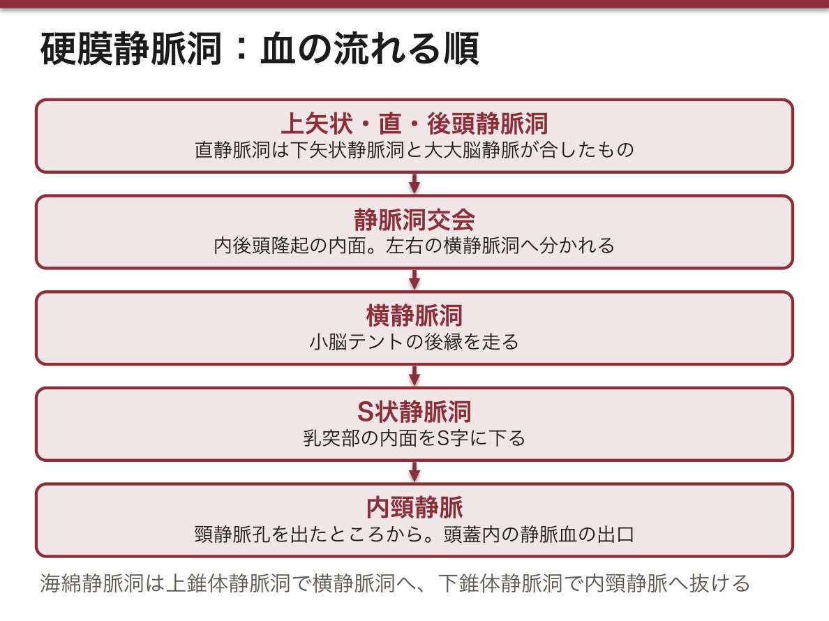
上のグループ(大脳鎌に沿う):Sinus sagittalis superior(大脳鎌の上縁)→ confluens sinuum(静脈洞交会)。Sinus sagittalis inferior(大脳鎌の下縁)→ 大大脳静脈と合して sinus rectus → 交会。Sinus occipitalis も交会へ。
交会から先:Sinus transversus(テント後縁)→ sinus sigmoideus(S状に曲がる)→ 頸静脈孔 → 内頸静脈。ここが唯一の出口である。
下のグループ:Sinus cavernosus(トルコ鞍の両側)— 前後の sinus intercavernosus で左右が繋がる。Sinus sphenoparietalis が前から注ぎ、後方へは sinus petrosus superior(→ 横洞)と inferior(→ 内頸静脈)で抜ける。
海綿静脈洞は「中を通るもの」が要点である。洞の中を通るのは 内頸動脈と VI 外転神経の2つだけ。外側壁に上から III・IV・V1・V2 が並ぶ。「中は2つ、壁は4つ」と覚える。
だから海綿静脈洞血栓症では、最初に外転神経麻痺(外転できない)が出やすい——洞の中を裸で走っており、圧に最も弱いからである。
弁が無いことが顔面の感染を危険にする。顔面静脈 → 上眼静脈 → 海綿静脈洞という逆流しうる経路があるため、鼻根から上口唇の三角(danger triangle)の癤を潰すと洞へ感染が届きうる。
クモ膜顆粒 granulationes arachnoideae は上矢状静脈洞に突き出す絨毛で、髄液はここから静脈血へ戻る。この吸収が落ちると交通性水頭症になる。
静脈洞には弁が無く、つぶれない。この2つが臨床のすべてを決める。
海綿静脈洞血栓症——顔面の感染(鼻・上唇の「危険三角」)が弁の無い顔面静脈を逆流して洞に達する。洞の中を通るのは内頸動脈と外転神経だけなので、最初に出るのは外転神経麻痺。ついで外側壁の III・IV・V1・V2 が落ちる。眼球突出・眼瞼腫脹・複視。
S状静脈洞は乳突蜂巣のすぐ内側を走る。中耳炎・乳様突起炎が波及してS状静脈洞血栓症を起こしうる。
骨折で洞が裂けると止血しにくい(壁が硬膜に固定されていて収縮しないため)。上矢状静脈洞が詰まれば両側傍矢状部の出血性梗塞となり、下肢優位の麻痺と痙攣を起こす。
English version
Core
The venous channels between the two layers of the dura. Their walls have neither smooth muscle nor valves, so they do not collapse and flow can reverse.
The upper group (along the falx): sinus sagittalis superior (the upper margin of the falx) → confluens sinuum. Sinus sagittalis inferior (the free lower margin) joins the great cerebral vein to form the sinus rectus → the confluence. The sinus occipitalis also reaches the confluence.
Beyond the confluence: sinus transversus (the posterior margin of the tentorium) → sinus sigmoideus → jugular foramen → internal jugular vein. This is the only exit.
The lower group: sinus cavernosus (on either side of the sella), joined across the midline by the anterior and posterior intercavernous sinuses. The sphenoparietal sinus enters it from in front; behind it drains by the superior petrosal sinus (→ transverse sinus) and the inferior petrosal sinus (→ internal jugular vein).
Going further
The point of the cavernous sinus is "what passes through it". Only two things pass through the interior: the internal carotid artery and the abducens nerve (VI). In the lateral wall lie, from above down, III, IV, V1 and V2. Remember it as "two inside, four in the wall".
So in cavernous sinus thrombosis an abducens palsy (inability to abduct) tends to come first — it runs naked through the interior of the sinus and is the most vulnerable to pressure.
The absence of valves is what makes facial infection dangerous. Because the route facial vein → superior ophthalmic vein → cavernous sinus can carry reflux, squeezing a boil in the triangle from the root of the nose to the upper lip (the danger triangle) can carry the infection to the sinus.
The granulationes arachnoideae are the villi projecting into the superior sagittal sinus, by which CSF returns to venous blood. When this absorption falls, communicating hydrocephalus results.
In the clinic
The sinuses have no valves and do not collapse. These two facts decide everything clinical.
Cavernous sinus thrombosis — infection of the face (the "danger triangle" of nose and upper lip) refluxes through the valveless facial vein and reaches the sinus. What passes through the interior of the sinus is only the internal carotid artery and the abducens nerve, so an abducens palsy comes first. Then III, IV, V1 and V2 in the lateral wall fail. Proptosis, swelling of the lids, diplopia.
The sigmoid sinus runs immediately inside the mastoid air cells, so otitis media and mastoiditis can spread to it and cause sigmoid sinus thrombosis.
A sinus torn by a fracture is hard to control (its wall is fixed to the dura and does not contract).
Occlusion of the superior sagittal sinus gives bilateral parasagittal haemorrhagic infarction, with leg-predominant weakness and seizures.
くも膜下槽 — 髄液が溜まる場所Cisterna subarachnoidea
クモ膜と軟膜のあいだの spatium subarachnoideum のうち、脳の表面と頭蓋底の形が離れて広くなった所を槽という。主なものは6つ。
| 槽 | 場所 |
|---|---|
| Cisterna cerebellomedullaris(大槽) | 小脳と延髄のあいだ。最大。Magendie 孔が開く |
| Cisterna pontocerebellaris | 橋の外側・橋小脳角 |
| Cisterna interpeduncularis | 両大脳脚のあいだ |
| Cisterna chiasmatica | 視交叉の周り |
| Cisterna venae magnae cerebri | 脳梁膨大の後下・松果体の上(四丘体槽) |
| Cisterna fossae lateralis cerebri | 外側溝の中(Sylvius 槽) |
脊髄側では cisterna lumbalis(腰槽・L2〜S2)が最大で、腰椎穿刺の目標である。
槽の位置と動脈瘤の好発部位はほぼ一致する。Willis 動脈輪とその枝はくも膜下腔を走るので、破裂すると血液は槽に沿って広がる。前交通動脈瘤 → chiasmatica・終板槽、後交通動脈瘤 → interpeduncularis、中大脳動脈瘤 → Sylvius 槽。CT で血液が溜まっている槽から、破れた動脈が推測できる。
橋小脳角は聴神経腫瘍の好発部位で、ここには VII・VIII が走る(前上方に V、下方に IX・X)。腫瘍が大きくなる順に、聴力低下 → 顔面のしびれ → 顔面神経麻痺 と症状が加わるのは、この解剖の順序そのものである。
動脈瘤は槽の中で破れる。Willis 動脈輪とその枝はくも膜下腔を走るので、出血した血液は槽の形に沿って広がる。CT でどの槽が高吸収かを見れば、破裂した動脈瘤の位置が推定できる。
前交通動脈瘤→ 交叉槽・終板槽。後交通動脈瘤→ 脚間槽(同側の動眼神経麻痺=瞳孔散大を伴う頭痛は破裂前の警告になる)。中大脳動脈瘤→ 外側溝(シルビウス裂)。
大槽(小脳延髄槽)は最大の槽で、Magendie 孔がここに開く。腰椎穿刺ができない場合の大槽穿刺の場でもあるが、延髄がすぐ前にあるため危険が大きい。
頭蓋内圧が高いときに腰椎穿刺をしてはならない。下から圧を抜くと、脳が槽を越えて落ち込む(脳ヘルニア)。先に眼底または CT で確かめる。
出典:イラスト解剖学 誌面578頁「脳ヘルニア」
English version
Core
Those parts of the subarachnoid space where the surface of the brain and the shape of the skull base draw apart and it widens are called cisterns. There are six chief ones.
| Cistern | Place |
|---|---|
| Cisterna cerebellomedullaris (cisterna magna) | Between cerebellum and medulla. The largest; the foramen of Magendie opens into it |
| Cisterna pontocerebellaris | Lateral to the pons — the cerebellopontine angle |
| Cisterna interpeduncularis | Between the two cerebral peduncles |
| Cisterna chiasmatica | Around the optic chiasm |
| Cisterna venae magnae cerebri | Behind and below the splenium, above the pineal (the quadrigeminal cistern) |
| Cisterna fossae lateralis cerebri | Within the lateral sulcus (the Sylvian cistern) |
On the spinal side the largest is the cisterna lumbalis (L2–S2), the target of lumbar puncture.
Going further
The positions of the cisterns coincide almost exactly with the sites of predilection of aneurysms. The circle of Willis and its branches run in the subarachnoid space, so when one ruptures the blood spreads along the cisterns. Anterior communicating aneurysm → chiasmatic and lamina terminalis cisterns; posterior communicating → interpeduncular; middle cerebral → Sylvian. From the cistern in which blood has collected on CT, the ruptured artery can be inferred.
The cerebellopontine angle is the site of predilection of acoustic neuroma, and VII and VIII run there (V anterosuperiorly, IX and X below). That the symptoms are added in the order hearing loss → facial numbness → facial palsy as the tumour enlarges is that anatomical order itself.
In the clinic
An aneurysm ruptures within a cistern. The circle of Willis and its branches run in the subarachnoid space, so the blood spreads into the shape of the cisterns. See which cistern is hyperdense on CT and the site of the ruptured aneurysm can be inferred.
- Anterior communicating aneurysm → chiasmatic and lamina terminalis cisterns.
- Posterior communicating aneurysm → interpeduncular cistern (a headache with an ipsilateral oculomotor palsy — a dilated pupil — is a warning before rupture).
- Middle cerebral aneurysm → the lateral (Sylvian) fissure.
The cisterna magna is the largest cistern, and the foramen of Magendie opens into it. It is also the site of cisternal puncture when lumbar puncture is impossible, but the medulla lies immediately in front and the risk is great.
Lumbar puncture must not be performed when the intracranial pressure is raised. Releasing pressure from below lets the brain descend past the cisterns (herniation). Check the fundus or the CT first.
脳脊髄液の循環 — 作る・流す・戻すCirculatio liquoris cerebrospinalis
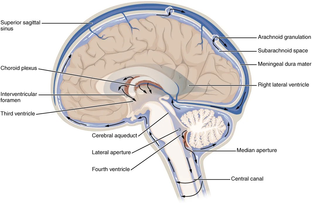
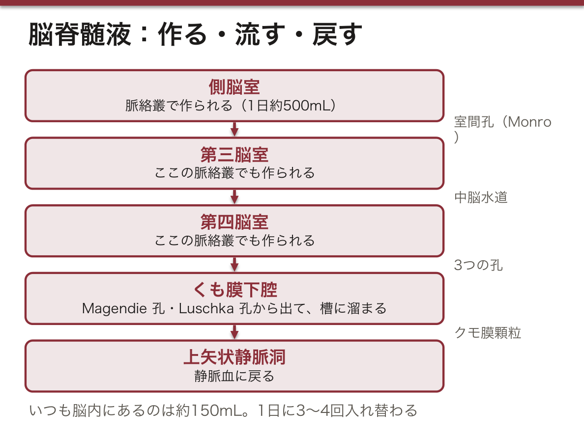
作る:脳室の脈絡叢 plexus choroideus(側脳室・第三脳室・第四脳室)。1日あたり約500mL 作られ、いつも脳内にあるのは約150mL——1日に3〜4回入れ替わる。
流す:側脳室 →(Monro 孔)→ 第三脳室 →(中脳水道)→ 第四脳室 →(Magendie 孔・Luschka 孔)→ くも膜下腔。
戻す:くも膜下腔 → クモ膜顆粒 → 上矢状静脈洞(=静脈血へ)。
役割は①衝撃の緩衝 ②浮力による脳の実効重量の軽減(1,400g → 約50g)③老廃物の排出。
「作る場所」と「戻す場所」が離れていることが、水頭症の分類を決める。経路の途中(孔・水道)で詰まれば非交通性(閉塞性)、くも膜下腔や顆粒の側で詰まれば交通性。詰まった場所より上流だけが拡大するので、CT の脳室拡大の分布から閉塞点が読める(側脳室だけ=Monro 孔、側脳室+第三脳室=水道、全部=交通性)。
脈絡叢は血液脳関門ではなく血液髄液関門を作る。毛細血管は有窓性だが、上衣細胞のあいだに閉鎖帯があり、そこが関門になっている。関門の場所が血管側ではなく上皮側にあるのが特徴で、組織学の内容とそのまま繋がる。
髄液は1日約500mL 作られ、脳内にあるのは約150mL——1日に3〜4回入れ替わる。産生・流通・吸収のどこが崩れても水頭症になる。
非交通性(閉塞性)水頭症——脳室系の中で詰まる。中脳水道狭窄・Luschka/Magendie 孔の閉塞・後頭蓋窩腫瘍。詰まった点より上流だけが拡大する。
交通性水頭症——脳室からくも膜下腔までは通じていて、その先(くも膜顆粒での吸収)が悪い。くも膜下出血後・髄膜炎後が代表である。
正常圧水頭症(NPH)——認知症・歩行障害・尿失禁の3徴で、髄液圧は正常。基本は交通性である。シャント術で改善しうる「治せる認知症」なので、高齢者のすり足歩行を見たら必ず思い出すこと。
出典:イラスト解剖学 誌面587頁「水頭症って?」
English version
Core
Production: the choroid plexus of the ventricles (lateral, third and fourth). About 500 mL a day is made, while about 150 mL is present at any time — it is replaced three or four times a day.
Flow: lateral ventricle → (foramen of Monro) → third ventricle → (cerebral aqueduct) → fourth ventricle → (foramina of Magendie and Luschka) → subarachnoid space.
Return: subarachnoid space → arachnoid granulations → superior sagittal sinus (that is, into venous blood).
Its roles: ① cushioning against impact ② reduction of the effective weight of the brain by buoyancy (1,400 g → about 50 g) ③ removal of waste.
Going further
That the place of production and the place of return are far apart decides the classification of hydrocephalus. A block along the route (a foramen, the aqueduct) is non-communicating (obstructive); a block on the side of the subarachnoid space or the granulations is communicating. Only what lies upstream of the block dilates, so the point of obstruction can be read from the distribution of ventricular dilatation on CT (lateral ventricles only = foramen of Monro; lateral plus third = aqueduct; all = communicating).
The choroid plexus forms not the blood–brain barrier but the blood–CSF barrier. The capillaries are fenestrated, but there are tight junctions between the ependymal cells and it is these that constitute the barrier. The barrier lies on the epithelial rather than the vascular side — a point that connects directly with the histology.
In the clinic
About 500 mL of CSF is made a day and about 150 mL is present — it turns over three or four times daily. A failure anywhere in production, flow or absorption gives hydrocephalus.
Non-communicating (obstructive) — a block within the ventricular system: aqueduct stenosis, obstruction of the foramina of Luschka or Magendie, a posterior fossa tumour. Only what lies upstream dilates.
Communicating — the route from ventricles to subarachnoid space is patent and the fault lies beyond it (absorption at the arachnoid granulations). After subarachnoid haemorrhage and after meningitis are the typical causes.
Normal pressure hydrocephalus (NPH) — the triad of dementia, gait disturbance and urinary incontinence, with a normal CSF pressure. It is fundamentally communicating. As a treatable dementia that may improve with shunting, always think of it on seeing a shuffling gait in an older person.
脊髄硬膜Dura mater spinalis
脊柱管の中で脊髄を包む硬膜。頭蓋の硬膜と決定的に違うのは、骨膜と癒着していないことで、そのため骨との間に 硬膜外腔 spatium epidurale(脂肪と内椎骨静脈叢が入る)がある。
上は大後頭孔で脳硬膜に続き、下は S2 の高さで袋状に終わって、終糸を包みながら尾骨に付く。
「硬膜外麻酔はこの外側、腰椎穿刺はこの内側」と言えれば足りる。標本では、脊髄をゆるく包む厚い一枚として示す。
English version
Core
The dura enclosing the cord within the vertebral canal. The decisive difference from the cranial dura is that it is not fused with the periosteum, so a spatium epidurale exists between it and the bone (containing fat and the internal vertebral venous plexus).
Above, it continues into the cranial dura at the foramen magnum; below, it ends as a sac at the level of S2 and, enclosing the filum terminale, attaches to the coccyx.
It is enough to be able to say "epidural anaesthesia is outside this, lumbar puncture inside it". On the specimen, show it as the single thick layer loosely enclosing the cord.
大脳鎌Falx cerebri
正中矢状面に立つ鎌の形をしたひだ。大脳縦裂に垂れ下がり、左右の大脳半球を隔てる。硬膜のひだの中でいちばん大きく、標本で最初に目に入る。
前端は鶏冠 crista galliに付き、後方は小脳テントに合流する。上縁に上矢状静脈洞、下縁(遊離縁)に下矢状静脈洞、テントとの合流線に直静脈洞——3つの洞の位置が、そのまま鎌の3つの縁である。
English version
Core
The sickle-shaped fold standing in the median sagittal plane, hanging into the longitudinal fissure and separating the two hemispheres. The largest of the dural folds and the first thing the eye meets on a specimen.
Its anterior end attaches to the crista galli and behind it joins the tentorium cerebelli.
The superior sagittal sinus lies in its upper margin, the inferior sagittal sinus in its free lower margin, and the straight sinus along its junction with the tentorium — the positions of three sinuses are precisely the three margins of the falx.
小脳鎌Falx cerebelli
小脳テントの下で、後頭骨内面の正中から左右の小脳半球の間へ入る小さな鎌。大脳鎌と同じ形だが、はるかに小さい。
後縁は内後頭稜に付き、その中を後頭静脈洞が走る。「大脳鎌の縮小版が小脳の間にもある」と押さえれば、テントの上下で対になっていることが見える。
English version
Core
The small sickle passing from the midline of the inner surface of the occipital bone, beneath the tentorium, between the two cerebellar hemispheres. The same shape as the falx cerebri but far smaller.
Its posterior margin attaches to the internal occipital crest, within which runs the occipital sinus.
Fix it as "a miniature falx cerebri also exists between the halves of the cerebellum", and the pairing above and below the tentorium becomes visible.
鞍隔膜Diaphragma sellae
トルコ鞍の上に水平に張る小さな膜で、下垂体窩の蓋になっている。中央に孔があり、そこを下垂体茎(漏斗 infundibulum)が通る。
4つのひだのうち唯一「鎌でもテントでもない」ので、標本では位置——トルコ鞍の真上——で示す。
English version
Core
The small membrane stretched horizontally over the sella turcica, forming the roof of the hypophyseal fossa. There is an opening at its centre through which the pituitary stalk (infundibulum) passes.
It is the only one of the four folds that is neither a falx nor a tentorium, so on a specimen show it by its position — directly above the sella turcica.
小脳テントTentorium cerebelli
大脳の後頭葉と小脳の間に、ほぼ水平に張るテント。後外側は後頭骨の横洞溝に、前外側は側頭骨錐体部の上縁に付く。
前縁は自由縁で、テント切痕 incisura tentorii という穴を作り、ここを中脳が通り抜ける。
「テント上 supratentorial」「テント下 infratentorial」の境がこれ。頭蓋内圧が上がったときに側頭葉内側が切痕へ落ち込むのがテント切痕ヘルニアで、動眼神経が巻き込まれる。
English version
Core
The tent stretched almost horizontally between the occipital lobe and the cerebellum, attached posterolaterally to the groove for the transverse sinus on the occipital bone and anterolaterally to the superior border of the petrous temporal bone.
Its anterior margin is free and forms the opening, the incisura tentorii, through which the midbrain passes. This is the boundary of "supratentorial" and "infratentorial".
Descent of the medial temporal lobe into the notch when the intracranial pressure rises is tentorial herniation, and the oculomotor nerve is caught up in it.
上矢状静脈洞Sinus sagittalis superior
大脳鎌の上縁、頭蓋冠の正中を前から後ろへ走る。前は鶏冠のあたりから始まり、後方で静脈洞交会に注ぐ。
ここにくも膜顆粒 granulationes arachnoideae が突き出していて、髄液が静脈血へ戻る出口になっている。「髄液の帰り道」として指せることが要る。
English version
Core
Runs from front to back in the midline of the vault, in the upper margin of the falx. It begins about the crista galli and drains behind into the confluence of the sinuses.
The arachnoid granulations project into it, and it is the exit by which CSF returns to venous blood. You must be able to point to it as "the way back for the CSF".
下矢状静脈洞Sinus sagittalis inferior
大脳鎌の下縁(遊離縁)を走る細い洞。後方で大大脳静脈(Galen 静脈)と合流して直静脈洞になる。
上矢状洞と対にして「鎌の上の縁と下の縁」で覚えると、標本の上で迷わない。
English version
Core
The slender sinus running in the free lower margin of the falx. Behind, it joins the great cerebral vein (of Galen) to form the straight sinus.
Learn it with the superior sagittal sinus as "the upper and the lower margin of the falx" and you will not lose your way on the specimen.
直静脈洞Sinus rectus
大脳鎌と小脳テントが合わさる線を、前上から後下へまっすぐ走る。名前のとおり曲がらない。
受けるのは下矢状静脈洞と大大脳静脈の2本で、後方の静脈洞交会へ注ぐ。「鎌とテントの継ぎ目」を指せば当たる。
English version
Core
Runs straight from anterosuperior to posteroinferior along the junction of falx and tentorium. As its name says, it does not bend.
It receives two vessels, the inferior sagittal sinus and the great cerebral vein, and drains behind into the confluence of the sinuses.
Point to the seam between falx and tentorium and you have it.
静脈洞交会Confluens sinuum
内後頭隆起の内面にある、洞の合流点。上矢状静脈洞・直静脈洞・後頭静脈洞が集まり、左右の横静脈洞へ分かれて出ていく。
左右対称に流れるとは限らず、上矢状洞は右へ、直洞は左へ流れることが多い。標本では位置——後頭部の正中、隆起の裏——で示す。
English version
Core
The meeting point of the sinuses on the inner surface of the internal occipital protuberance. The superior sagittal, straight and occipital sinuses gather there and leave as the two transverse sinuses.
The flow is not necessarily symmetrical: the superior sagittal sinus commonly drains to the right and the straight sinus to the left.
On a specimen show it by its position — the midline of the occiput, behind the protuberance.
後頭静脈洞Sinus occipitalis
小脳鎌の中を、静脈洞交会から大後頭孔の縁へ下る細い洞。大後頭孔のまわりで静脈叢をつくり、S状洞や椎骨静脈叢とつながる。
4つの洞が集まる交会のうち唯一「下へ向かう」ものなので、方向で見分けられる。
English version
Core
The slender sinus descending within the falx cerebelli from the confluence to the margin of the foramen magnum, where it forms a venous plexus continuous with the sigmoid sinus and the vertebral venous plexus.
It is the only one of the four sinuses meeting at the confluence that runs downwards, and so is recognised by its direction.
横静脈洞Sinus transversus
静脈洞交会から左右に水平へ走る。走る場所は小脳テントの後外側の付着縁で、骨には後頭骨の横洞溝として跡が残る。
外側の端で向きを変え、S状静脈洞に続く。「テントの後ろの付け根を横に」と覚える。
English version
Core
Runs horizontally to each side from the confluence, along the posterolateral attached margin of the tentorium, leaving its trace on the occipital bone as the groove for the transverse sinus. At its lateral end it changes direction and continues as the sigmoid sinus.
Remember it as "transversely along the posterior root of the tentorium".
S状静脈洞Sinus sigmoideus
横静脈洞の続きで、側頭骨乳突部の内面をS字に曲がりながら下る。頸静脈孔を出たところで内頸静脈になる。
頭蓋内の静脈血が外へ出る、最後の一本。中耳・乳突蜂巣のすぐ内側を走るため、中耳炎がここへ及ぶと敗血症性血栓症になる。
English version
Core
The continuation of the transverse sinus, curving in an S down the inner surface of the mastoid part of the temporal bone. On leaving the jugular foramen it becomes the internal jugular vein. The last single channel by which intracranial venous blood leaves.
It runs immediately inside the middle ear and mastoid air cells, so otitis media spreading to it causes septic thrombosis.
上錐体静脈洞Sinus petrosus superior
側頭骨錐体部の上縁(小脳テントが付く線)を走り、海綿静脈洞から横静脈洞〜S状静脈洞へつなぐ。
English version
Core
Runs along the superior border of the petrous temporal bone (the line of attachment of the tentorium), connecting the cavernous sinus with the transverse and sigmoid sinuses.
下錐体静脈洞Sinus petrosus inferior
錐体部の後下縁(後頭骨との縫合)を走り、海綿静脈洞から直接頸静脈孔・内頸静脈へ落ちる。
上下ひとそろいで「海綿洞から後ろへ抜ける2本」。上は横洞へ、下は内頸静脈へ、と行き先で区別する。
English version
Core
Runs along the posteroinferior border of the petrous bone (the suture with the occipital), dropping directly from the cavernous sinus to the jugular foramen and the internal jugular vein.
Take the two as a set — the two channels leaving the cavernous sinus backwards. Distinguish them by destination: the superior to the transverse sinus, the inferior to the internal jugular vein.
海綿静脈洞Sinus cavernosus
トルコ鞍の左右に広がる、海綿状に区切られた洞。左右は鞍の前後で連絡して輪をつくる。
要はここを通るもの。洞の中を 内頸動脈と外転神経(VI)が通り、外側壁に動眼神経(III)・滑車神経(IV)・眼神経(V1)・上顎神経(V2)が上から順に並ぶ。
顔面の静脈は弁を持たず、眼静脈を通じてここへ逆流しうる。鼻や上唇の感染がここへ及ぶ経路が、いわゆる危険三角である。
English version
Core
The sinus spreading on either side of the sella turcica, divided internally like a sponge. The two sides communicate in front of and behind the sella to form a ring.
The point is what passes through it. Within the sinus run the internal carotid artery and the abducens nerve (VI); in its lateral wall lie, from above down, the oculomotor (III), trochlear (IV), ophthalmic (V1) and maxillary (V2) nerves.
The veins of the face have no valves and can reflux into it through the ophthalmic veins. The route by which infection of the nose or upper lip reaches it is the so-called danger triangle.
小脳延髄槽(大槽)Cisterna cerebellomedullaris
小脳の下面と延髄の背側の間にある、くも膜下槽で最大のもの。大槽 cisterna magna とも呼ぶ。
第四脳室の正中孔(Magendie 孔)がここへ開き、髄液がくも膜下腔へ出る最初の場所になる。後頭下穿刺の標的でもある。
English version
Core
The cistern between the inferior surface of the cerebellum and the dorsal medulla, the largest of the subarachnoid cisterns, also called the cisterna magna.
The median aperture (of Magendie) of the fourth ventricle opens into it, so it is the first place at which CSF enters the subarachnoid space. It is also the target of suboccipital puncture.
橋小脳槽Cisterna pontocerebellaris
橋の外側面と小脳の間に広がる槽(橋槽の外側部)。ここが小脳橋角 angulus pontocerebellaris にあたる。
顔面神経(VII)と内耳神経(VIII)が通るため、聴神経腫瘍ができる場所として臨床でも名前が出る。
English version
Core
The cistern between the lateral surface of the pons and the cerebellum (the lateral part of the pontine cistern). This is the cerebellopontine angle.
The facial (VII) and vestibulocochlear (VIII) nerves pass through it, so it is named clinically as the site of acoustic neuroma.
脚間槽Cisterna interpeduncularis
左右の大脳脚の間(脚間窩)を満たす槽。動眼神経(III)がここから出るほか、脳底動脈の分岐部と後大脳動脈が入っている。
綴りは interpeduncularis(l は1つ)。
English version
Core
The cistern filling the space between the cerebral peduncles (the interpeduncular fossa). The oculomotor nerve (III) emerges from it, and it contains the bifurcation of the basilar artery and the posterior cerebral arteries.
The spelling is interpeduncularis, with one l.
交叉槽Cisterna chiasmatica
視交叉のまわり、トルコ鞍の上に広がる槽。前方で終板槽へ、後方で脚間槽へ続く。
脳底面の標本では、視交叉を目印にその周囲として示せばよい。
English version
Core
The cistern around the optic chiasm, above the sella turcica, continuing forwards into the lamina terminalis cistern and backwards into the interpeduncular cistern.
On a specimen of the base of the brain it is enough to take the chiasm as the landmark and show the space around it.
大大脳静脈槽Cisterna venae magnae cerebri
脳梁膨大の後下、小脳の上面と四丘体の間にある槽。四丘体槽 cisterna quadrigeminalis ともいう。
名のとおり大大脳静脈(Galen 静脈)を入れ、松果体もここに顔を出す。
English version
Core
The cistern behind and below the splenium, between the upper surface of the cerebellum and the quadrigeminal plate; also called the cisterna quadrigeminalis.
As its name says it contains the great cerebral vein (of Galen), and the pineal gland also faces into it.
まとめ
| 脳硬膜と、その4つのひだ | 脳硬膜は2葉からなる。外葉=頭蓋骨の内骨膜、内葉=髄膜固有の層。 |
|---|---|
| 硬膜静脈洞 — 血の流れる順に覚える | 硬膜の2葉のあいだの静脈路。壁に平滑筋も弁も無いので、つぶれず、逆流しうる。 |
| くも膜下槽 — 髄液が溜まる場所 | クモ膜と軟膜のあいだの spatium subarachnoideum のうち、脳の表面と頭蓋底の形が離れて広くなった所を槽という。 |
| 脳脊髄液の循環 — 作る・流す・戻す | 作る:脳室の脈絡叢 plexus choroideus(側脳室・第三脳室・第四脳室)。 |
| 脊髄硬膜 | 脊柱管の中で脊髄を包む硬膜。頭蓋の硬膜と決定的に違うのは、骨膜と癒着していないことで、そのため骨との間に 硬膜外腔 spatium epidurale(脂肪と内椎骨静脈叢が入る)が… |
| 大脳鎌 | 正中矢状面に立つ鎌の形をしたひだ。大脳縦裂に垂れ下がり、左右の大脳半球を隔てる。 |
| 小脳鎌 | 小脳テントの下で、後頭骨内面の正中から左右の小脳半球の間へ入る小さな鎌。 |
| 鞍隔膜 | トルコ鞍の上に水平に張る小さな膜で、下垂体窩の蓋になっている。 |
| 小脳テント | 大脳の後頭葉と小脳の間に、ほぼ水平に張るテント。 |
| 上矢状静脈洞 | 大脳鎌の上縁、頭蓋冠の正中を前から後ろへ走る。 |
| 下矢状静脈洞 | 大脳鎌の下縁(遊離縁)を走る細い洞。後方で大大脳静脈(Galen 静脈)と合流して直静脈洞になる。 |
| 直静脈洞 | 大脳鎌と小脳テントが合わさる線を、前上から後下へまっすぐ走る。 |
| 静脈洞交会 | 内後頭隆起の内面にある、洞の合流点。上矢状静脈洞・直静脈洞・後頭静脈洞が集まり、左右の横静脈洞へ分かれて出ていく。 |
| 後頭静脈洞 | 小脳鎌の中を、静脈洞交会から大後頭孔の縁へ下る細い洞。 |
| 横静脈洞 | 静脈洞交会から左右に水平へ走る。走る場所は小脳テントの後外側の付着縁で、骨には後頭骨の横洞溝として跡が残る。 |
| S状静脈洞 | 横静脈洞の続きで、側頭骨乳突部の内面をS字に曲がりながら下る。 |
| 上錐体静脈洞 | 側頭骨錐体部の上縁(小脳テントが付く線)を走り、海綿静脈洞から横静脈洞〜S状静脈洞へつなぐ。 |
| 下錐体静脈洞 | 錐体部の後下縁(後頭骨との縫合)を走り、海綿静脈洞から直接頸静脈孔・内頸静脈へ落ちる。 |
| 海綿静脈洞 | トルコ鞍の左右に広がる、海綿状に区切られた洞。 |
| 小脳延髄槽(大槽) | 小脳の下面と延髄の背側の間にある、くも膜下槽で最大のもの。 |
| 橋小脳槽 | 橋の外側面と小脳の間に広がる槽(橋槽の外側部)。 |
| 脚間槽 | 左右の大脳脚の間(脚間窩)を満たす槽。動眼神経(III)がここから出るほか、脳底動脈の分岐部と後大脳動脈が入っている。 |
| 交叉槽 | 視交叉のまわり、トルコ鞍の上に広がる槽。 |
| 大大脳静脈槽 | 脳梁膨大の後下、小脳の上面と四丘体の間にある槽。 |
このノートは医学生が授業の内容を自分の言葉でまとめたもので、診療の判断に使うものではありません。誤りに気づいたらお知らせください。