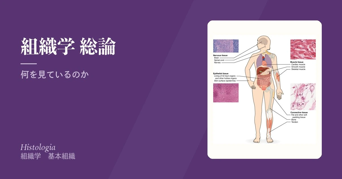
組織学は「顕微鏡の中の解剖学」だが、暗記の科目ではない。切片は三次元の構造を二次元に切った断面であり、同じ器官でも切る向きが変われば別物に見える。だから組織学でまずやるべきは、像を覚えることではなく「いま何をどの向きで切ったのか」を復元する訓練である。
体を作る組織は4つしかない。上皮・結合・筋・神経。どんな器官もこの4つの組み合わせでできている。胃も腎臓も皮膚も、上皮が表面を覆い、結合組織が支え、筋が動かし、神経が制御する——この型を先に入れておくと、器官組織学が「新しい暗記」ではなく「型の当てはめ」になる。
組織学の学習は、標本を見て同定できることが中心になる。国試は組織そのものより病理の土台として問う。この章の要点は両方に効く。
- 組織
- ディフェロン — 組織を「幹細胞からの系列」として見る
- 実質
- 細胞外基質
- 基底膜
- HE染色 — なぜ青と赤なのか
- 切片は断面である — 像の読み違えを防ぐ
- 何が見えて、何が見えないか — 分解能という限界
- 腫瘍の名前は、どの組織から出たかで決まっている
- 組織学という学問 — 何を目的に、どの基礎医学とつながるか
組織Textus
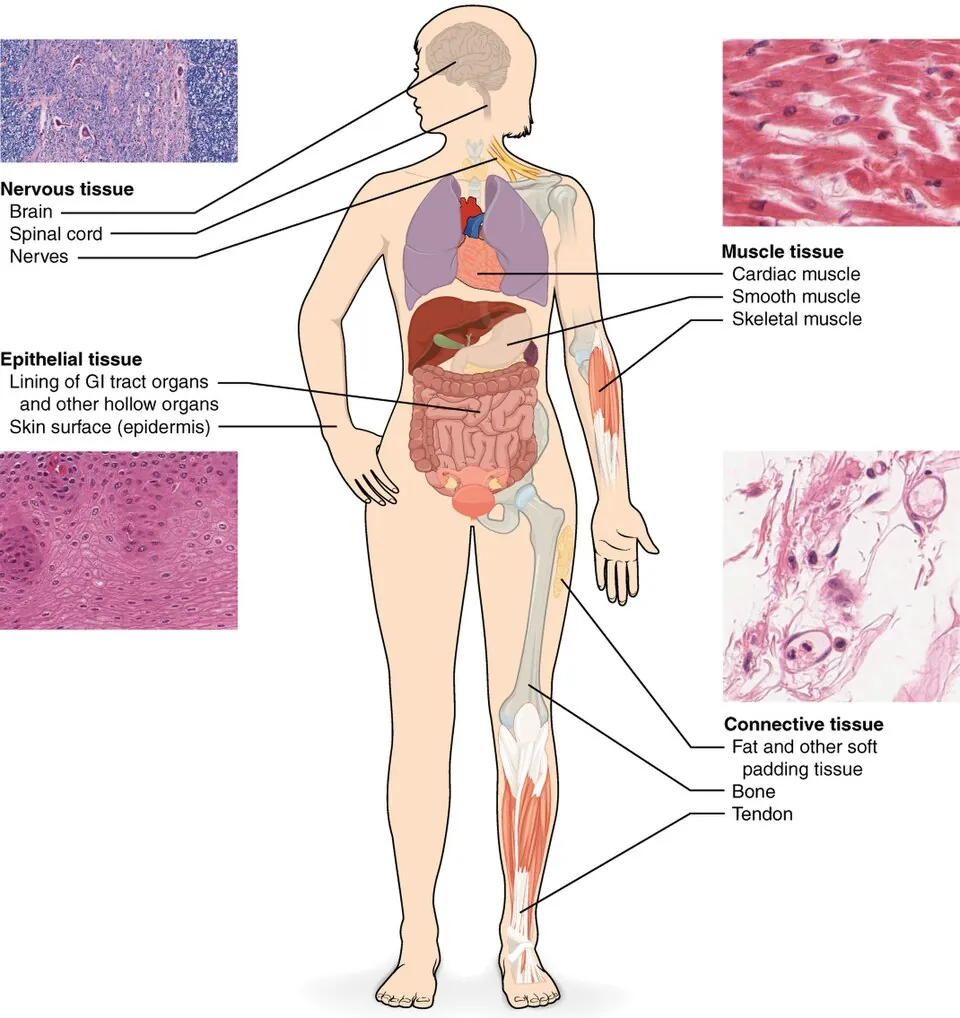
同じ働きをする細胞と細胞外基質の集まり。体には4種類しかない——上皮組織・結合組織・筋組織・神経組織。
器官はこの4つの層構造としてできている。たとえば消化管はどの高さでも粘膜(上皮+結合組織)→粘膜下層(結合組織)→筋層(筋)→漿膜(上皮)で、神経叢が2か所に挟まる。食道と大腸で違うのは上皮の種類と筋の厚さだけである。
4つの区別の本質は細胞と細胞外基質のどちらが主役かにある。上皮は細胞がびっしり詰まり基質がほぼ無い。結合組織は逆に基質が主役で細胞はまばら。筋と神経は上皮寄りだが、収縮と伝導という特殊化に振り切っている。
発生学的にも整理できる。上皮は三胚葉すべてから生じる(外胚葉=表皮、内胚葉=消化管、中胚葉=腎尿細管・体腔の中皮)。結合組織と筋は基本的に中胚葉、神経は外胚葉。「上皮=外胚葉」と覚えると腎臓と中皮で必ず崩れる。
4種類しかないから、腫瘍の分類も4種類で足りる。上皮由来=癌腫(carcinoma)、非上皮(間葉)由来=肉腫(sarcoma)。免疫染色でサイトケラチン陽性なら癌腫、ビメンチン陽性なら肉腫——転移巣しか見つからないときに原発を推定する手がかりになる。
発生頻度も転移経路も違う。癌腫は圧倒的に多く、まずリンパ行性に広がる。肉腫は稀で、血行性に肺へ飛ぶ。「上皮か非上皮か」という組織学の最初の区分が、そのまま検索すべき転移先を決める。
器官が4つの層でできていることは、炎症の深さの評価にそのまま使える。潰瘍性大腸炎は粘膜にとどまり、Crohn 病は全層に及ぶ——層で考えれば、穿孔や瘻孔がどちらで起こるかが導ける。
English version
Core
A collection of cells and extracellular matrix performing a common function. There are only four: epithelial, connective, muscular and nervous tissue. Organs are built as layered arrangements of these four.
The digestive tract, for example, has the same plan at every level: mucosa (epithelium plus connective tissue) → submucosa (connective tissue) → muscularis (muscle) → serosa (epithelium), with a nerve plexus in two of the intervals. Between oesophagus and colon, only the type of epithelium and the thickness of the muscle differ.
Going further
The essence of the fourfold division is whether the cells or the matrix predominate. Epithelium is packed with cells and has almost no matrix; connective tissue is the reverse, matrix-dominated with sparse cells. Muscle and nerve resemble epithelium in cellularity but have committed themselves entirely to contraction and to conduction.
It can also be organised embryologically. Epithelium arises from all three germ layers — ectoderm for the epidermis, endoderm for the gut, mesoderm for the renal tubules and the mesothelium of the body cavities. Connective tissue and muscle are essentially mesodermal, nervous tissue ectodermal. Remembering "epithelium equals ectoderm" will always fail on the kidney and the mesothelium.
In the clinic
Because there are only four tissues, four categories suffice for naming tumours. Epithelial origin gives a carcinoma; non-epithelial, mesenchymal origin gives a sarcoma. Immunohistochemistry follows the same logic: cytokeratin positive means carcinoma, vimentin positive means sarcoma — the clue to the primary site when only a metastasis has been found.
Incidence and route of spread differ as well. Carcinomas are far commoner and spread first by lymphatics; sarcomas are rare and spread haematogenously to the lung. The very first distinction in histology — epithelial or not — decides where to look for metastases.
That organs are built in layers is directly usable in assessing the depth of inflammation. Ulcerative colitis is confined to the mucosa, Crohn disease is transmural — thinking in layers tells you which of the two produces perforation and fistula.
ディフェロン — 組織を「幹細胞からの系列」として見る
ディフェロン(differon、組織発生系列)は、東ヨーロッパの組織学で使われる考え方で、1つの系統の細胞を、幹細胞から成熟した細胞まで並べた全体を指す。
幹細胞 → 前駆細胞(半幹細胞・決定された細胞)→ 芽細胞 → 成熟途中の細胞 → 成熟(分化)した細胞
組織は1つ以上のディフェロンの集まりとして説明される。たとえば表皮は角化細胞のディフェロンが主体で、そこにメラノサイトと Langerhans 細胞のディフェロンが加わる。英米の教科書では同じことを幹細胞の階層(stem cell → transit-amplifying cell → terminally differentiated cell)と呼ぶ。
入れ替わり方で、組織の細胞集団は3つに分けられる。
| 集団 | 性質 | 例 |
|---|---|---|
| 更新する集団 | 幹細胞が常に分裂し、成熟した細胞は捨てられ続ける | 表皮、腸の上皮(3〜5日で入れ替わる)、血液 |
| 増えうる集団 | ふだんは G0 で休むが、失われると分裂を再開する | 肝細胞、尿細管上皮、線維芽細胞 |
| 静止した集団 | 成熟後はほとんど分裂しない | 神経細胞、心筋細胞 |
病理学で習う不安定細胞・安定細胞・永久細胞と同じ分け方である。
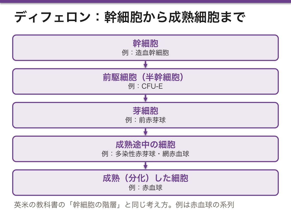
この見方の利点は、どの段の細胞があるかで、その組織が治る力を予測できることにある。幹細胞と増殖する段がそろっていれば、失われても同じ組織で埋め戻せる(再生)。分裂する段が無ければ、空いた場所は結合組織で埋まる(瘢痕)。
また、どの段で止まったか・どの段が増えすぎたかで病気を整理できる。血液のディフェロンでは、幹細胞が減れば汎血球減少、途中で止まれば未熟な細胞の増加(白血病)になる。
抗癌薬と放射線が口内炎・下痢・骨髄抑制・脱毛を起こすのは、更新する集団の分裂中の段を叩くからである。肝臓の一部を切り取っても元の大きさに戻るので生体肝移植が成り立つ。心筋梗塞のあとが瘢痕になるのは、心筋細胞が静止した集団だからである。
止まった成熟を先へ進める治療もある。急性前骨髄球性白血病に対する全トランス型レチノイン酸は、ディフェロンを次の段へ押し出す分化誘導療法である。
English version
Core
A differon (histogenetic series) is an idea used in Eastern European histology. It means the whole series of one cell line, from stem cell to mature cell.
stem cell → progenitor (semi-stem, committed cell) → blast → maturing cell → mature (differentiated) cell
A tissue is described as a set of one or more differons. The epidermis, for example, is mainly the keratinocyte differon, with the melanocyte and Langerhans-cell differons added. English-language textbooks call the same thing the stem-cell hierarchy (stem cell → transit-amplifying cell → terminally differentiated cell).
By how they renew, cell populations fall into three groups.
| Population | Behaviour | Examples |
|---|---|---|
| Renewing | stem cells divide all the time; mature cells are lost continuously | epidermis, gut epithelium (renewed in 3–5 days), blood |
| Expanding | usually rest in G0 but resume dividing after loss | hepatocytes, tubular epithelium, fibroblasts |
| Static | hardly divide after maturation | neurons, cardiomyocytes |
This is the same as the labile, stable and permanent cells of pathology.
Going further
The value of this view is that the stages present predict how a tissue heals. If stem cells and dividing stages are present, losses are refilled with the same tissue (regeneration). If no dividing stage exists, the gap fills with connective tissue (scar).
Disease can also be sorted by which stage stopped or which stage grew too much. In the blood differon, losing stem cells gives pancytopenia; a block halfway gives an excess of immature cells (leukaemia).
In the clinic
Chemotherapy and radiation cause mouth ulcers, diarrhoea, marrow suppression and hair loss because they hit the dividing stages of renewing populations. Living-donor liver transplantation works because a partly removed liver grows back. A myocardial infarct heals with a scar because cardiomyocytes are a static population.
Some treatments push blocked maturation forward. All-trans retinoic acid for acute promyelocytic leukaemia is differentiation therapy, moving the differon to its next stage.
実質Parenchyma
器官の仕事をしている部分。肝臓なら肝細胞、腎臓ならネフロン、脳なら神経細胞。
対になるのが間質 stromaで、支持・栄養・防御を担う結合組織と血管である。
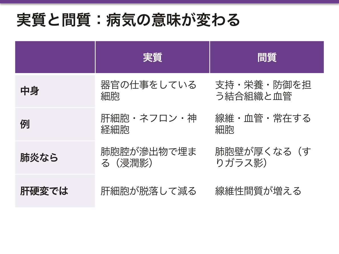
この区別は組織学の用語というより病理を読むための道具である。「実質性か間質性か」で病態の意味がまるで違う。
- 実質性肺炎=肺胞腔が滲出物で埋まる(細菌性・大葉性肺炎)。ガス交換の場そのものが潰れる
- 間質性肺炎=肺胞壁が厚くなる。肺胞腔は空いているのに拡散が落ちる
だから画像も所見も違う。実質性は浸潤影・気管支呼吸音、間質性はすりガラス影・捻髪音・拘束性換気障害。同じ「肺炎」で正反対の生理を示すのは、この区別があるからである。
肝硬変は「実質が減って間質が増える」病態そのもの。肝細胞が脱落し、線維性間質が置き換わる。だから合成能の低下(アルブミン↓・PT延長)と門脈圧亢進が同時に来る。前者は実質の喪失、後者は間質の増生による血流障害である。
English version
Core
The part of an organ that does its work: hepatocytes in the liver, the nephron in the kidney, the neurons in the brain. Its counterpart is the stroma, the connective tissue and vessels that support, nourish and defend.
Going further
This distinction is less a histological term than a tool for reading pathology. Whether a process is parenchymal or interstitial changes its meaning entirely.
- Alveolar (parenchymal) pneumonia — the alveolar spaces fill with exudate; the site of gas exchange itself is obliterated. Bacterial, lobar pneumonia.
- Interstitial pneumonia — the alveolar walls thicken; the airspaces remain open, yet diffusion falls.
The imaging and the physical signs differ accordingly: consolidation with bronchial breathing in the first, ground-glass shadowing with fine crackles and a restrictive defect in the second. Two conditions called "pneumonia" show opposite physiology, and this distinction is why.
In the clinic
Cirrhosis is precisely the state in which parenchyma is lost and stroma increases. Hepatocytes drop out and fibrous stroma replaces them. That is why failure of synthesis (falling albumin, prolonged prothrombin time) and portal hypertension arrive together: the first from loss of parenchyma, the second from obstruction to flow by the proliferating stroma.
細胞外基質Matrix extracellularis
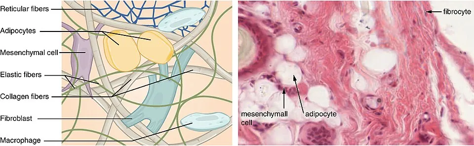
細胞の外を埋める、細胞が自分で作って外に出した構造物。線維成分(コラーゲン・弾性線維・細網線維)と基質(プロテオグリカン・糖タンパク・水)からなる。
単なる詰め物ではなく、細胞の形・移動・分化・生死を決める足場である。
細胞は基質とインテグリンで繋がっており、この接着が切れるとアノイキス(足場喪失アポトーシス)で死ぬ。正常細胞が本来いる場所を離れて生きられないのはこの仕組みによる。癌の転移とは、この安全装置を外した細胞が起こす現象である。
基質を壊す酵素がMMP(マトリックスメタロプロテアーゼ)で、生理的には創傷治癒・組織再構築に働くが、腫瘍浸潤でも同じ酵素が使われる。
細胞外基質が壊れることが、浸潤と転移の本体である。基底膜を破って下の結合組織へ入った時点で「浸潤癌」となり、それ以前は上皮内癌(非浸潤癌)——転移しない。この1枚の膜を越えたかどうかが、治療も予後も分ける。
癌細胞はマトリックスメタロプロテアーゼ(MMP)で基質を溶かして進む。
コラーゲンの異常は全身病になる。Ehrlers–Danlos 症候群(皮膚の過伸展・関節の過可動・血管の破綻)、骨形成不全症(I 型コラーゲン・易骨折性・青色強膜)、Marfan 症候群(フィブリリン——弾性線維の骨格。大動脈解離・水晶体亜脱臼・高身長)。基質の成分ごとに、壊れる臓器が決まっている。
線維化はコラーゲンの過剰沈着である。肝硬変・肺線維症・ケロイドはいずれも「修復の行き過ぎ」で、Masson トリクローム染色で青く染めて評価する。
English version
Core
The material filling the space outside cells, made by the cells themselves and exported. It consists of fibres — collagen, elastic and reticular — and ground substance — proteoglycans, glycoproteins and water.
It is not mere packing. It is the scaffold that determines a cell’s shape, migration, differentiation and survival.
Going further
Cells are attached to the matrix through integrins, and if that attachment is broken the cell dies by anoikis — apoptosis from loss of anchorage. This is why a normal cell cannot survive away from where it belongs, and metastasis is what happens when a cell disables that safeguard.
The enzymes that degrade the matrix are the matrix metalloproteinases (MMPs). Physiologically they serve wound healing and tissue remodelling; tumour invasion uses the very same enzymes.
In the clinic
Destruction of the extracellular matrix is the substance of invasion and metastasis. Once the basement membrane is breached and the tumour enters the connective tissue beneath, it is an invasive carcinoma; before that it is carcinoma in situ and does not metastasise. Whether that single membrane has been crossed decides both treatment and prognosis.
Defects of collagen become systemic disease, and the component that fails determines the organ that suffers: Ehlers–Danlos syndrome (hyperextensible skin, hypermobile joints, vascular rupture), osteogenesis imperfecta (type I collagen; fragile bones, blue sclerae), Marfan syndrome (fibrillin, the scaffold of elastic fibres; aortic dissection, ectopia lentis, tall stature).
Fibrosis is excessive deposition of collagen. Cirrhosis, pulmonary fibrosis and keloid are all repair carried too far, and all are assessed by Masson trichrome, which stains the collagen blue.
基底膜Membrana basalis
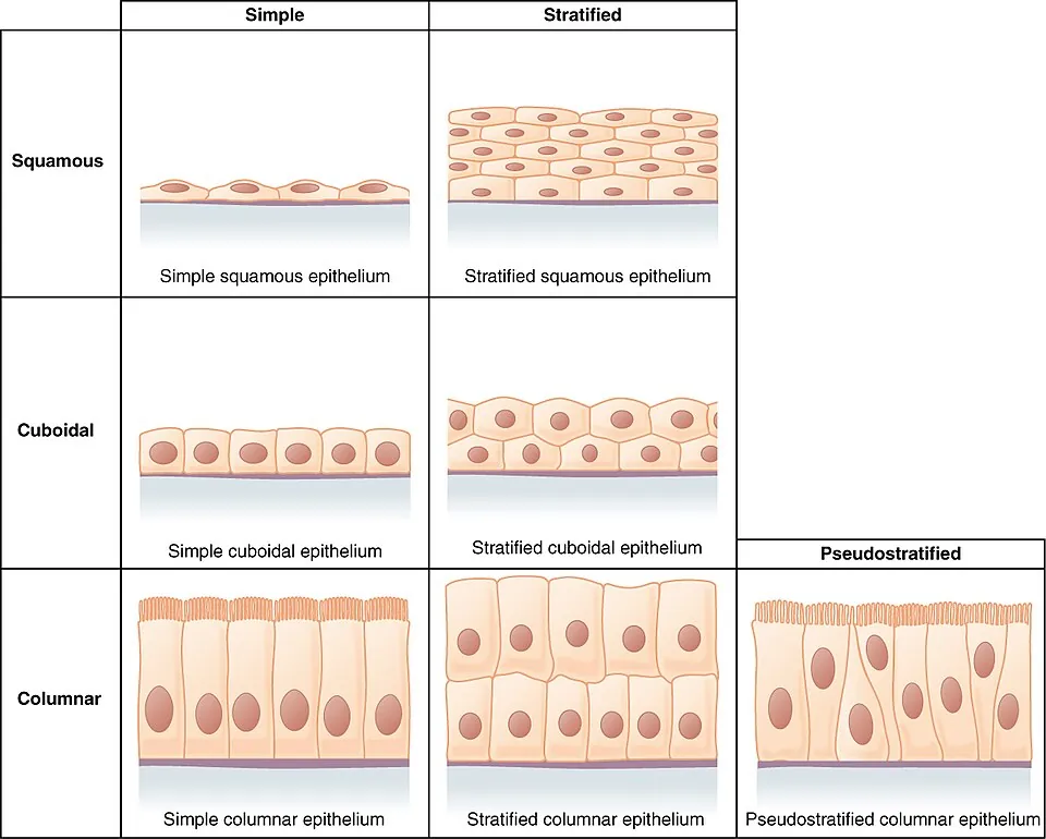
上皮の底面と結合組織のあいだにある薄い膜。IV型コラーゲンとラミニンが骨格。光学顕微鏡ではPAS染色で赤紫に染まる。
働きは3つ——上皮を固定する/物質の通過を選ぶ(濾過)/上皮の再生の足場になる。
基底膜は上皮だけでなく、筋線維・シュワン細胞・脂肪細胞も個別に包んでいる。「上皮の下だけにある」と覚えると末梢神経と筋の再生が理解できなくなる。神経が切れても再生できるのは、シュワン細胞の基底膜が管として残り、軸索がその中を伸びるからである。
癌の診断で最も重要な線がこれ。基底膜を破っていなければ上皮内癌(carcinoma in situ)で転移しない。破れば浸潤癌になり、リンパ管・血管に到達しうる。上皮内には血管もリンパ管も無いためである。
Goodpasture 症候群——IV型コラーゲンα3鎖に対する自己抗体が、糸球体基底膜と肺胞基底膜を同時に攻撃する。同じ分子が両方にあるから、腎炎と肺出血が同時に起きる。Alport 症候群は同じIV型コラーゲンでも別のα鎖(多くはX連鎖のα5=COL4A5)の遺伝子変異で、基底膜が構造的に作れず、血尿+感音難聴+眼症状が揃う。自己抗体で壊すのが Goodpasture、最初から作れないのが Alport という対比で覚える。
English version
Core
A thin membrane between the base of an epithelium and the connective tissue beneath. Its framework is type IV collagen and laminin. By light microscopy it stains magenta with PAS.
It has three functions: to anchor the epithelium, to select what passes (filtration), and to serve as the scaffold for regeneration.
Going further
The basement membrane invests not only epithelium but also individual muscle fibres, Schwann cells and adipocytes. Remembering it as "the layer under epithelium" makes the regeneration of peripheral nerve and muscle incomprehensible. A severed nerve can regenerate because the Schwann cell basement membrane remains as a tube and the axon grows inside it.
This is also the single most important line in cancer diagnosis. While the basement membrane is intact the lesion is carcinoma in situ and cannot metastasise; once breached it is invasive and can reach lymphatics and blood vessels — because there are no vessels of either kind within an epithelium.
In the clinic
Goodpasture syndrome — autoantibodies against the α3 chain of type IV collagen attack the glomerular and alveolar basement membranes simultaneously. The same molecule is present in both, so nephritis and pulmonary haemorrhage occur together.
Alport syndrome involves the same type IV collagen but a different chain, usually the X-linked α5 (COL4A5), and the defect is genetic: the membrane cannot be built properly, giving haematuria with sensorineural deafness and ocular signs.
Hold the two together as a contrast: Goodpasture destroys it with antibody, Alport was never able to build it.
HE染色 — なぜ青と赤なのか
標本の基本染色。ヘマトキシリンが青紫、エオジンが赤(ピンク)。
- 青紫に染まる=好塩基性。酸性の(負に帯電した)ものが塩基性色素を引きつける。→ 核(DNA)・粗面小胞体(RNA)・軟骨基質
- 赤に染まる=好酸性(エオジン好性)。塩基性のものが酸性色素を引きつける。→ 細胞質のタンパク・コラーゲン・ミトコンドリア・赤血球
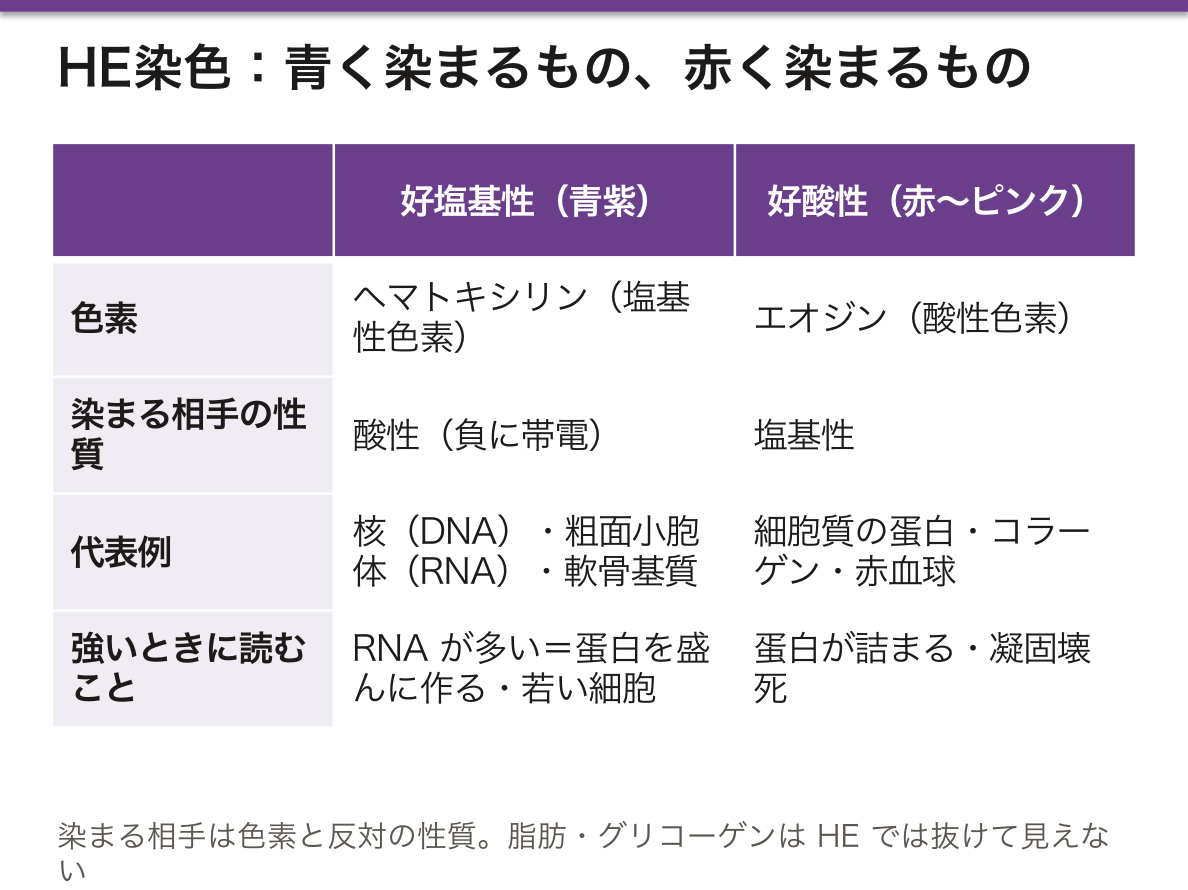
「好塩基性=塩基に染まる」ではなく「塩基性色素に染まる=それ自体は酸性」という逆転が毎回混乱の元になる。染まる相手が反対の性質と覚える。
この理屈が分かると像が読める。タンパク合成が盛んな細胞(形質細胞・膵腺房細胞)は粗面小胞体が多く、細胞質まで青みを帯びる。逆にミトコンドリアだらけの細胞(心筋・腎近位尿細管・胃壁細胞)は細胞質が濃い赤になる。染色の色が、そのままオルガネラの構成を語っている。
染まり方の変化が、そのまま病態を示す。
好塩基性が強い(青い) — RNA が多い=蛋白を盛んに作っている。形質細胞(抗体を作る)、低分化な悪性腫瘍、再生している細胞。「青い細胞が増えていたら、何かを作っているか、若いか」と読む。
好酸性が強い(赤い) — 蛋白が詰まっている。壊死した細胞(凝固壊死)は核が消えて細胞質だけが赤く残る——心筋梗塞の early changeがこれである。アポトーシス小体も丸く好酸性に見える。
HE で染まらないものに注意する。脂肪・グリコーゲン・石灰は処理の過程で溶け出したり抜けたりする。脂肪を見たいなら凍結切片+Sudan III、グリコーゲンなら PAS を追加する。「見えないこと」は「無いこと」ではない。
English version
Core
The routine stain. Haematoxylin gives blue-purple, eosin gives red or pink.
- Blue-purple = basophilic. Acidic, negatively charged material attracts the basic dye → nucleus (DNA), rough endoplasmic reticulum (RNA), cartilage matrix.
- Red = acidophilic (eosinophilic). Basic material attracts the acidic dye → cytoplasmic protein, collagen, mitochondria, erythrocytes.
Going further
The recurring source of confusion is the inversion: basophilic does not mean "stained by base" but "stained by a basic dye, and therefore itself acidic". Remember that a structure is stained by the dye of opposite character.
Once the logic is clear the image can be read. Cells synthesising protein intensively — plasma cells, pancreatic acinar cells — are rich in rough ER, so even the cytoplasm takes on a blue tint. Conversely cells packed with mitochondria — cardiac muscle, the renal proximal tubule, gastric parietal cells — have deeply red cytoplasm. The colour is already telling you the organelle content.
In the clinic
A change in staining is itself a statement about the process.
- Strongly basophilic (blue) — abundant RNA, so the cell is actively making protein: plasma cells producing antibody, poorly differentiated malignancy, regenerating cells. Read blue cells as "making something, or young".
- Strongly acidophilic (red) — packed with protein. A necrotic cell in coagulative necrosis loses its nucleus and remains as red cytoplasm alone — this is the early change of myocardial infarction. Apoptotic bodies also appear round and eosinophilic.
Beware of what H&E does not show. Fat, glycogen and calcium are dissolved or lost during processing. To see fat use a frozen section with Sudan III; for glycogen add PAS. Not being visible is not the same as not being there.
切片は断面である — 像の読み違えを防ぐ
顕微鏡で見えるのは三次元の構造を数μmで切った1枚の断面。ここから元の形を復元するのが組織学の作業である。
- 管を斜めに切ると楕円や、輪が入れ子になった像になる
- 核が見えない細胞は、核の無い高さで切れただけのことが多い(本当に無核なのは成熟赤血球と血小板)
- 絨毛を横に切ると粘膜の中に浮いた島に見える
実際の標本には作製過程のアーチファクトも混ざる。固定不良による細胞の縮み(上皮と基底膜のあいだの人工的な隙間)、刃こぼれによる線状の傷、折れ込み、気泡。これを病変と読まないことが、標本を同定するときの実際のつまずきどころになる。
見分ける手掛かりは再現性である。生体の構造なら別の切片・別の高さでも同じ形が繰り返し出るが、アーチファクトはその1枚にしか無い。
断面を三次元に戻し損なうと、誤診になる。
「核が見えない細胞」は死んでいるとは限らない——核の無い高さで切れただけである。本当に核が無いのは赤血球と角質細胞、核が消えているのは壊死で、周囲に炎症細胞が集まっているかで区別する。
腺管が斜めに切れると、腺の中に腺があるように見える。これを癌の篩状構造(cribriform pattern)と読み違えない。連続切片で追えば解決する。
生検は標本抽出である。陰性は「無い」ことの証明にならない——狙った場所を外しただけかもしれない。臨床所見と食い違う病理結果は、再検するのが原則である。腫瘍の辺縁や壊死部を採ると診断がつかないので、どこを採るかが結果を決める。
English version
Core
What the microscope shows is a single slice, a few micrometres thick, through a three-dimensional structure. The work of histology is to reconstruct the original form from it.
- A tube cut obliquely appears as an ellipse, or as rings nested inside one another.
- A cell with no visible nucleus has usually simply been cut at a level above or below it. The only truly anucleate cells are the mature erythrocyte and the platelet.
- A villus cut transversely appears as an island floating within the mucosa.
Going further
Real sections also contain artefacts of preparation: shrinkage from poor fixation, producing an artificial gap between epithelium and basement membrane; linear scores from a damaged blade; folds; air bubbles. Reading these as lesions is a real pitfall when identifying specimens.
The way to tell them apart is reproducibility. A genuine structure recurs in other sections and at other levels; an artefact exists only on that one slide.
In the clinic
Failing to restore the third dimension leads to misdiagnosis.
- A cell without a visible nucleus is not necessarily dead — it may simply have been cut at the wrong level. Truly anucleate are erythrocytes and corneocytes; a nucleus that has disappeared indicates necrosis, and the presence of surrounding inflammatory cells distinguishes the two.
- Obliquely cut glands appear as glands within glands. Do not read this as the cribriform pattern of carcinoma; serial sections resolve it.
A biopsy is a sample. A negative result does not prove absence — the target may simply have been missed. A pathology result that conflicts with the clinical picture should as a rule be repeated. Sampling the margin or a necrotic area yields no diagnosis, so where the biopsy is taken determines the answer.
何が見えて、何が見えないか — 分解能という限界Microscopium
| 道具 | 分解能 | 見えるもの |
|---|---|---|
| 肉眼 | 約 0.2 mm | 器官・肉眼的病変 |
| 光学顕微鏡 | 約 0.2 μm | 細胞・核・ミトコンドリア(かろうじて点として) |
| 透過型電子顕微鏡 | 約 0.2 nm | 膜・小器官の内部構造・ウイルス・細胞間接着装置 |
| 走査型電子顕微鏡 | 数 nm | 表面の立体像 |
光学顕微鏡の限界は、光の波長で決まる——可視光の半分より小さいものは分けられない。
だから光顕で「見えている」小器官は、限られる——細胞膜そのもの・粗面小胞体の膜・線毛の 9+2 構造は、光顕では見えない。
見えないものは、染色で「その存在を推定する」ことになる——好塩基性の細胞質=リボソームが多い、など。
「染色像は、見えないものの間接的な影である」という理解が、組織学の読み方そのものになる。核が青いのは DNA のリン酸、細胞質が赤いのは蛋白の塩基性アミノ酸を見ている(この章の HE 染色の項)——「色は、分子の性質を電荷として拾っている」。
だから電子顕微鏡でしか決まらない診断がある(糸球体疾患の章)——「沈着物が基底膜のどの層にあるかは、光顕では分けられない」。
線毛の異常も同じになる(線毛・鞭毛の章)——「原発性線毛機能不全症の確定に、電子顕微鏡が使われてきた」。
免疫染色は、分解能ではなく特異性で見る道具になる(免疫組織化学の項)——「小さくて見えなくても、そこに在ることは示せる」。
共焦点顕微鏡や超解像顕微鏡は、光学の限界の内側と外側を押し広げている——「限界がどこにあり、なぜそこなのかを言えることが、道具を選ぶ力になる」。
見たいものの大きさで、道具を選ぶ。
光学顕微鏡で足りないときに電子顕微鏡が要る。実際に現役なのは腎生検である。糸球体基底膜の厚さと、免疫複合体の沈着部位(上皮下・内皮下・メサンギウム)は電子顕微鏡でしか決まらず、それが病型を決める。微小変化型ネフローゼは光学顕微鏡で「正常に見える」——足細胞の突起の消失は電子顕微鏡で初めて見える。
繊毛の異常(Kartagener 症候群)もダイニン腕の欠如を電子顕微鏡で確認する。
ウイルスは光学顕微鏡では見えない。見えるのはウイルスが作った結果——封入体(CMV のフクロウの目、ヘルペスのすりガラス様核)や細胞の変化(HPV のコイロサイト)である。「見えないものを、見える痕跡から読む」のが組織診断の作法になる。
English version
Core
| Instrument | Resolution | What can be seen |
|---|---|---|
| Naked eye | about 0.2 mm | Organs, macroscopic lesions |
| Light microscope | about 0.2 µm | Cells, nuclei, mitochondria (barely, as dots) |
| Transmission EM | about 0.2 nm | Membranes, internal structure of organelles, viruses, junctions |
| Scanning EM | a few nm | Three-dimensional surface images |
The limit of the light microscope is set by the wavelength of light — nothing smaller than about half the wavelength of visible light can be resolved. So the cell membrane itself, the membrane of the rough ER and the 9+2 axoneme cannot be seen by light microscopy; their presence has to be inferred from staining — basophilic cytoplasm meaning abundant ribosomes, and so on.
Going further
Understanding that a stained image is the indirect shadow of what cannot be seen is itself the method of reading histology. The nucleus is blue because of the phosphate of DNA and the cytoplasm red because of the basic amino acids of protein: colour is picking up the electrical character of molecules.
Hence some diagnoses can only be made by electron microscopy — in which layer of the glomerular basement membrane a deposit lies cannot be resolved by light microscopy. The same applies to ciliary defects, where electron microscopy has been used to confirm primary ciliary dyskinesia.
Immunohistochemistry sees by specificity rather than by resolution: even when something is too small to be seen, its presence can be demonstrated. Confocal and super-resolution microscopy push at the boundary from both sides. Being able to say where the limit lies, and why it lies there, is what allows the right instrument to be chosen.
In the clinic
Choose the instrument by the size of what you need to see. Electron microscopy is required when light microscopy is not enough, and the living example is renal biopsy: the thickness of the glomerular basement membrane and the site of immune complex deposition — subepithelial, subendothelial or mesangial — can be determined only by electron microscopy, and it is the site that defines the disease.
Minimal change disease looks normal by light microscopy; effacement of the podocyte foot processes appears only on electron microscopy. Ciliary disorders such as Kartagener syndrome are confirmed by the absence of dynein arms on EM.
Viruses cannot be seen by light microscopy. What is seen is what the virus has produced — inclusion bodies (the owl’s eye of CMV, the ground-glass nucleus of herpes) or cellular change (the koilocyte of HPV). Reading the invisible from its visible traces is the discipline of histological diagnosis.
腫瘍の名前は、どの組織から出たかで決まっている
腫瘍名は勝手に付いているのではなく、由来する組織+良性か悪性かの規則で組まれている。名前が読めれば、どの組織を見ればよいかが分かる。
| 良性 | 組織名+-oma | 脂肪腫・平滑筋腫・軟骨腫・腺腫(adenoma)・乳頭腫 |
| 悪性・上皮性 | carcinoma(癌腫) | 腺癌・扁平上皮癌・尿路上皮癌。リンパ行性転移が多い |
| 悪性・非上皮性 | sarcoma(肉腫) | 骨肉腫・脂肪肉腫・平滑筋肉腫。血行性転移が多い |
だから「癌」と「がん」を書き分ける。癌=上皮性悪性腫瘍(carcinoma)、がん=悪性腫瘍全般という使い分けが、日本語の医学では定着している。
⚠️ 規則を外れる名前があり、そこが試験でも実務でも問われる。
| -oma だが悪性 | リンパ腫・黒色腫・精上皮腫・骨髄腫・神経膠腫・中皮腫 | いずれも悪性。名前だけでは良性に見える |
| 腫瘍ではないのに -oma | 血腫・肉芽腫・過誤腫・粥腫 | 語尾は「かたまり」を意味するにすぎない |
| 人名が付く | Wilms 腫瘍・Ewing 肉腫・Hodgkin リンパ腫 | 由来が分かる前に記載されたもの |
由来が分からない腫瘍もある。分化が進みすぎて(低分化・未分化)、HE 染色だけでは上皮性か非上皮性かも言えないことがある。そこで免疫染色で由来を決める——サイトケラチンが陽性なら上皮性、ビメンチンなら間葉系、S-100 と HMB-45 なら黒色腫、白血球共通抗原ならリンパ腫。
「名前を決めること」は分類のためではなく、治療のためである。上皮性なら手術と化学療法の組み立てが違い、リンパ腫なら手術ではなく化学療法が中心になり、肉腫なら広範な切除が要る。由来を間違えると、治療の枠組みごと間違う。
患者に「悪性リンパ腫」と伝えるとき、名前に『腫』が付くから軽い、という誤解が起こりうる。言葉を補って説明する。
English version
Core
Tumour names are not arbitrary. They are built from the tissue of origin plus whether the tumour is benign or malignant. If the name can be read, it tells you which tissue to examine.
| Category | Suffix | Examples |
|---|---|---|
| Benign | tissue name + -oma | lipoma, leiomyoma, chondroma, adenoma, papilloma |
| Malignant, epithelial | carcinoma | adenocarcinoma, squamous cell carcinoma, urothelial carcinoma. Spreads mainly by lymphatics |
| Malignant, non-epithelial | sarcoma | osteosarcoma, liposarcoma, leiomyosarcoma. Spreads mainly by blood |
Going further
⚠️ There are names that break the rule, and they are asked about in examinations and matter in practice.
- -oma but malignant: malignant lymphoma, melanoma, seminoma, myeloma, glioma, mesothelioma. The name alone makes them look benign.
- -oma but not a tumour at all: haematoma, granuloma, hamartoma, atheroma. The suffix means no more than "a mass".
- Named after a person: Wilms tumour, Ewing sarcoma, Hodgkin lymphoma — described before their origin was known.
Some tumours have no recognisable origin. When differentiation is lost, H&E alone may not even say whether the tumour is epithelial. Immunohistochemistry then decides: cytokeratin for epithelial, vimentin for mesenchymal, S-100 and HMB-45 for melanoma, leukocyte common antigen for lymphoma.
Naming exists for treatment, not for classification. An epithelial tumour is approached differently by surgery and chemotherapy; a lymphoma is treated chiefly by chemotherapy rather than surgery; a sarcoma requires wide excision. Mistake the origin and the whole framework of treatment is wrong.
In the clinic
When telling a patient they have a malignant lymphoma, the name itself can mislead: in Japanese the character 腫 suggests something mild. Explain in words rather than relying on the name.
組織学という学問 — 何を目的に、どの基礎医学とつながるかHistologia
組織学(ギリシャ語の histos=組織、logos=学)は、正常な細胞・組織・器官の発生・構造(微細構造を含む)・働きを調べる学問である。ヨーロッパの医学部では「組織学・細胞学・発生学」を1つの科目として学ぶことが多く、中身は4つに分かれる。
| 分野 | 対象 |
|---|---|
| 細胞学 | 細胞の構造と働き(小器官・核・細胞周期) |
| 発生学 | 受精から器官ができるまで |
| 一般組織学 | 4つの基本組織(上皮・結合・筋・神経) |
| 器官組織学(顕微解剖学) | 器官ごとの組織の組み合わせ |
主な方法は、光学顕微鏡(HE 染色)・組織化学(PAS や Sudan のように、組織の中で物質を化学的に見分ける)・免疫組織化学(抗体で蛋白の場所を示す)・電子顕微鏡・細胞と組織の培養である。
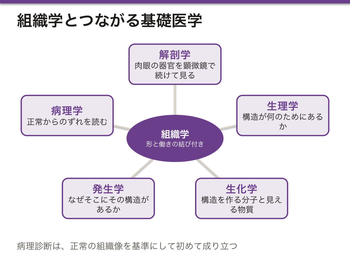
組織学の目的は、標本の名前を当てることではなく、形と働きの結び付きを理解することである。そのために、ほかの基礎医学と次のようにつながる。
- 解剖学——肉眼で見た器官の中身を、顕微鏡の倍率で続けて見る
- 生理学——その構造が何のためにあるかを説明する(例:糸球体の構造と濾過)
- 生化学・細胞生物学——構造を作る分子と、組織化学で見える物質
- 発生学——なぜその構造がそこにあるのかを説明する
- 病理学——正常からのずれを読む学問。組織学はその基準になる
日本の医学部では組織学を解剖学の一部として、発生学を別に教えることが多いが、扱う中身は同じである。
病理診断は、正常の組織像を知っていて初めて成り立つ。生検・手術標本・細胞診・術中の凍結切片はどれも、「この場所にあるはずの組織と比べて何が違うか」を読む作業である。CBT や国試で組織像の問題が病理と一緒に出るのも、組織学が病理の前提だからである。
English version
Core
Histology (Greek histos = tissue, logos = study) studies the development, structure (including fine structure) and function of normal cells, tissues and organs. In European medical schools, "histology, cytology and embryology" is often one subject, with four parts.
| Field | Object |
|---|---|
| Cytology | structure and function of cells (organelles, nucleus, cell cycle) |
| Embryology | from fertilisation to the formation of organs |
| General histology | the four basic tissues (epithelial, connective, muscle, nervous) |
| Organ histology (microscopic anatomy) | how tissues combine in each organ |
The main methods are light microscopy (H&E), histochemistry (identifying substances chemically in tissue, as with PAS or Sudan), immunohistochemistry (showing where a protein is with antibodies), electron microscopy, and cell and tissue culture.
Going further
The aim of histology is not to guess the name of a slide but to understand how form and function are linked. For this it connects to other basic sciences:
- Anatomy — continues looking inside the organs seen by eye, at microscope magnification.
- Physiology — explains what a structure is for (e.g., glomerular structure and filtration).
- Biochemistry and cell biology — the molecules that build structures, and the substances histochemistry shows.
- Embryology — explains why a structure is where it is.
- Pathology — reads departures from normal; histology is its reference.
Japanese medical schools often teach histology as part of anatomy and embryology separately, but the content is the same.
In the clinic
Pathological diagnosis works only if you know the normal picture. Biopsies, surgical specimens, cytology and intraoperative frozen sections are all read as "what differs from the tissue that should be here?". Histology images appear with pathology in the CBT and the national exam because histology is the basis of pathology.
まとめ
| 組織 | 同じ働きをする細胞と細胞外基質の集まり。 |
|---|---|
| ディフェロン — 組織を「幹細胞からの系列」として見る | ディフェロン(differon、組織発生系列)は、東ヨーロッパの組織学で使われる考え方で、1つの系統の細胞を、幹細胞から成熟した細胞まで並べた全体を指す。 |
| 実質 | 器官の仕事をしている部分。肝臓なら肝細胞、腎臓ならネフロン、脳なら神経細胞。 |
| 細胞外基質 | 細胞の外を埋める、細胞が自分で作って外に出した構造物。 |
| 基底膜 | 上皮の底面と結合組織のあいだにある薄い膜。 |
| HE染色 — なぜ青と赤なのか | 標本の基本染色。ヘマトキシリンが青紫、エオジンが赤(ピンク)。 |
| 切片は断面である — 像の読み違えを防ぐ | 顕微鏡で見えるのは三次元の構造を数μmで切った1枚の断面。 |
| 何が見えて、何が見えないか — 分解能という限界 | 道具分解能見えるもの肉眼約 0.2 mm器官・肉眼的病変光学顕微鏡約 0.2 μm細胞・核・ミトコンドリア(かろうじて点として)透過型電子顕微鏡約 0.2 nm膜・小器官の内部構造… |
| 腫瘍の名前は、どの組織から出たかで決まっている | 腫瘍名は勝手に付いているのではなく、由来する組織+良性か悪性かの規則で組まれている。 |
| 組織学という学問 — 何を目的に、どの基礎医学とつながるか | 組織学(ギリシャ語の histos=組織、logos=学)は、正常な細胞・組織・器官の発生・構造(微細構造を含む)・働きを調べる学問である。 |
このノートは医学生が授業の内容を自分の言葉でまとめたもので、診療の判断に使うものではありません。誤りに気づいたらお知らせください。