X線は、医学で最も古く、今も最も多く使われている画像の道具である。この章では、X線管の中で X線がどう生まれるか(制動放射と特性X線)、そのスペクトルを管電圧・管電流・濾過でどう動かすか、体を通るときにどう弱まって白黒の画像になるかを順に追い、透視・血管造影・乳房撮影、CT の原理と CT 値、最後にリニアックによる放射線治療までをつなげる。
要の2文:「X線はレンズで曲げられないので、画像はすべて影絵である」「白く写るのは、X線をよく吸収する原子番号の大きい原子が多いところ」。相互作用の詳しい話と放射線の単位・防護は電離放射線の章にある。
- X線とは何か — 曲げられない光だから「影絵」で写す
- X線管 — 電子を加速してぶつけ、エネルギーの99%は熱になる
- 制動放射と特性X線 — なめらかな山と鋭いピーク
- スペクトルを動かすもの — 管電圧・管電流・濾過
- 減弱とコントラスト — 白く写るもの・黒く写るもの・造影剤
- 透視・血管造影・乳房撮影 — 動きを見る・管を見る・柔らかいものを見る
- CT の原理と CT 値 — 多方向の影から断面を計算する
- 放射線治療とリニアック — X線で腫瘍を壊す
X線とは何か — 曲げられない光だから「影絵」で写す
X線(X-rays)は、紫外線よりさらに波長の短い電磁波である。質量も電荷も持たず、光速で進む。波長はおよそ 0.01〜10 nm で、光子1個のエネルギーは
E = hf = hc/λ (覚え方:E[keV] ≈ 1.24 / λ[nm])
たとえば波長 1 nm の X線は振動数 f = c/λ = 3×10⁸/10−9 = 3×10¹⁷ Hz、エネルギーは約 1.2 keV。診断に使う X線は数十〜100 keV 程度で、原子から電子をはじき出せる(電離できる)エネルギーを持つ。
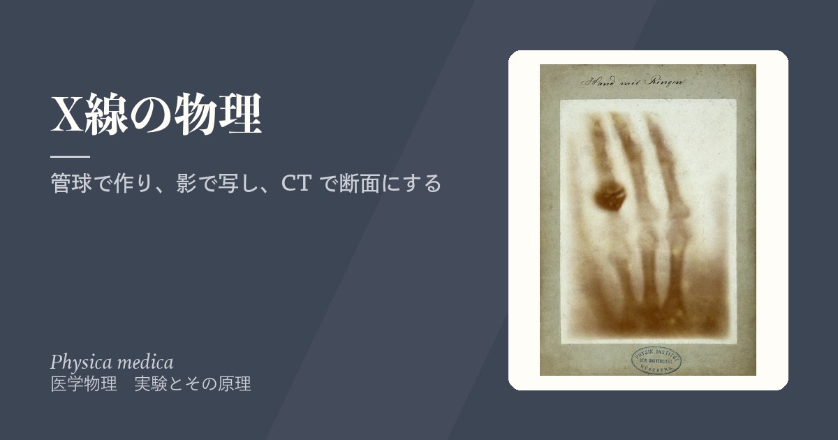
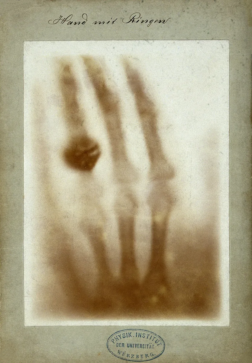
1895年、Röntgen(レントゲン)は陰極線(真空管の中を飛ぶ電子の流れ)の実験中に、離れた蛍光板が光るのに気づき、正体不明の線として「X線」と名づけた。磁場で曲がらず、陰極線よりはるかによく物を通り抜けた。夫人の手の骨を写した写真は最も有名な初期の X線像で、彼は1901年に最初のノーベル物理学賞を受けた。
X線は、ふつうの物質ではほとんど屈折も反射もしない。屈折率がほぼ 1 なので、レンズで集めて像を結ばせることができない。だから X線撮影は、カメラのように「像を結ぶ」のではなく、点に近い線源から出た X線が体を通り抜けた後の「影」を検出器に落とす投影(影絵)になる。
影絵なので、幾何学がそのまま画像に効く。①物体が検出器から離れるほど影は拡大する。②線源(焦点)が大きいほど影の縁がぼける(半影)。③線源を遠ざけるほど拡大とぼけは減る。X線管の焦点をできるだけ小さくし、線源と検出器の距離を一定に決めて撮るのはこのためである。
電波は電子を軌道間で動かすにはエネルギーが小さすぎて体を素通りし、可視光はちょうど吸収されやすいので体を通らない。X線が体を通り抜けるのは、逆にエネルギーが大きすぎるからで、それでもときどき原子から電子をたたき出して吸収される。この「ときどき」の割合が組織ごとに違うことが、画像になる。
胸部単純X線写真は、ふつう背中から前へ(PA)撮る。心臓は体の前寄りにあるので、検出器を胸の前に当てれば心臓が検出器に近くなり、拡大が小さくなる。ベッドの上で前から撮る(AP)ポータブル撮影では心臓が検出器から遠くなり、心陰影が大きく写る。心胸郭比(CTR)を評価するときに、撮影方向を必ず確かめるのはこのためである。
English version
Core
X-rays are electromagnetic waves with an even shorter wavelength than ultraviolet. They have no mass and no charge and travel at the speed of light. The wavelength is roughly 0.01–10 nm, and the energy of one photon is
E = hf = hc/λ (handy form: E[keV] ≈ 1.24 / λ[nm])
For example, an X-ray of wavelength 1 nm has frequency f = c/λ = 3×10⁸/10−9 = 3×10¹⁷ Hz and energy about 1.2 keV. Diagnostic X-rays are tens of keV up to about 100 keV — enough to knock electrons out of atoms (to ionise).
In 1895 Röntgen, while working with cathode rays (streams of electrons in a vacuum tube), noticed that a fluorescent screen some distance away glowed. He named the unknown rays "X-rays". They were not bent by a magnetic field and passed through matter far better than cathode rays. His picture of his wife’s hand bones is the best-known early X-ray image, and in 1901 he received the first Nobel Prize in Physics.
Going further
X-rays are hardly refracted or reflected by ordinary matter. The refractive index is almost 1, so a lens cannot focus them into an image. So X-ray imaging does not "form an image" like a camera. It is a projection: X-rays from an almost point-like source pass through the body and drop a "shadow" on the detector.
Because it is a shadow, geometry acts directly on the image. (1) The farther an object is from the detector, the more its shadow is enlarged. (2) The larger the source (focal spot), the more blurred the shadow’s edge (penumbra). (3) Moving the source farther away reduces both enlargement and blur. This is why X-ray tubes have a focal spot as small as possible and images are taken at a fixed source-to-detector distance.
Radio waves have too little energy to move electrons between orbits, so they pass through the body. Visible light is just right to be absorbed, so it does not pass. X-rays pass through for the opposite reason — they have too much energy — yet now and then they knock an electron out of an atom and are absorbed. The "now and then" differs between tissues, and that difference becomes the image.
In the clinic
A chest X-ray is usually taken from back to front (PA). The heart lies toward the front of the chest, so with the detector against the front, the heart is close to the detector and enlarged less. In a portable AP film taken from the front in bed, the heart is farther from the detector, and the heart shadow looks larger. This is why you must always check the projection when judging the cardiothoracic ratio (CTR).
X線管 — 電子を加速してぶつけ、エネルギーの99%は熱になる
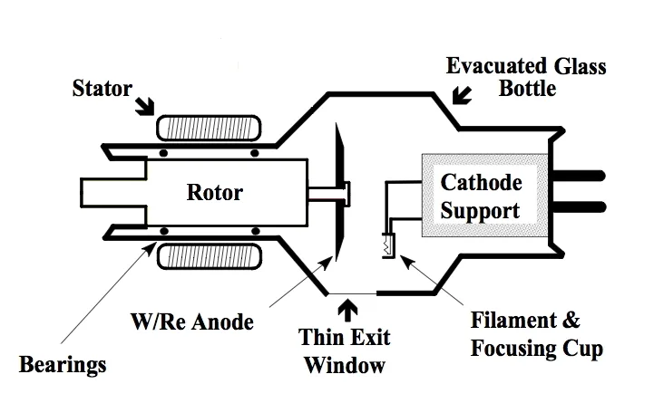
X線管(X-ray tube)は、真空にしたガラスまたは金属の容器の中に、陰極(cathode)と陽極(anode)を向かい合わせたものである。
①陰極のフィラメント(タングステンの細い線)に電流を流して熱すると、表面から電子が飛び出す(熱電子放出, thermionic emission)。②陰極と陽極のあいだに管電圧(数十〜百数十 kV)をかけると、電子は陽極に向かって加速される。100 kV で加速された電子は 100 keV の運動エネルギーを持つ。③電子は陽極のターゲット(多くはタングステン)に衝突して急に減速し、そのエネルギーの一部が X線になる。④X線は管の窓から外へ出る。
管を流れる電子の流れが管電流(mA)で、フィラメントの温度で調節する。管電流 × 撮影時間(mAs)が、ターゲットに当たる電子の総数を決める。
電子のエネルギーのうち X線になるのは 1% 程度で、残りの 99% 前後は熱になる。X線管の設計の大部分は、この熱をどう逃がすかの工夫である。
なぜターゲットはタングステンなのか。X線の発生効率は、ターゲットの原子番号 Z と管電圧にほぼ比例して上がる(効率の目安 ≈ 10−9 × Z × 電圧[V]。タングステン Z = 74、100 kV で約 0.7%)。タングステンは Z が大きいうえに、融点が約 3,400 °C と金属で最も高いので、集中して当たる電子の熱に耐えられる。「X線をよく出す」と「熱で溶けない」を両立できる材料だからである。
回転陽極:陽極を円盤にして高速で回すと、電子が当たる場所が次々に入れかわり、熱が円周全体に分散する。焦点(電子の当たる点)は小さいほど画像がシャープになるが、小さいほど熱が集中する——画質と熱の負荷はトレードオフで、回転陽極はその妥協点を大きく広げた。
発生する X線の向きにも偏りがあり、エネルギーの高い電子ほど X線を前方(電子の進む向き)に出しやすく、低いエネルギーでは横方向に出やすい。診断用の管では、電子の流れに対して横向きに取り出している。
English version
Core
An X-ray tube is a vacuum container of glass or metal with a cathode and an anode facing each other.
(1) Current heats the cathode filament (a thin tungsten wire), and electrons leave its surface (thermionic emission). (2) A tube voltage of tens to over a hundred kV between cathode and anode accelerates the electrons toward the anode. An electron accelerated through 100 kV has 100 keV of kinetic energy. (3) The electrons hit the anode target (usually tungsten) and slow down suddenly; part of their energy becomes X-rays. (4) The X-rays leave through the tube window.
The flow of electrons through the tube is the tube current (mA), set by the filament temperature. Tube current × exposure time (mAs) sets the total number of electrons hitting the target.
Only about 1% of the electrons’ energy becomes X-rays; the remaining 99% or so becomes heat. Most of X-ray tube design is about getting rid of this heat.
Going further
Why tungsten? The efficiency of X-ray production rises roughly in proportion to the atomic number Z of the target and the tube voltage (rule of thumb: efficiency ≈ 10−9 × Z × voltage in V; for tungsten, Z = 74, at 100 kV about 0.7%). Tungsten has a high Z and also the highest melting point of any metal, about 3,400 °C, so it survives the concentrated heat of the electron beam. It is a material that both "makes X-rays well" and "does not melt".
Rotating anode: if the anode is a disc spinning fast, the spot hit by the electrons keeps changing and the heat spreads around the whole rim. A smaller focal spot gives a sharper image, but concentrates the heat — image quality and heat load are a trade-off, and the rotating anode greatly widened the compromise.
The direction of X-ray emission is also uneven: higher-energy electrons tend to emit X-rays forward (in the electron’s direction), lower energies more to the side. Diagnostic tubes take the beam out sideways to the electron flow.
制動放射と特性X線 — なめらかな山と鋭いピーク
X線管から出る X線のスペクトル(エネルギーごとの強さ)は、なめらかに広がる連続スペクトルの上に、鋭いピークがいくつか立った形をしている。2つは生まれ方がまったく違う。
| 制動放射(制動X線) bremsstrahlung | 特性X線 characteristic X-rays | |
|---|---|---|
| 何が起きるか | 電子が原子核のそばを通るとき、核の正電荷に引かれて曲がり、減速した分のエネルギーを光子として出す | 電子が原子の内殻(K殻など)の電子をたたき出し、空いた席に外側の殻の電子が落ちるとき、殻のエネルギー差を光子として出す |
| スペクトル | 連続(0 から最大値まで) | 線(ピーク):決まったエネルギーだけ |
| 何で決まるか | 最大エネルギーは管電圧で決まる | エネルギーはターゲットの元素で決まる(管電圧によらない) |
| 例(タングステン) | 100 kV なら最大 100 keV | Kα 約 59 keV、Kβ 約 67 keV |
「Bremsstrahlung」はドイツ語で「ブレーキの放射」の意味である。
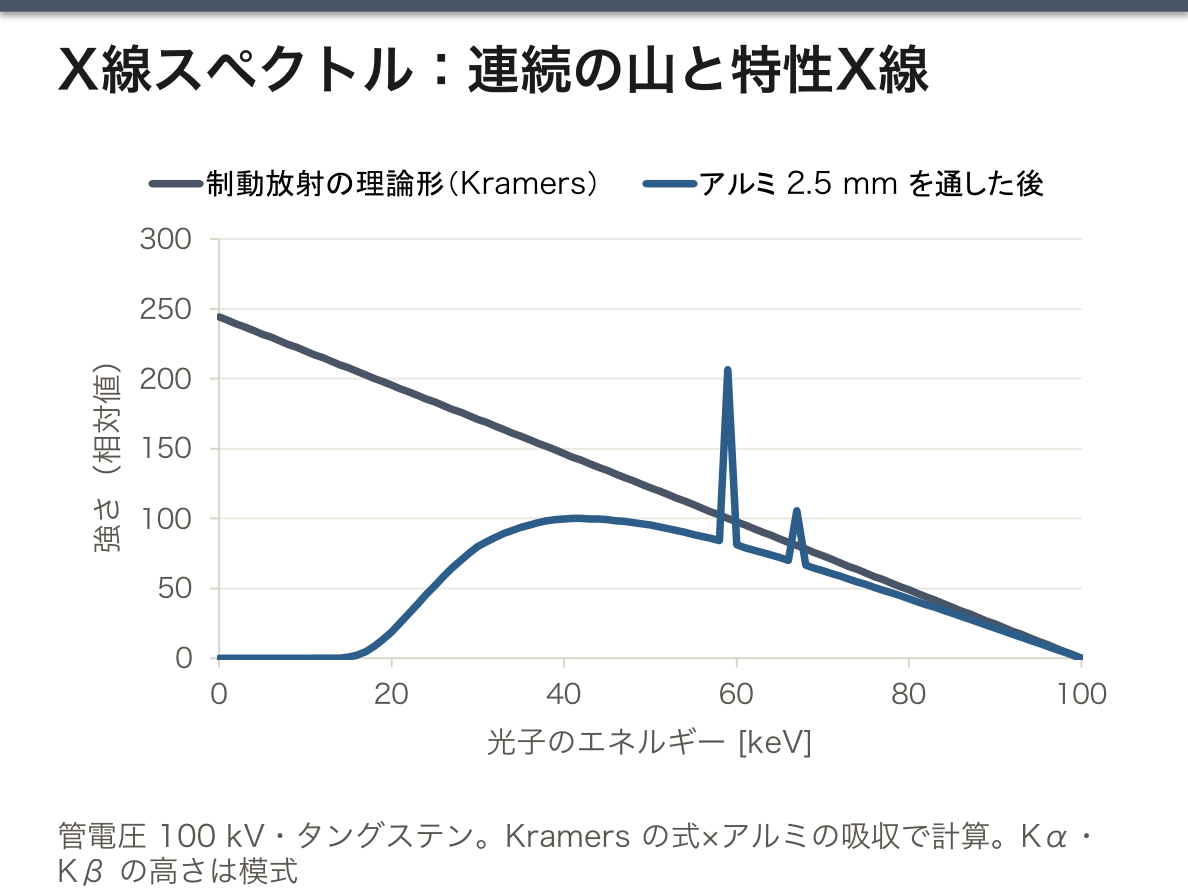
制動放射のスペクトルに上限がある理由。電子1個が持つエネルギーは eV(V:管電圧)である。ほとんどの電子は何度も小さく減速してエネルギーを少しずつ失うので、いろいろなエネルギーの光子が混ざった連続スペクトルになる。しかし、1回の衝突で全エネルギーを1個の光子に渡したときが最大で、それ以上にはならない。だから最大エネルギーは Emax = eV、最短波長は λmin = hc/eV(λmin[nm] ≈ 1.24/V[kV])になる(Duane–Hunt の法則)。100 kV の管から 100 keV を超える X線は出ない。
特性X線が「元素の指紋」になる理由。K殻・L殻の電子の結合エネルギーは元素ごとに決まった値なので、L殻から K殻へ落ちるときに出る光子(Kα)、M殻から落ちるときの光子(Kβ)のエネルギーも元素ごとに決まる。ただし、まず K殻の電子をたたき出せなければ始まらないので、入ってくる電子のエネルギーが K殻の結合エネルギー(タングステンで約 69.5 keV)を超えないと K系列の特性X線は出ない。管電圧を 60 kV に下げると、タングステンの Kα のピークは消える。
低エネルギー側で連続スペクトルが 0 に落ちるのは、そのあたりの柔らかい X線がターゲット自身・管の窓・フィルタで吸収されてしまうからである。
English version
Core
The spectrum of an X-ray tube (intensity at each energy) is a smooth, broad continuous spectrum with a few sharp peaks on top. The two come from completely different processes.
| Bremsstrahlung | Characteristic X-rays | |
|---|---|---|
| What happens | An electron passing near a nucleus is pulled by its positive charge and bends; the energy lost in slowing down leaves as a photon | An electron knocks out an inner-shell electron (e.g. K shell); an outer-shell electron drops into the gap and the energy difference between shells leaves as a photon |
| Spectrum | Continuous (from 0 to a maximum) | Lines (peaks): only fixed energies |
| Set by | Maximum energy set by the tube voltage | Energy set by the target element (not by tube voltage) |
| Example (tungsten) | At 100 kV, maximum 100 keV | Kα about 59 keV, Kβ about 67 keV |
"Bremsstrahlung" is German for "braking radiation".
Going further
Why bremsstrahlung has an upper limit. Each electron has energy eV (V: tube voltage). Most electrons slow down in many small steps and lose energy bit by bit, so photons of many energies mix into a continuous spectrum. But the maximum is when one collision gives all the energy to a single photon, and it cannot go higher. So the maximum energy is Emax = eV and the shortest wavelength is λmin = hc/eV (λmin[nm] ≈ 1.24/V[kV]) — the Duane–Hunt law. A 100 kV tube cannot produce X-rays above 100 keV.
Why characteristic X-rays are an "element fingerprint". The binding energies of K- and L-shell electrons are fixed for each element, so the photon from an L-to-K drop (Kα) and from an M-to-K drop (Kβ) also have fixed energies for each element. But first a K-shell electron must be knocked out, so K-series characteristic X-rays appear only when the incoming electron energy exceeds the K-shell binding energy (about 69.5 keV for tungsten). Lower the tube voltage to 60 kV and the tungsten Kα peak disappears.
The continuous spectrum falls to zero at the low-energy end because those soft X-rays are absorbed by the target itself, the tube window and the filter.
スペクトルを動かすもの — 管電圧・管電流・濾過
撮影する人が操作できるのは主に3つで、それぞれスペクトルの違う部分を動かす。
| 操作 | スペクトルへの効果 | 画像・被曝への効果 |
|---|---|---|
| 管電圧(kV)を上げる | 最大エネルギーが上がり、山全体が高エネルギー側へ伸びる。強さも大きく増える(おおよそ kV の2乗) | 透過力が増す。コントラストは下がる |
| 管電流(mA)・時間を増やす | 形は変わらず、高さだけが比例して増える | 光子の数が増えて画像のざらつき(ノイズ)が減る。被曝は比例して増える |
| 濾過(アルミ板など)を厚くする | 低エネルギー側が削られ、平均エネルギーが上がる(線質硬化) | 画像に役立たない皮膚の被曝が減る |
よく言われる対比は「kV は質(エネルギー)、mA は量(数)」。連続 X線の平均エネルギーは、最大エネルギーのおよそ 1/3〜1/2 である。
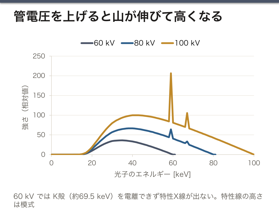
なぜ濾過が必要なのか。低エネルギーの X線は体の表面近くですぐに吸収されてしまい、検出器まで届かない=画像に何も寄与しないのに、皮膚の線量だけを増やす。アルミの板を通すと、こうした柔らかい成分が優先的に吸収され(光電効果は低エネルギーほど起きやすい)、透過力のある成分が残る。これを線質硬化(beam hardening)という。診断用の装置には一定以上の濾過が義務づけられている。
スペクトルから何が分かるか。①連続スペクトルの端(最大エネルギー)から管電圧が分かる——装置の電圧が表示どおりか点検できる。②特性X線のピークの位置から元素が分かる。Moseley は1913年、Kα 線の振動数の平方根が (Z − 1) に比例することを見つけ、元素の並びを原子量ではなく原子番号で決めるべきことを示した。この原理は今も、試料に X線を当てて出てくる特性X線から成分元素を調べる蛍光X線分析として使われている。
ターゲットの元素を変えると、使えるピークも変わる。乳房撮影では、モリブデン(Kα 約 17.5 keV)などをターゲットにして、柔らかい乳腺組織の差が出やすい低いエネルギー帯に X線を集中させる。
撮影条件は「何を見たいか」で決まる。胸部写真は肺の血管影と肋骨の重なりを両方見たいので、高めの管電圧(100 kV 以上)で骨のコントラストを抑える。乳房撮影は脂肪と乳腺のわずかな差や微小石灰化を見たいので、30 kV 前後の低い管電圧を使う。小児では mAs を下げ、濾過を足して被曝を減らす——いずれも上の表の使い分けである。
English version
Core
The operator mainly controls three things, and each moves a different part of the spectrum.
| Change | Effect on the spectrum | Effect on image and dose |
|---|---|---|
| Raise the tube voltage (kV) | The maximum energy rises and the whole curve stretches to higher energy. Intensity also rises strongly (roughly kV squared) | More penetration; lower contrast |
| Increase tube current (mA) and time | Shape unchanged; only the height rises in proportion | More photons, less graininess (noise); dose rises in proportion |
| Add filtration (e.g. aluminium) | The low-energy side is cut; the mean energy rises (beam hardening) | Less skin dose that does nothing for the image |
The usual contrast: "kV is quality (energy), mA is quantity (number)". The mean energy of the continuous spectrum is roughly 1/3 to 1/2 of the maximum.
Going further
Why filtration is needed. Low-energy X-rays are absorbed close to the body surface, never reach the detector and add nothing to the image, yet they increase skin dose. An aluminium plate absorbs these soft photons first (the photoelectric effect is more likely at low energy), leaving the penetrating part. This is beam hardening. Diagnostic machines must have at least a set amount of filtration.
What the spectrum tells us. (1) The end of the continuous spectrum (the maximum energy) gives the tube voltage — a way to check that the machine’s voltage is as shown. (2) The positions of the characteristic peaks identify the element. In 1913 Moseley found that the square root of the Kα frequency is proportional to (Z − 1), showing that elements should be ordered by atomic number, not atomic mass. The same principle is used today in X-ray fluorescence analysis, which finds the elements in a sample from the characteristic X-rays it gives off when irradiated.
Changing the target element changes the available peaks. Mammography uses targets such as molybdenum (Kα about 17.5 keV) to concentrate X-rays in the low-energy band where differences in soft breast tissue show best.
In the clinic
Exposure settings depend on what you want to see. A chest film should show both lung vessels and overlapping ribs, so a higher tube voltage (100 kV or more) is used to tone down bone contrast. Mammography needs to show small differences between fat and gland and tiny calcifications, so a low voltage around 30 kV is used. In children, mAs is lowered and filtration added to cut dose — all uses of the table above.
減弱とコントラスト — 白く写るもの・黒く写るもの・造影剤
X線が体を通ると、吸収(光電効果)と散乱(コンプトン散乱)によって弱まる(減弱, attenuation)。細い線束なら I = I0e−μx(μ:線減弱係数、x:厚さ)で、相互作用そのものは電離放射線の章で扱った。
μ は ①原子番号(光電効果はおよそ Z3 に比例)、②密度、③X線のエネルギー(低いほど大きい)で決まる。軟部組織は H・C・N・O という小さな原子でできているので X線をあまり吸収せず、骨は Ca(Z = 20)や P を含むので強く吸収する。
単純写真では、通り抜けた X線が多いほど黒く、少ないほど白く写る。基本の濃度は5段階で、空気(黒)→ 脂肪 → 水・軟部組織 → 骨 → 金属(白)。ただし写るのは「μ × 厚さ」の積なので、厚さも濃さに効く。
コントラストを落とすのが散乱線である。散乱した X線は向きがばらばらで、像のない「かぶり」を全体に足す。照射野を必要な範囲に絞る(コリメーション)ことと、検出器の前に鉛の薄板を並べたグリッド(anti-scatter grid)を置いて斜めに来る散乱線を止めることで対策する。
造影剤は、軟部組織どうしの差がないところに人工的に差をつくる。代表は硫酸バリウム(Ba, Z = 56。消化管)とヨード造影剤(I, Z = 53。血管・尿路・CT)。どちらも原子番号が大きいだけでなく、K殻の結合エネルギー(K吸収端)がヨードで約 33 keV、バリウムで約 37 keV と、診断用 X線の平均エネルギーのすぐ下にある。光子のエネルギーが K吸収端を超えたところで光電効果が急に起きやすくなるので、ちょうど診断に使う X線をよく吸収する——これが2つが選ばれている物理的な理由である。造影剤は放射性ではない。
造影剤に求められる性質は、反応しにくく毒性が低いこと、体に吸収・蓄積されず、速やかに排泄されることである。
エネルギーによる減弱の違いを逆に利用するのが骨密度測定(DXA)である。2種類のエネルギーの X線で撮ると、骨と軟部組織とでエネルギーによる減弱の変わり方が違うので、2本の式から軟部組織の影響を消して骨塩量(g/cm2)を出せる。被曝は胸部写真よりさらに少ない。
消化管穿孔が疑われるときにバリウムは使わない。腹腔に漏れたバリウムは吸収されず、重い腹膜炎を起こすためで、代わりに水溶性のヨード造影剤を使う。ヨード造影剤では、アレルギー様の反応と腎機能への影響に注意し、使用前に腎機能(eGFR など)と過去の反応歴を確かめる。注入中に血管の外へ漏れる(血管外漏出)と腕が腫れて痛むので、注入部の観察が欠かせない。
DXA は骨粗鬆症の診断の基準になる検査で、若年成人の平均との差を標準偏差で表した T スコアが −2.5 以下なら骨粗鬆症と判定する(WHO の基準)。腰椎と大腿骨近位部で測るのが標準である。
English version
Core
As X-rays pass through the body they weaken by absorption (the photoelectric effect) and scattering (Compton scattering) — attenuation. For a narrow beam, I = I0e−μx (μ: linear attenuation coefficient, x: thickness); the interactions themselves are covered in the ionizing radiation chapter.
μ depends on (1) atomic number (the photoelectric effect goes roughly as Z3), (2) density, and (3) X-ray energy (larger at low energy). Soft tissue is made of small atoms — H, C, N, O — and absorbs little; bone contains Ca (Z = 20) and P and absorbs strongly.
On a plain film, more X-rays getting through means darker; fewer means whiter. There are five basic densities: air (black) → fat → water/soft tissue → bone → metal (white). But what shows is the product "μ × thickness", so thickness also affects density.
Scattered radiation lowers contrast. Scattered X-rays go in all directions and add an even "fog" with no image. Two fixes: narrow the beam to the needed area (collimation), and place a grid of thin lead strips in front of the detector (anti-scatter grid) to stop scatter arriving at an angle.
Going further
Contrast media create a difference where soft tissues have none. The main ones are barium sulfate (Ba, Z = 56; gut) and iodinated contrast (I, Z = 53; vessels, urinary tract, CT). Both have high atomic numbers, and their K-shell binding energy (K-edge) — about 33 keV for iodine and 37 keV for barium — lies just below the mean energy of diagnostic X-rays. Just above the K-edge the photoelectric effect suddenly becomes much more likely, so they absorb exactly the X-rays used for diagnosis — the physical reason these two are chosen. Contrast media are not radioactive.
A contrast agent should be unreactive and low in toxicity, should not be absorbed or retained, and should be excreted quickly.
Bone densitometry (DXA) turns the energy dependence of attenuation to use. With X-rays of two energies, bone and soft tissue change their attenuation differently with energy, so two equations remove the soft tissue and give bone mineral (g/cm2). The dose is even lower than a chest film.
In the clinic
Barium is not used when a gut perforation is suspected. Barium that leaks into the abdomen is not absorbed and causes severe peritonitis, so a water-soluble iodinated agent is used instead. With iodinated contrast, watch for allergy-like reactions and effects on kidney function; check renal function (e.g. eGFR) and any past reaction beforehand. If contrast leaks out of the vein during injection (extravasation), the arm swells and hurts, so the injection site must be watched.
DXA is the reference test for diagnosing osteoporosis. A T-score of −2.5 or below (standard deviations from the young-adult mean) means osteoporosis (WHO criteria). Measurement at the lumbar spine and proximal femur is standard.
透視・血管造影・乳房撮影 — 動きを見る・管を見る・柔らかいものを見る
透視(fluoroscopy)は、X線を出し続けて(または短いパルスをくり返して)体の中の動きを実時間でモニターに映す方法である。昔は蛍光板を直接見ていたが、今は X線を光に変えて増幅するイメージインテンシファイアや、平面検出器(フラットパネル)を使う。造影剤と組み合わせて、飲み込み・消化管の動き・カテーテルの位置を見ながら検査や治療を進める。
血管造影(angiography)は、カテーテルを動脈(大腿動脈や橈骨動脈)から目的の血管まで進め、ヨード造影剤を注入しながら短い間隔で連続撮影する。冠動脈造影では狭窄や閉塞の場所が分かり、そのまま治療(カテーテル治療)へ移れる。静脈を写すものは静脈造影、乳管・唾液腺の管に造影剤を入れるものは乳管造影・唾液腺造影と呼ぶ。
乳房撮影(mammography)は乳房専用の X線撮影で、30 kV 前後の低い管電圧を使う。乳房を板で圧迫して薄く広げて撮る。
デジタルサブトラクション血管造影(DSA):造影剤を入れる前の画像(マスク像)を、入れた後の画像から引き算する。骨や軟部組織は両方の画像に同じように写っているので消え、造影剤の入った血管だけが残る。患者が動くとずれが生じて消え残りが出る(モーションアーチファクト)。
乳房撮影で低い管電圧を使う理由:乳腺・脂肪・腫瘍はどれも軟部組織で、μ の差がわずかである。光電効果は低エネルギーほど起きやすく、原子番号の差が強く出るので、低エネルギーにすると小さな差が大きなコントラストになる。圧迫の理由:①厚さが減れば必要な線量が減る、②散乱線が減ってコントラストが上がる、③組織の重なりがほどけ、動きによるぼけも減る——どれも物理で説明できる。
透視の被曝は「時間」で決まる。撮影1枚と違い、透視は出している時間の分だけ被曝が積み重なる。パルス透視(必要なコマだけ X線を出す)、最終画像の保持(ペダルを離しても最後の像を表示しておく)、照射野を絞ることが、患者と術者の被曝を減らす基本になる。
長時間のカテーテル治療では、皮膚に確定的影響が出ることがある。同じ場所に長く X線を当て続けると皮膚の線量が数 Gy に達し、数週間後に紅斑・脱毛、重いと潰瘍が出る。施術中に線量を表示し、角度を変えて照射部位を分散させる。また、透視の散乱線は患者から出るので、術者は防護衣・防護眼鏡を使い、患者から一歩離れることが有効である。
乳房撮影の画像では、形の不規則な腫瘤と微小石灰化(細かい白い点の集まり)が乳癌を疑う所見になる。乳腺が密な若い女性では腫瘤が乳腺に隠れやすいので、超音波検査を組み合わせることが多い。
English version
Core
Fluoroscopy keeps the X-rays on (or repeats short pulses) to show movement inside the body in real time on a monitor. Once a fluorescent screen was viewed directly; now an image intensifier, which turns X-rays into light and amplifies it, or a flat-panel detector is used. Combined with contrast, it lets doctors watch swallowing, gut movement or the position of a catheter while testing or treating.
In angiography, a catheter is passed from an artery (femoral or radial) to the target vessel, and iodinated contrast is injected while images are taken in quick succession. Coronary angiography shows where a narrowing or block is, and can move straight on to catheter treatment. Imaging veins is venography; putting contrast into the ducts of the breast or salivary glands is galactography or sialography.
Mammography is X-ray imaging only for the breast, using a low tube voltage around 30 kV. The breast is pressed flat between plates.
Going further
Digital subtraction angiography (DSA): the image taken before contrast (the mask) is subtracted from the image after contrast. Bone and soft tissue appear the same in both, so they vanish, and only the vessels with contrast remain. If the patient moves, the images no longer match and leftovers appear (motion artefact).
Why mammography uses low voltage: gland, fat and tumour are all soft tissue, and their μ differ only a little. The photoelectric effect is stronger at low energy and shows atomic-number differences strongly, so low energy turns small differences into large contrast. Why compression: (1) thinner tissue needs less dose, (2) less scatter means more contrast, (3) overlapping tissue spreads out and motion blur falls — all explained by physics.
Fluoroscopy dose depends on time. Unlike a single image, dose keeps adding up for as long as the beam is on. Pulsed fluoroscopy (X-rays only for the needed frames), last-image hold (the last image stays on screen after the pedal is released) and narrowing the beam are the basics for cutting dose to patient and operator.
In the clinic
Long catheter procedures can cause deterministic effects on the skin. If X-rays hit one spot for a long time, skin dose can reach several Gy, causing redness and hair loss after a few weeks, and ulcers in severe cases. Dose is shown during the procedure, and the beam angle is changed to spread the entry site. Also, scatter in fluoroscopy comes from the patient, so the operator should wear a lead apron and protective glasses and step back from the patient.
On a mammogram, an irregularly shaped mass and microcalcifications (clusters of fine white dots) suggest breast cancer. In young women with dense breasts, a mass can hide behind gland tissue, so ultrasound is often added.
CT の原理と CT 値 — 多方向の影から断面を計算する
単純写真は、体の厚み全体の減弱が1枚の影に重なったものである。前後に重なった構造は区別できず、濃度も大まかに5段階しか分からない。CT(computed tomography, コンピュータ断層撮影)は、これを根本から解決した。
原理:X線管と、反対側に並んだ検出器の列が体のまわりを回り、薄い扇形の X線で同じ断面をあらゆる方向から測る。各方向の測定値は、その線に沿った μ の合計である。多数の方向の合計から、断面の各点(画素)の μ を逆算する——これが画像再構成で、代表的な方法がフィルタ補正逆投影法(フーリエ変換を使う)である。1970年代初めに Hounsfield が装置を実用化し、理論を先に示した Cormack とともに1979年のノーベル生理学・医学賞を受けた。
CT 値(Hounsfield unit, HU)は、水を基準にした μ の相対値である。
CT 値 = 1000 × (μ − μ水) / (μ水 − μ空気)
| 組織 | CT 値(HU, 目安) |
|---|---|
| 空気 | −1000 |
| 脂肪 | −100 前後 |
| 水 | 0 |
| 軟部組織・筋 | +20〜+60 程度 |
| 新しい出血(凝血) | +50〜+80 程度 |
| 骨 | 数百〜+1000 以上 |
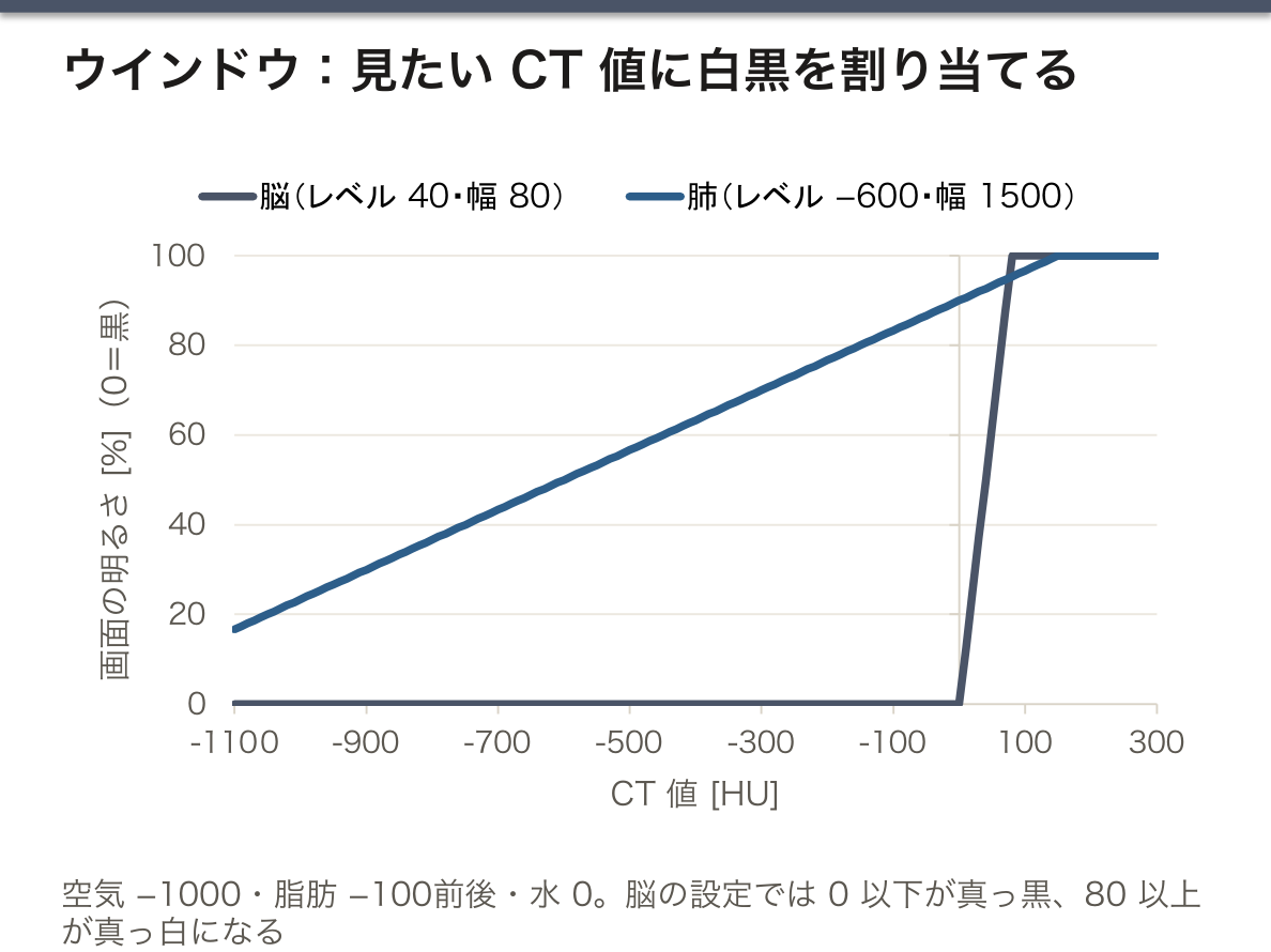
ウインドウ:CT 値は −1000 から +1000 以上まで2000段階以上あるが、人の目はそんなに多くの灰色を見分けられない。そこで、見たい範囲の CT 値だけを白から黒に割り当てる。中心の値をウインドウレベル、幅をウインドウ幅と呼ぶ。脳を見るときは狭い幅(例:レベル 40・幅 80 前後)で灰白質と白質のわずかな差を強調し、肺を見るときはレベルを大きく負にして幅を広げる。同じデータでも、ウインドウを変えるとまったく違う画像になる。
ヘリカル(らせん)CT:X線管が回り続けたまま寝台を一定の速さで動かすと、体に対して X線はらせんを描く。1枚ずつ止めて撮る方式と違い、範囲全体を切れ目のない立体データとして一気に集められ、短時間で撮れて呼吸のずれも減る。検出器を体軸方向に何列も並べた多列検出器 CT(MDCT)で、さらに速く薄くなった。立体データからは、任意の断面(MPR)・3D 画像・腸の内腔を内視鏡のように表示する仮想内視鏡(CT コロノグラフィなど)を作れる。
単純写真との違いのまとめ:CT は ①重なりのない断面、②わずかな濃度差を数値で区別できる、③立体データが得られる。代わりに被曝が大きい(胸部単純写真 1枚 約 0.02 mSv に対し、頭部 CT 約 2 mSv、胸部 CT はその数倍)。
脳卒中が疑われたら、まず頭部 CT を撮る。新しい出血は CT 値が高く(白く)、発症直後からはっきり写る。一方、脳梗塞は発症後数時間は CT でほとんど変化が見えないことが多い。だからCT で「出血ではない」ことを確かめてから、血栓溶解療法などを考える——CT 値の物理が、そのまま救急の手順になっている。
CT の被曝は小さくないので、とくに小児や若年者では本当に必要か(正当化)と必要最小限の線量か(最適化)を考える。日本は人口あたりの CT 装置の台数が多く、医療被曝が多い国とされている。
English version
Core
A plain film is the attenuation of the whole body thickness piled into one shadow. Structures in front of and behind each other cannot be told apart, and density is known only in about five rough steps. CT (computed tomography) solved this at the root.
Principle: an X-ray tube and a row of detectors opposite it rotate around the body, measuring the same slice from every direction with a thin fan of X-rays. Each measurement is the sum of μ along that line. From the sums in many directions, the μ of each point (pixel) in the slice is worked back — this is image reconstruction, and a typical method is filtered back projection (using Fourier transforms). In the early 1970s Hounsfield built a working scanner, and he shared the 1979 Nobel Prize in Physiology or Medicine with Cormack, who had earlier set out the theory.
The CT number (Hounsfield unit, HU) is μ relative to water:
CT number = 1000 × (μ − μwater) / (μwater − μair)
| Tissue | CT number (HU, approx.) |
|---|---|
| Air | −1000 |
| Fat | around −100 |
| Water | 0 |
| Soft tissue, muscle | about +20 to +60 |
| Fresh blood (clot) | about +50 to +80 |
| Bone | several hundred to over +1000 |
Going further
Window: CT numbers run from −1000 to over +1000 — more than 2000 steps — but the eye cannot tell that many greys apart. So only the range of CT numbers you want to see is mapped from white to black. The centre value is the window level, and the range is the window width. For the brain a narrow width (e.g. level about 40, width about 80) brings out the small difference between grey and white matter; for the lungs the level is set strongly negative and the width wide. The same data look completely different with a different window.
Helical (spiral) CT: if the table moves at a steady speed while the tube keeps rotating, the X-rays trace a spiral around the body. Unlike stopping for each slice, the whole region is collected as one continuous volume, quickly and with less breathing mismatch. Multi-detector CT (MDCT), with many detector rows along the body axis, made it faster and thinner still. From volume data one can build any plane (MPR), 3D images, and a virtual endoscopy that shows the gut lumen as if through an endoscope (e.g. CT colonography).
Summary of the difference from plain films: CT gives (1) slices without overlap, (2) small density differences told apart as numbers, (3) volume data. In exchange, the dose is larger (about 0.02 mSv for one chest film, about 2 mSv for a head CT, several times more for a chest CT).
In the clinic
When a stroke is suspected, a head CT comes first. Fresh blood has a high CT number (white) and shows clearly from the start. A cerebral infarct, by contrast, often shows almost no change on CT for the first few hours. So CT is used to confirm "it is not a bleed" before thrombolysis is considered — the physics of CT numbers becomes the emergency pathway.
CT dose is not small, so, especially in children and young people, ask whether it is really needed (justification) and whether the dose is as low as needed (optimisation). Japan has many CT scanners per head of population and is regarded as a country with high medical exposure.
放射線治療とリニアック — X線で腫瘍を壊す
放射線治療(radiotherapy)は、手術・薬物療法と並ぶがん治療の柱である。電離放射線が細胞の DNA を傷つけ、分裂できなくしたり死なせたりすることを利用する。
現在の外部照射の中心は直線加速器(リニアック, linear accelerator)である。電子をマイクロ波で加速管の中を一直線に加速して数 MeV〜20 MeV 程度まで上げ、①タングステンのターゲットに当てて高エネルギーの X線(制動放射)を作るか、②電子線のまま取り出して浅い病変に当てる。診断用 X線管と同じ「電子をぶつけて X線を作る」原理だが、エネルギーが百倍ほど高い。
装置の照射部(ガントリー)は患者のまわりを回り、腫瘍に向けて多くの方向から照射する。各方向のビームが重なる腫瘍には高い線量が集まり、通り道の正常組織は1方向分の線量ですむ。照射野の形は、何十枚もの細い金属の板を動かすマルチリーフコリメータで腫瘍の形に合わせる。
放射線が細胞を傷つける2つの経路:①直接作用——放射線(から生じた電子)が DNA そのものを切る。②間接作用——体の大部分を占める水が電離してヒドロキシルラジカル(・OH)などのラジカルができ、それが DNA を傷つける。X線では間接作用の寄与が大きい。酸素があると DNA の傷が「固定」されて修復されにくくなるので、酸素の少ない(低酸素の)腫瘍は放射線が効きにくい。
分割照射の理由:治療は1回 2 Gy 前後を何十回にも分けて行うことが多い。正常細胞の多くは照射と照射のあいだに傷を修復できるが、腫瘍細胞は修復が不十分なことが多い。分けて当てるほど、正常組織と腫瘍の差が積み重なる。分裂の盛んな細胞ほど放射線に弱いこと(Bergonié–Tribondeau の法則)も腫瘍に有利に働く。
高エネルギー X線は皮膚を守る:数 MeV の X線が体に入ると、はじき出された電子は前方へ数 mm〜数 cm 走ってからエネルギーを渡す。そのため線量が最大になるのは皮膚ではなく少し深いところ(6 MV で約 1.5 cm)になり、皮膚の線量は低く抑えられる(ビルドアップ)。
CT と放射線治療は、同じ発想の裏表である。CT は多方向から X線を通して断面の μ を「読み取る」。リニアックは多方向から X線を当てて腫瘍に線量を「書き込む」。実際、治療計画は CT 画像の上で腫瘍と避けたい臓器を描き、各方向のビームの形と強さを計算して決める(強度変調放射線治療, IMRT)。
診断の X線にもリスクはあるが、その大きさを知っておくことが大事である。低線量の発がんリスクは、しきい値がなく線量に比例すると仮定して見積もる(直線しきい値なしモデル)。この仮定で見積もると、胸部写真1枚の追加の致死がんリスクは百万分の1のけた、胸部 CT では数千分の1のけたとされる。「X線はこわい」でも「X線は無害」でもなく、検査で得られる利益と比べて判断するための数字である。
English version
Core
Radiotherapy is one of the pillars of cancer treatment, alongside surgery and drug therapy. It uses the fact that ionising radiation damages a cell’s DNA, so the cell cannot divide or dies.
Modern external-beam therapy centres on the linear accelerator (linac). Electrons are accelerated in a straight line by microwaves in an accelerating tube to a few MeV up to about 20 MeV. Then either (1) they hit a tungsten target to make high-energy X-rays (bremsstrahlung), or (2) the electron beam itself is used for shallow lesions. It is the same principle as a diagnostic tube — "hit a target with electrons to make X-rays" — but with about a hundred times more energy.
The treatment head (gantry) rotates around the patient and irradiates the tumour from many directions. Where the beams overlap, in the tumour, the dose adds up; normal tissue along each path gets only one beam’s share. The field is shaped to the tumour by a multileaf collimator, dozens of thin metal leaves that move.
Going further
Two ways radiation damages cells: (1) direct action — the radiation (or electrons it sets free) breaks the DNA itself. (2) indirect action — water, which makes up most of the body, is ionised to form radicals such as the hydroxyl radical (·OH), which then damage DNA. For X-rays the indirect part is large. Oxygen "fixes" DNA damage so it is harder to repair, so tumours low in oxygen (hypoxic) respond less to radiation.
Why fractionation: treatment is often given as about 2 Gy at a time, split over dozens of sessions. Many normal cells repair the damage between sessions, while tumour cells often repair poorly. The more the dose is split, the more the difference between normal tissue and tumour builds up. That rapidly dividing cells are more sensitive (the Bergonié–Tribondeau law) also works in favour of treating tumours.
High-energy X-rays spare the skin: when X-rays of a few MeV enter the body, the electrons they set free travel forward a few mm to a few cm before giving up their energy. So the dose peaks not at the skin but a little deeper (about 1.5 cm for 6 MV), keeping the skin dose low (build-up).
In the clinic
CT and radiotherapy are two sides of the same idea. CT sends X-rays from many directions to "read" μ in a slice. A linac sends X-rays from many directions to "write" dose into a tumour. In practice, the treatment plan is drawn on CT images — the tumour and the organs to avoid — and the shape and strength of each beam are calculated (intensity-modulated radiotherapy, IMRT).
Diagnostic X-rays also carry risk, and it matters to know how big it is. Cancer risk at low doses is estimated by assuming no threshold and a risk proportional to dose (the linear no-threshold model). On that assumption, the added risk of fatal cancer is said to be of the order of one in a million for a chest film and one in a few thousand for a chest CT. These numbers are not for "X-rays are scary" or "X-rays are harmless", but for weighing the risk against the benefit of the test.
まとめ
| X線とは何か — 曲げられない光だから「影絵」で写す | X線(X-rays)は、紫外線よりさらに波長の短い電磁波である。 |
|---|---|
| X線管 — 電子を加速してぶつけ、エネルギーの99%は熱になる | X線管(X-ray tube)は、真空にしたガラスまたは金属の容器の中に、陰極(cathode)と陽極(anode)を向かい合わせたものである。 |
| 制動放射と特性X線 — なめらかな山と鋭いピーク | X線管から出る X線のスペクトル(エネルギーごとの強さ)は、なめらかに広がる連続スペクトルの上に、鋭いピークがいくつか立った形をしている。 |
| スペクトルを動かすもの — 管電圧・管電流・濾過 | 撮影する人が操作できるのは主に3つで、それぞれスペクトルの違う部分を動かす。 |
| 減弱とコントラスト — 白く写るもの・黒く写るもの・造影剤 | X線が体を通ると、吸収(光電効果)と散乱(コンプトン散乱)によって弱まる(減弱, attenuation)。 |
| 透視・血管造影・乳房撮影 — 動きを見る・管を見る・柔らかいものを見る | 透視(fluoroscopy)は、X線を出し続けて(または短いパルスをくり返して)体の中の動きを実時間でモニターに映す方法である。 |
| CT の原理と CT 値 — 多方向の影から断面を計算する | 単純写真は、体の厚み全体の減弱が1枚の影に重なったものである。 |
| 放射線治療とリニアック — X線で腫瘍を壊す | 放射線治療(radiotherapy)は、手術・薬物療法と並ぶがん治療の柱である。 |
このノートは医学生が授業の内容を自分の言葉でまとめたもので、診療の判断に使うものではありません。誤りに気づいたらお知らせください。