ヘモグロビン症は2つの型に分かれる。質の異常(構造異常症)と量の異常(サラセミア)である。前者の代表が鎌状赤血球症、後者が地中海貧血で、どちらもマラリア流行地域で頻度が高い——ヘテロ接合体がマラリアに抵抗性を持つためである。
1個のアミノ酸置換が分子の溶解度を変え、それが血管閉塞という全身の病態になる——分子から臨床までが一直線に繋がる、生化学の代表的な題材である。
- 鎌状赤血球症 — 溶解度が下がるということ
- サラセミア — 作れない鎖と、余る鎖
- 酸素解離曲線 — 曲線の形そのものが、機能になっている
- HbA1cとグリコアルブミン — それぞれ、何日分を見ているか
- 鉄が 3 価になると、酸素を運べない — メトヘモグロビン血症
- 胎児ヘモグロビンを、もう一度作らせる
鎌状赤血球症 — 溶解度が下がるということ
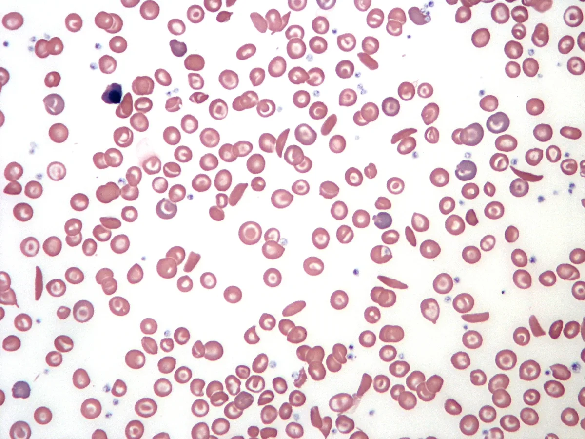
β鎖6番目のグルタミン酸がバリンに置換(点変異、ミスセンス)。
- グルタミン酸は親水性、バリンは疎水性 → 脱酸素化したときに分子どうしが疎水結合で重合する
- 重合したヘモグロビンSが繊維を作り、赤血球が鎌状に変形する
- 変形した赤血球が微小血管を閉塞 → 疼痛発作・脾梗塞・急性胸部症候群・脳梗塞
- 慢性溶血 → 貧血・胆石・下腿潰瘍
誘因は「脱酸素化を進める状況」——低酸素・脱水・アシドーシス・感染・寒冷・高所。
- 脾機能低下により莢膜を持つ細菌に弱い → 肺炎球菌ワクチンとペニシリン予防投与
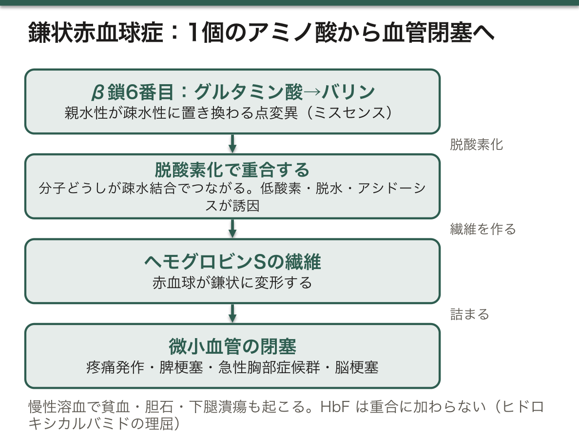
ヒドロキシカルバミド(ヒドロキシ尿素)が効く理屈が、この病気の理解の核心である。この薬は胎児ヘモグロビン(HbF、α₂γ₂)の産生を増やす。
- HbFは重合に加わらない
- だからHbFの割合が上がると、ヘモグロビンSの重合が起きにくくなる
- 疼痛発作と急性胸部症候群が明らかに減る
「胎児期の遺伝子をもう一度読ませる」という治療で、近年はゲノム編集でHbFの抑制因子(BCL11A)を壊す治療が承認されている——遺伝子治療が実際に届いた最初の一群になった。
ヘテロ接合体(鎌状赤血球形質)は通常無症状だが、腎髄質だけは例外である。髄質は生理的に低酸素・高浸透圧・低pH——重合に最も適した環境なので、ヘテロでも腎乳頭壊死と血尿を起こしうる。「無症状のはずの保因者で血尿」は、機序を知らなければ説明できない。
マラリア抵抗性は、感染した赤血球が鎌状化して早期に脾で除去されることなどによる。致死的な病気の遺伝子が高頻度で維持されているのは、ヘテロ接合体に生存上の利点があったからである——平衡選択の教科書的な例で、集団遺伝学の意味を最もよく示す。
English version
Core
Glutamate at position 6 of the β chain is replaced by valine (a point mutation, missense).
- Glutamate is hydrophilic and valine hydrophobic → on deoxygenation the molecules polymerise through hydrophobic bonds.
- Polymerised haemoglobin S forms fibres and the red cell deforms into a sickle.
- The deformed cells occlude the microvasculature → pain crises, splenic infarction, acute chest syndrome, stroke.
- Chronic haemolysis → anaemia, gallstones, leg ulcers.
The triggers are situations that advance deoxygenation — hypoxia, dehydration, acidosis, infection, cold, altitude.
Hyposplenism gives vulnerability to encapsulated bacteria → pneumococcal vaccination and penicillin prophylaxis.
Going further
The reasoning by which hydroxycarbamide (hydroxyurea) works is the core of understanding this disease. The drug increases the production of fetal haemoglobin (HbF, α₂γ₂).
- HbF does not join the polymer.
- So as the proportion of HbF rises, the polymerisation of haemoglobin S becomes less likely.
- Pain crises and acute chest syndrome are clearly reduced.
A treatment consisting of "having a gene of fetal life read once again", and recently a treatment using genome editing to destroy a suppressor of HbF (BCL11A) has been approved — the first group in which gene therapy actually arrived.
The heterozygote (sickle cell trait) is usually asymptomatic, but the renal medulla is the exception. The medulla is physiologically hypoxic, hyperosmolar and of low pH — the environment most favourable to polymerisation, so even a heterozygote may have papillary necrosis and haematuria. "Haematuria in a carrier who should be asymptomatic" cannot be explained without the mechanism.
Malaria resistance arises partly because infected red cells sickle and are removed early in the spleen. That the gene of a lethal disease is maintained at a high frequency is because the heterozygote had a survival advantage — the textbook example of balancing selection, and the best illustration of what population genetics means.
サラセミア — 作れない鎖と、余る鎖
グロビン鎖の産生量が減る疾患群。質ではなく量の病気である。
| αサラセミア | βサラセミア | |
|---|---|---|
| 遺伝子 | 4個(2対)。主に欠失 | 2個。主に点変異 |
| 余る鎖 | β鎖 → HbH(β₄)/胎児期はγ₄(Bart) | α鎖 → 不安定で赤芽球を壊す |
| 重症型 | 4個欠失で胎児水腫(子宮内死亡) | Cooley貧血。生後半年から発症 |
- 共通所見:小球性低色素性貧血・標的赤血球・骨髄の代償性拡大(頬骨突出・毛髪立像)・肝脾腫
- 鉄欠乏性貧血との鑑別——サラセミアでは赤血球数は保たれ、フェリチンは低くない
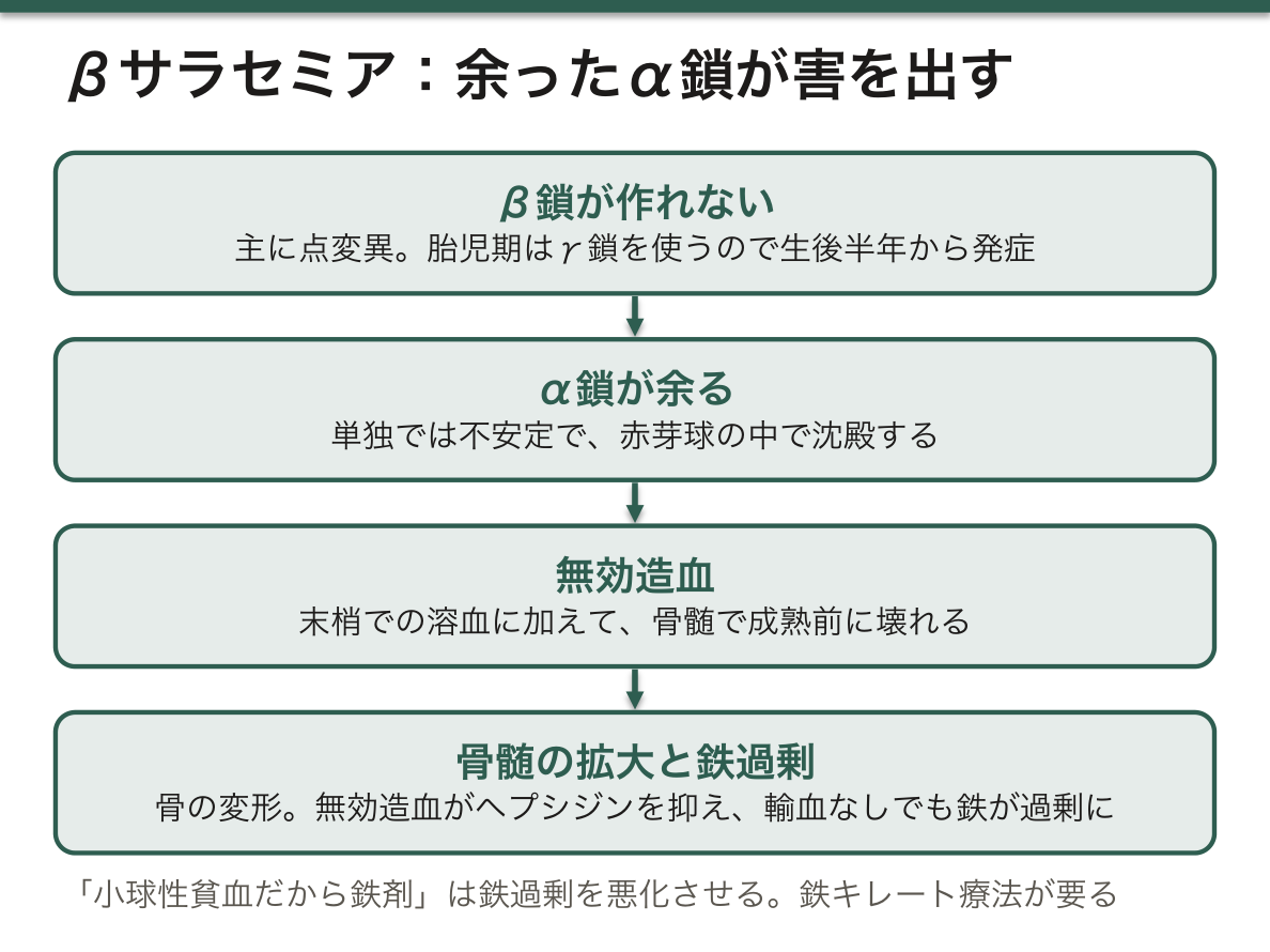
βサラセミアが生後すぐには発症しないのは、胎児期はγ鎖を使っており、β鎖への切り替えが生後数か月かけて起こるためである。αサラセミアは胎児期から症状が出る——α鎖は胎児ヘモグロビンにも必要だからである。発症時期の違いが、どの鎖の病気かを教える。
「余る鎖が害を出す」という視点が、症状の重さを説明する。βサラセミアでは余ったα鎖が単独では不安定で、赤芽球の中で沈殿し、細胞を壊す。だから末梢での溶血だけでなく、骨髄で成熟前に壊れる(無効造血)。
- 無効造血が骨髄を拡大させる → 骨の変形
- 無効造血がヘプシジンを抑える → 輸血していなくても鉄が過剰に吸収される
だから鉄過剰症が、輸血の有無に関わらず起きる——鉄キレート療法が要る。ここは実務で重要で、「小球性貧血だから鉄剤」と反射的に処方すると、鉄過剰を悪化させる。
Mentzer指数(平均赤血球容積 ÷ 赤血球数)が鑑別の目安になる。13未満ならサラセミア、13を超えれば鉄欠乏——サラセミアでは赤血球の数そのものは多いからである。
日本では稀だが、欧州では地中海沿岸出身者に一定の頻度がある。出身地を問診で確認することが、そのまま鑑別に効く場面がある。
English version
Core
A group of diseases in which the amount of globin chain produced is reduced. A disease of quantity, not quality.
| α-Thalassaemia | β-Thalassaemia | |
|---|---|---|
| Genes | Four (two pairs). Chiefly deletions | Two. Chiefly point mutations |
| The chain left over | β chain → HbH (β₄) / in fetal life γ₄ (Bart’s) | α chain → unstable, destroying the erythroblast |
| Severe form | Deletion of all four gives hydrops fetalis (intrauterine death) | Cooley’s anaemia. Presents from six months of age |
Shared findings: a microcytic hypochromic anaemia, target cells, compensatory expansion of the marrow (prominent cheekbones, hair-on-end), hepatosplenomegaly.
Distinction from iron deficiency anaemia — in thalassaemia the red cell count is preserved and the ferritin is not low.
Going further
β-Thalassaemia does not present immediately after birth because the γ chain is used in fetal life and the switch to the β chain takes some months after birth. α-Thalassaemia shows from fetal life — because the α chain is needed in fetal haemoglobin too. The difference in the age at onset tells which chain is at fault.
The view that the chain left over does the harm explains the severity. In β-thalassaemia the surplus α chain is unstable alone, precipitates within the erythroblast and destroys the cell. So there is not only haemolysis in the periphery but destruction before maturity in the marrow (ineffective erythropoiesis).
- Ineffective erythropoiesis expands the marrow → deformity of bone.
- Ineffective erythropoiesis suppresses hepcidin → iron is absorbed in excess even without transfusion.
Hence iron overload occurs with or without transfusion — iron chelation is required. This matters in practice: prescribe iron reflexively because it is a microcytic anaemia and the iron overload is made worse.
The Mentzer index (mean corpuscular volume ÷ red cell count) is the guide in the differential. Below 13 means thalassaemia, above 13 iron deficiency — because in thalassaemia the number of red cells is itself high.
Rare in Japan, but in Europe it occurs at a definite frequency among people of Mediterranean origin. Confirming the region of origin in the history can bear directly on the differential.
酸素解離曲線 — 曲線の形そのものが、機能になっている
ヘモグロビンは4量体で、1つ酸素が付くと次が付きやすくなる(協同性)。だから曲線が S 字になる。
P50(酸素飽和度 50% になる酸素分圧)は約 27 mmHg。
| 右にずれる(酸素を離しやすい) | 左にずれる(酸素を離しにくい) |
|---|---|
| 体温↑・二酸化炭素↑・pH↓(アシドーシス)・2,3-BPG↑ | 体温↓・二酸化炭素↓・pH↑・2,3-BPG↓ |
| 運動している筋・発熱 | 胎児ヘモグロビン・一酸化炭素中毒・保存血 |
ミオグロビンは単量体なので協同性が無く、双曲線になる——低い分圧でも酸素を放さない。
Bohr 効果——組織で二酸化炭素と酸が増え、酸素が離れやすくなる。
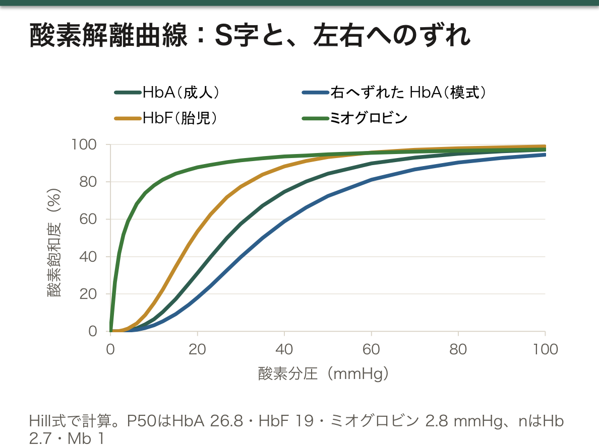
S 字であることが、「肺で満載し、組織で大きく手放す」を同時に可能にしている。高い分圧の側は平らなので、多少の低下では飽和度が落ちない。低い分圧の側は急なので、少しの低下で大量に放出できる——「1つの曲線の、上と下で役割が違う」。
だから平らな部分の意味が、臨床でそのまま出る。PaO₂ が 100 から 70 mmHg へ落ちても SpO₂ はほとんど変わらないが、60 を割ると急に落ちる(呼吸の章)——「SpO₂ が下がり始めた時点で、すでに崖の縁にいる」。
2,3-BPG は、解糖系の側枝で作られる調節分子になる。高地や慢性の低酸素で増えて曲線を右へ動かし、組織への受け渡しを助ける(解糖の章)——「赤血球の中の代謝が、酸素の配り方を変えている」。
胎児ヘモグロビン(HbF)は 2,3-BPG と結合しにくいので、曲線が左にある(胎児の生理の章)——「母体から酸素を奪い取れるのは、この差による」。
保存血では 2,3-BPG が減って左へずれる(輸血の章)——「大量輸血の直後は、酸素を運べても渡しにくい」。
一酸化炭素中毒では、2つの害が重なる。結合部位を奪うだけでなく、残った部位の親和性を上げて曲線を左へ動かす(中毒の章)——「SpO₂ が正常に見えるので、パルスオキシメータを信じない」。
English version
Core
Haemoglobin is a tetramer, and once one oxygen binds the next binds more readily (cooperativity). Hence the curve is sigmoid.
P50 (the oxygen tension at 50 per cent saturation) is about 27 mmHg.
| Shifts right (releases oxygen more readily) | Shifts left (releases it less readily) |
|---|---|
| Temperature ↑, CO₂ ↑, pH ↓ (acidosis), 2,3-BPG ↑ | Temperature ↓, CO₂ ↓, pH ↑, 2,3-BPG ↓ |
| Working muscle, fever | Fetal haemoglobin, carbon monoxide poisoning, stored blood |
Myoglobin is a monomer with no cooperativity and gives a hyperbola — it does not release oxygen even at a low tension.
The Bohr effect — carbon dioxide and acid increase in the tissue and oxygen is released more readily.
Going further
- Being sigmoid makes it possible at once to "load fully in the lung and let go largely in the tissue". The high-tension side is flat, so saturation does not fall with a modest drop. The low-tension side is steep, so a small drop releases a great deal — the upper and lower parts of one curve having different roles.
- Hence the meaning of the flat part appears directly in the clinic. PaO₂ falling from 100 to 70 mmHg scarcely changes the SpO₂, but below 60 it falls suddenly (the chapter on respiration) — by the time the SpO₂ has begun to fall, one is already at the edge of the cliff.
- 2,3-BPG is a regulatory molecule made in a side branch of glycolysis. It increases at altitude and in chronic hypoxia, moves the curve right and assists delivery to the tissue (the chapter on glycolysis) — metabolism inside the red cell changing how oxygen is distributed.
- Fetal haemoglobin (HbF) binds 2,3-BPG poorly, so its curve is to the left (the chapter on fetal physiology) — this difference is how it takes oxygen from the mother.
- In stored blood the 2,3-BPG falls and it shifts left (the chapter on transfusion) — immediately after a massive transfusion, oxygen can be carried and is not readily handed over.
- In carbon monoxide poisoning two harms coincide. It takes the binding sites and raises the affinity of the remaining sites, moving the curve left (the chapter on poisoning) — the SpO₂ looks normal, so do not trust the pulse oximeter.
HbA1cとグリコアルブミン — それぞれ、何日分を見ているか
糖化は酵素を介さない反応(非酵素的糖化)——グルコースが蛋白のアミノ基と結合する。血糖の高さと、その蛋白が生きている時間の積で決まる。
| 指標 | 反映する期間 | 基準 |
|---|---|---|
| HbA1c | 過去 1〜2 か月(赤血球の寿命 120 日) | 6.5% 以上で糖尿病型 |
| グリコアルブミン | 過去 2〜3 週(アルブミンの半減期 約20 日) | — |
| 1,5-アンヒドログルシトール | 過去数日 | 尿糖が出ると下がる |
HbA1c が実際より低く出る場面——溶血・出血・鉄剤やエリスロポエチンの投与開始後・肝硬変・妊娠(赤血球が若いと、糖化する時間が足りない)。
高く出る場面——鉄欠乏性貧血(未治療)・腎不全・脾摘後。
「赤血球が何日生きているか」が、そのまま指標の妥当性になる。血糖ではなく、赤血球の寿命の話として読む必要がある(赤血球の章)——「血糖が同じでも、赤血球が若ければ HbA1c は低く出る」。
だから溶血性貧血のある糖尿病では、グリコアルブミンを使う(糖尿病の各論の章)——「使えない指標を、別の物差しで置き換える」。
期間の違いは、治療の評価にそのまま使える。血糖の変動が大きい人や、治療を変えた直後は、短い物差しのほうが早く動く——「2週間後に効果を見たいなら、HbA1c では間に合わない」。
妊娠では 2 つの理由で低く出る。赤血球の産生が増えて平均年齢が下がり、血漿量も増える(妊娠糖尿病の章)——「妊娠糖尿病の診断に HbA1c を使わないのは、この理由による」。
同じ非酵素的糖化が、長期の合併症も作っている。糖化が進むと不可逆な終末糖化産物(AGEs)になり、受容体(RAGE)を介して炎症と線維化を起こす(糖化の章)——「検査の指標と、合併症の機序が同じ反応から出ている」。
English version
Core
Glycation is a non-enzymatic reaction — glucose binds an amino group of a protein. It is set by the product of the blood glucose and the time that protein lives.
| Index | Period reflected | Standard |
|---|---|---|
| HbA1c | The past 1–2 months (red cell lifespan 120 days) | 6.5 per cent or more is the diabetic range |
| Glycated albumin | The past 2–3 weeks (albumin half-life about 20 days) | — |
| 1,5-Anhydroglucitol | The past few days | Falls when glucose appears in the urine |
Situations in which HbA1c reads lower than the truth — haemolysis, bleeding, after starting iron or erythropoietin, cirrhosis, pregnancy (with young red cells there is not enough time for glycation).
Situations in which it reads high — iron deficiency anaemia (untreated), renal failure, after splenectomy.
Going further
- How many days the red cell lives is directly the validity of the index. It has to be read as a matter of red cell lifespan rather than of blood glucose (the chapter on the red cell) — with the same blood glucose, young red cells give a low HbA1c.
- Hence in diabetes with haemolytic anaemia, glycated albumin is used (the chapter on the particulars of diabetes) — replacing an unusable index with another measure.
- The difference in period is directly useful in judging treatment. In someone with wide glycaemic swings, or just after a change of treatment, the shorter measure moves sooner — if the effect is to be seen in two weeks, HbA1c is not in time.
- In pregnancy it reads low for two reasons. Red cell production increases and the mean age falls, and the plasma volume increases too (the chapter on gestational diabetes) — this is why HbA1c is not used to diagnose gestational diabetes.
- The same non-enzymatic glycation also creates the long-term complications. Glycation advances to irreversible advanced glycation end-products (AGEs), causing inflammation and fibrosis through the receptor (RAGE) (the chapter on glycation) — the index of the test and the mechanism of the complications coming out of the same reaction.
鉄が 3 価になると、酸素を運べない — メトヘモグロビン血症
ヘモグロビンの鉄は 2 価でなければ酸素を結合できない。酸化されて 3 価になったものがメトヘモグロビンで、酸素を運べないうえ、残りのサブユニットの酸素親和性を上げて放させなくする。だから解離曲線が左へずれ、組織はいっそう酸素を受け取れない。
体は常に少量を作っており(1〜2%)、NADH-メトヘモグロビン還元酵素(シトクロム b5 還元酵素)が戻している。
| 先天性 | 還元酵素の欠損/異常ヘモグロビン M | 生下時からのチアノーゼ。元気なのに青い |
| 後天性(大半) | 酸化する物質に曝露 | 亜硝酸・ダプソン・局所麻酔薬・スルホンアミド・アニリン系色素・井戸水の硝酸塩 |
診断の決め手は、数値の食い違いである。
- チアノーゼがあるのに、動脈血の酸素分圧は正常——溶けている酸素は普通にあるため
- 酸素を投与しても改善しない
- パルスオキシメータが 85% 前後に張り付く——メトヘモグロビンが 2 つの波長を同程度に吸収するので、実際の値がいくつでも 85% 付近を示す。下がりも上がりもしないのが特徴になる
- 採血した血が、暗いチョコレート色で、空気に触れても赤くならない
確定は共酸素測定(CO オキシメトリ)で、血液ガス分析装置でメトヘモグロビン分画を直接測る。
治療はメチレンブルー。NADPH-メトヘモグロビン還元酵素の電子の受け渡し役として働き、鉄を 2 価へ戻す。
⚠️ G6PD 欠損症ではメチレンブルーが効かないうえ、かえって溶血を起こす。メチレンブルーが働くには NADPH が要るが、それを作るのがペントースリン酸経路だからである。この場合はアスコルビン酸・交換輸血・高気圧酸素を用いる。
乳児は特に弱い。胎児ヘモグロビンは酸化されやすく、還元酵素の活性も低い。井戸水で調乳した粉ミルクによる中毒(blue baby syndrome)が知られている。
酸素を上げても SpO2 が 85% から動かない患者を見たら、機器の不良と決めつける前に、メトヘモグロビン血症を考える。
English version
Core
The iron of haemoglobin must be divalent to bind oxygen. Oxidised to trivalent it is methaemoglobin, which cannot carry oxygen and, besides, raises the oxygen affinity of the remaining subunits so that they will not release it. So the dissociation curve shifts left and the tissue receives still less oxygen.
The body makes a small amount constantly (1–2 per cent), and NADH-methaemoglobin reductase (cytochrome b5 reductase) converts it back.
| Congenital | Deficiency of the reductase / haemoglobin M | Cyanosis from birth. Blue and yet well |
| Acquired (most cases) | Exposure to an oxidising substance | Nitrites, dapsone, local anaesthetics, sulphonamides, aniline dyes, nitrate in well water |
Going further
What settles the diagnosis is the discrepancy between the numbers.
- There is cyanosis and the arterial oxygen tension is normal — because the dissolved oxygen is ordinary.
- Giving oxygen does not improve it.
- The pulse oximeter sticks around 85 per cent — methaemoglobin absorbs the two wavelengths about equally, so whatever the true value it reads near 85 per cent. That it neither falls nor rises is the characteristic.
- The blood drawn is a dark chocolate colour and does not turn red on exposure to air.
Confirmation is by co-oximetry, measuring the methaemoglobin fraction directly on a blood gas analyser.
Treatment is methylene blue. It acts as the carrier of electrons for NADPH-methaemoglobin reductase and returns the iron to the divalent state.
⚠️ In G6PD deficiency methylene blue does not work and causes haemolysis instead. For methylene blue to work NADPH is needed, and what makes it is the pentose phosphate pathway. In that case use ascorbic acid, exchange transfusion or hyperbaric oxygen.
Infants are especially vulnerable. Fetal haemoglobin is readily oxidised and the reductase activity is low. Poisoning from formula made up with well water (blue baby syndrome) is well known.
In the clinic
Seeing a patient whose SpO2 will not move from 85 per cent however much oxygen is given, think of methaemoglobinaemia before concluding the machine is faulty.
胎児ヘモグロビンを、もう一度作らせる
グロビン遺伝子は発生の時期に応じて切り替わる。ε → γ → β と読まれる遺伝子が移り、生後 6 か月ごろに HbF(α2γ2)から HbA(α2β2)へ入れ替わる。
この切り替えが、鎌状赤血球症とβサラセミアの発症時期を決めている。どちらもβ鎖の病気なので、HbF が主役のあいだは症状が出ない。生後半年ごろから発症するのはこのためである。
逆に言えば、γ鎖をもう一度作らせれば治療になる。実際、HbF が高く残る体質(遺伝性高 HbF 症)を併せ持つ患者は、症状が軽い——この自然の観察が、治療戦略の出発点になった。
γ遺伝子を止めているのは、BCL11A という転写抑制因子である。だからこれを外せば HbF が戻る。
| ヒドロキシカルバミド | HbF を増やす(機序は完全には解明されていない) | 安価で経口。鎌状赤血球症の標準治療。血管閉塞発作と急性胸部症候群を減らす |
| 遺伝子編集 | 造血幹細胞の BCL11A の赤血球特異的な調節領域をCRISPR で壊す | 鎌状赤血球症とβサラセミアで承認された。自家細胞を編集して戻すため、拒絶が無い |
| 遺伝子付加 | 正常なβ様グロビン遺伝子をベクターで入れる | 同じくすでに実用化されている |
編集の設計が巧妙である。BCL11A は他の細胞でも重要な働きを持つので、丸ごと壊すわけにはいかない。そこで赤血球系でだけ働くエンハンサーを狙って壊す——「どこで発現するか」を決めている部分だけを外すという設計になっている。
⚠️ 課題は残っている。
- 移植前処置に大量の化学療法が要る——不妊と二次発癌の危険
- 極めて高額で、患者の大半がいる地域(アフリカ・南アジア)に届かない——医療の公平性の問題として議論されている
- 長期の安全性はまだ追跡中である
「遺伝子を治す」時代の最初の実例が、生化学の教科書に載っていた病気だった——1949 年に Pauling が「分子病」と呼んだ鎌状赤血球症が、分子のレベルで治療されるところまで来ている。
β鎖の異常症が生後半年から発症する理由を説明できるかは、国試でも問われる。HbF の切り替えで答える。
English version
Core
The globin genes switch according to the stage of development. The genes read move ε → γ → β, and around six months of age HbF (α2γ2) is replaced by HbA (α2β2).
This switch decides the age at onset of sickle cell disease and β-thalassaemia. Both being diseases of the β chain, there are no symptoms while HbF is the principal haemoglobin. This is why they present from about six months.
Put the other way, having the γ chain made again is a treatment. Indeed, patients who also carry a constitution in which HbF remains high (hereditary persistence of fetal haemoglobin) have milder disease — this observation of nature was the starting point of the therapeutic strategy.
Going further
What silences the γ gene is a transcriptional repressor called BCL11A. So removing it brings HbF back.
| Hydroxycarbamide | Increases HbF (the mechanism is not fully elucidated) | Cheap and oral. The standard treatment of sickle cell disease. It reduces vaso-occlusive crises and acute chest syndrome |
| Genome editing | CRISPR destroys the erythroid-specific regulatory region of BCL11A in haematopoietic stem cells | Approved for sickle cell disease and β-thalassaemia. The cells are the patient’s own, edited and returned, so there is no rejection |
| Gene addition | A vector puts in a normal β-like globin gene | Likewise already in practical use |
The design of the editing is ingenious. BCL11A has important roles in other cells, so it cannot be destroyed wholesale. Instead an enhancer that works only in the erythroid lineage is targeted — a design that removes only the part deciding "where it is expressed".
⚠️ Problems remain.
- Heavy chemotherapy is needed as conditioning — risks of infertility and secondary cancer.
- Extremely expensive, and it does not reach the regions where most patients are (Africa, South Asia) — discussed as a problem of equity in medicine.
- Long-term safety is still being followed.
The first instance of an age of "curing genes" was a disease that was in the biochemistry textbook — sickle cell disease, which Pauling called a "molecular disease" in 1949, has come to be treated at the level of the molecule.
In the clinic
Whether one can explain why a disease of the β chain presents from six months of age is asked in the national examination too. Answer with the switch of HbF.
まとめ
| 鎌状赤血球症 — 溶解度が下がるということ | β鎖6番目のグルタミン酸がバリンに置換(点変異、ミスセンス)。 |
|---|---|
| サラセミア — 作れない鎖と、余る鎖 | グロビン鎖の産生量が減る疾患群。質ではなく量の病気である。 |
| 酸素解離曲線 — 曲線の形そのものが、機能になっている | ヘモグロビンは4量体で、1つ酸素が付くと次が付きやすくなる(協同性)。 |
| HbA1cとグリコアルブミン — それぞれ、何日分を見ているか | 糖化は酵素を介さない反応(非酵素的糖化)——グルコースが蛋白のアミノ基と結合する。 |
| 鉄が 3 価になると、酸素を運べない — メトヘモグロビン血症 | ヘモグロビンの鉄は 2 価でなければ酸素を結合できない。 |
| 胎児ヘモグロビンを、もう一度作らせる | グロビン遺伝子は発生の時期に応じて切り替わる。 |
このノートは医学生が授業の内容を自分の言葉でまとめたもので、診療の判断に使うものではありません。誤りに気づいたらお知らせください。