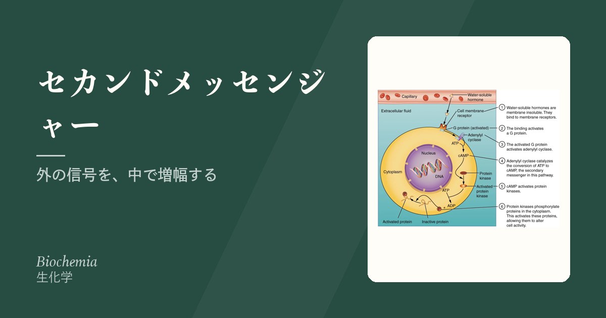
膜受容体に届いた信号は、そのままでは細胞の中へ入れない。膜の内側で別の小分子を作り、それが中を走る——これがセカンドメッセンジャーである。
意味は2つある。増幅(1個のホルモンが数万個の分子を動かす)と、分岐(1つの信号を複数の経路へ配る)である。3つの系統を押さえれば、薬理学の大半が読める。
- 環状AMPと環状GMP — 増やす薬と、壊させない薬
- イノシトールリン脂質とカルシウム — 2つに分かれる信号
- 細胞内カルシウムは、なぜ4桁も低く保たれているのか
- JAK-STAT — サイトカインの信号が、最短で核へ届く
- カルシウム信号を薬で止める — カルシニューリン阻害薬
- G タンパク質そのものが壊れると、病気になる
環状AMPと環状GMP — 増やす薬と、壊させない薬
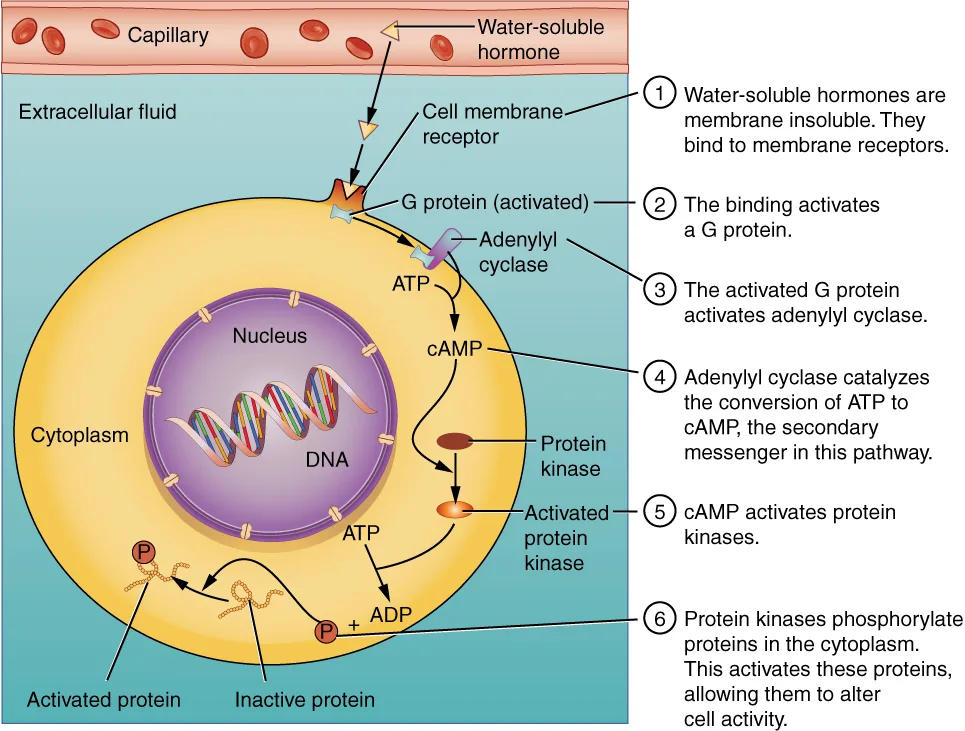
環状AMP系:受容体 → Gs/Gi → アデニル酸シクラーゼ → 環状AMP → プロテインキナーゼA
- Gsで増え、Giで減る
- 分解するのはホスホジエステラーゼ
環状GMP系:一酸化窒素 → 可溶性グアニル酸シクラーゼ → 環状GMP → プロテインキナーゼG → 平滑筋弛緩
| 薬・毒素 | 作用点 | 結果 |
|---|---|---|
| コレラ毒素 | Gsを活性化しっぱなしにする | 腸管からの分泌が止まらず水様下痢 |
| 百日咳毒素 | Giを働かなくする | 環状AMPが増える |
| テオフィリン | ホスホジエステラーゼを阻害 | 気管支拡張 |
| シルデナフィル | ホスホジエステラーゼ5を阻害 | 環状GMPが残る |
| ミルリノン | ホスホジエステラーゼ3を阻害 | 強心+血管拡張 |
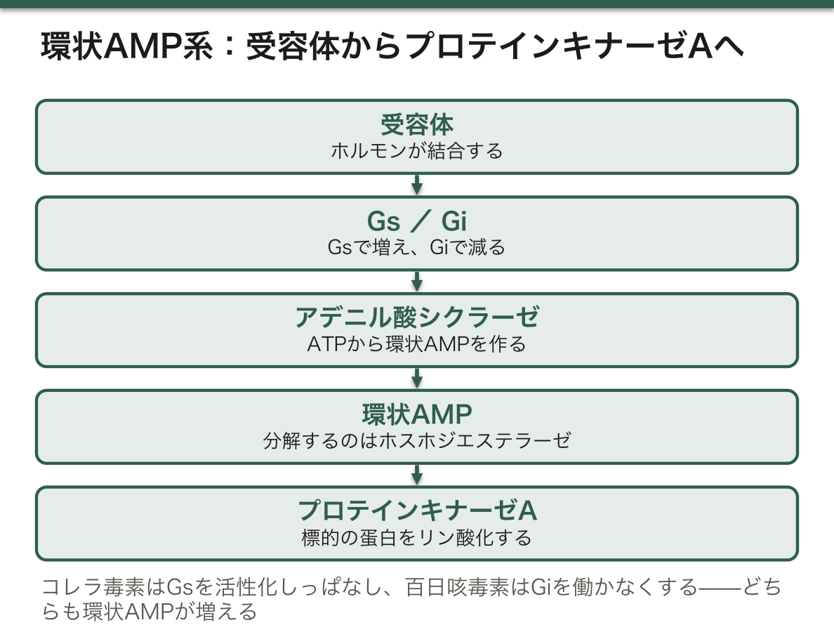
コレラ毒素と百日咳毒素は、どちらも環状AMPを増やすのに、機序が正反対である。コレラ毒素はアクセルを踏みっぱなしにし、百日咳毒素はブレーキを壊す。Gタンパク質の阻害と活性化の区別を体で覚えるのに、この対比はよくできている。
コレラの治療が「経口補水」である理由も生化学から出る。環状AMPが増えても、Na⁺/グルコース共輸送体は影響を受けない。だからブドウ糖と塩を一緒に飲ませれば、ナトリウムが吸収され、水がついてくる。点滴が無い環境で無数の命を救ったのは、この一点の理解である。
ホスホジエステラーゼには複数の型があり、選択性が薬の性格を決める。
- 3型——心筋と血管平滑筋。ミルリノン。強心薬だが長期使用で死亡率が上がるので急性期に限る
- 4型——炎症細胞。ロフルミラスト(慢性閉塞性肺疾患)、アプレミラスト(乾癬)
- 5型——陰茎海綿体と肺動脈。シルデナフィル。肺高血圧にも使う
硝酸薬とホスホジエステラーゼ5阻害薬の併用が禁忌である理由は、片方が環状GMPを増やし、もう片方が分解を止める——同じ経路の入口と出口を同時に押さえるので、血管拡張が制御不能になる。胸痛で救急を受診した患者に硝酸薬を使う前に、勃起不全治療薬の服用歴を確認するのは、この生化学が根拠である。
English version
Core
The cyclic AMP system: receptor → Gs/Gi → adenylate cyclase → cyclic AMP → protein kinase A
- Gs raises it, Gi lowers it.
- It is degraded by phosphodiesterase.
The cyclic GMP system: nitric oxide → soluble guanylate cyclase → cyclic GMP → protein kinase G → relaxation of smooth muscle
| Drug or toxin | Point of action | Result |
|---|---|---|
| Cholera toxin | Leaves Gs permanently activated | Secretion from the gut does not stop: watery diarrhoea |
| Pertussis toxin | Disables Gi | Cyclic AMP increases |
| Theophylline | Inhibits phosphodiesterase | Bronchodilatation |
| Sildenafil | Inhibits phosphodiesterase 5 | Cyclic GMP remains |
| Milrinone | Inhibits phosphodiesterase 3 | Inotropy plus vasodilatation |
Going further
Cholera and pertussis toxins both raise cyclic AMP and their mechanisms are opposite. Cholera toxin holds the accelerator down; pertussis toxin breaks the brake. This contrast is well made for learning in the body the distinction between inhibiting and activating a G protein.
The reason the treatment of cholera is oral rehydration also comes out of the biochemistry. Cyclic AMP may rise, and the Na⁺/glucose cotransporter is unaffected. So give glucose and salt together and the sodium is absorbed and water follows. It saved countless lives where there were no drips, on this single point of understanding.
There are several types of phosphodiesterase, and the selectivity decides the character of the drug.
- Type 3 — myocardium and vascular smooth muscle. Milrinone. An inotrope, but mortality rises with long use, so it is limited to the acute phase.
- Type 4 — inflammatory cells. Roflumilast (chronic obstructive pulmonary disease), apremilast (psoriasis).
- Type 5 — corpus cavernosum and pulmonary artery. Sildenafil. Also used in pulmonary hypertension.
A nitrate with a phosphodiesterase 5 inhibitor is contraindicated because one raises cyclic GMP and the other stops its degradation — holding down the entrance and the exit of the same pathway at once, the vasodilatation becomes uncontrollable. Asking a patient presenting with chest pain about drugs for erectile dysfunction before giving a nitrate rests on this biochemistry.
イノシトールリン脂質とカルシウム — 2つに分かれる信号
Gq → ホスホリパーゼC → 膜のホスファチジルイノシトール二リン酸を2つに割る
- イノシトール三リン酸——水溶性。小胞体へ行き、Ca²⁺を放出させる
- ジアシルグリセロール——膜に残る。プロテインキナーゼCを活性化(Ca²⁺も要る)
カルシウムは最も汎用されるセカンドメッセンジャーである。
- 細胞内濃度は細胞外の1万分の1に保たれている——だから少し入るだけで大きな信号になる
- 受け手はカルモジュリン(多くの酵素を制御)とトロポニンC(骨格筋・心筋の収縮)
- 汲み出すのは細胞膜Ca²⁺-ATPase・筋小胞体Ca²⁺-ATPase・Na⁺/Ca²⁺交換体
「細胞内カルシウムを極端に低く保つ」ことに、細胞は多大なコストを払っている。その理由は2つある。
- 信号として使うため——背景が低いほど、少量の流入が明確な信号になる
- リン酸と結合して沈殿するのを避けるため——細胞内にはリン酸が多く、カルシウムが高いとリン酸カルシウムとして析出する
細胞死でカルシウムが決定的な役割を果たすのもここから来る。虚血でATPが枯れるとポンプが止まり、カルシウムが流入する。するとカルシウム依存性のプロテアーゼ(カルパイン)とホスホリパーゼが活性化して、細胞を内側から分解する。再灌流障害の中心的な機序である。
平滑筋と骨格筋で収縮の機構が違う点も、この経路の理解に効く。
| Ca²⁺の受け手 | 制御 | |
|---|---|---|
| 骨格筋・心筋 | トロポニンC | トロポミオシンがずれてアクチンが露出する |
| 平滑筋 | カルモジュリン | ミオシン軽鎖キナーゼがミオシンをリン酸化する |
だから平滑筋は環状AMPで弛緩する——プロテインキナーゼAがミオシン軽鎖キナーゼをリン酸化して不活化するためである。β2刺激薬が気管支を広げるのはこの経路で、心筋では環状AMPが収縮を強めるのと逆向きになる。同じセカンドメッセンジャーが組織で逆の効果を持つ——受け手の分子が違うからである。
English version
Core
Gq → phospholipase C → splits the membrane phosphatidylinositol bisphosphate into two
- Inositol trisphosphate — water-soluble. It goes to the endoplasmic reticulum and releases Ca²⁺.
- Diacylglycerol — remains in the membrane. It activates protein kinase C (Ca²⁺ is needed too).
Calcium is the most widely used second messenger.
- The intracellular concentration is kept at one ten-thousandth of the extracellular — so a small influx makes a large signal.
- The receivers are calmodulin (regulating many enzymes) and troponin C (contraction of skeletal and cardiac muscle).
- What pumps it out: the plasma membrane Ca²⁺-ATPase, the sarcoplasmic reticulum Ca²⁺-ATPase, the Na⁺/Ca²⁺ exchanger.
Going further
The cell pays a great cost to keep intracellular calcium extremely low. There are two reasons.
- To use it as a signal — the lower the background, the more a small influx is a clear signal.
- To avoid precipitating with phosphate — there is much phosphate within the cell, and a high calcium precipitates as calcium phosphate.
That calcium plays a decisive part in cell death comes from here too. When ATP is exhausted in ischaemia the pumps stop and calcium floods in. Then calcium-dependent proteases (calpains) and phospholipases are activated and dismantle the cell from within. The central mechanism of reperfusion injury.
That contraction works differently in smooth and in striated muscle also tells in understanding this pathway.
| The receiver of Ca²⁺ | Control | |
|---|---|---|
| Skeletal and cardiac muscle | Troponin C | Tropomyosin shifts and actin is exposed |
| Smooth muscle | Calmodulin | Myosin light chain kinase phosphorylates myosin |
Hence smooth muscle relaxes with cyclic AMP — because protein kinase A phosphorylates and inactivates myosin light chain kinase. This is the pathway by which a β2 agonist dilates the bronchus, and it runs opposite to cyclic AMP strengthening contraction in the myocardium. The same second messenger having opposite effects in different tissues — because the receiving molecule differs.
細胞内カルシウムは、なぜ4桁も低く保たれているのか
細胞外は約 1〜2 mmol/L、細胞質は約 100 nmol/L——1万倍の差を、絶えずエネルギーを使って維持している。
下げる仕組み——細胞膜の Ca²⁺-ATPase・Na⁺/Ca²⁺ 交換体・小胞体の SERCA・ミトコンドリア。
上げる仕組み——電位依存性 Ca²⁺ チャネル・IP₃ 受容体・リアノジン受容体。
受け取るのはカルモジュリンなどの結合蛋白で、そこから CaM キナーゼ・カルシニューリン・ミオシン軽鎖キナーゼへ伝わる。
低く保つ理由は2つ——①わずかな流入で大きな比率の変化になる(感度) ②リン酸と結合して沈殿するのを防ぐ。
「静かな背景があるから、小さな信号が読める」という設計が、この項の要点になる。もし細胞内が最初から高ければ、10 倍にするのに膨大な量が要る(ナトリウムポンプの項)——「感度を買うために、コストを払い続けている」。
だから ATP が尽きると、カルシウムが細胞内に溢れる(細胞傷害の章)——「プロテアーゼ・ホスホリパーゼ・エンドヌクレアーゼが一斉に動き、不可逆の傷害になる」。
カルシニューリンは、免疫抑制薬の標的になっている(免疫抑制薬の章)——「シクロスポリンとタクロリムスが、T細胞の IL-2 産生を止める」。
リアノジン受容体の異常が、悪性高熱症になる(麻酔の合併症の章)——「ダントロレンが、この放出チャネルを止める薬になる」。
心筋では、少量の流入が大量の放出を引き起こす(カルシウム誘発性カルシウム放出)(筋の収縮の章)——「だから心筋は細胞外カルシウムに依存し、骨格筋は依存しない」。
English version
Core
Outside the cell about 1–2 mmol/L, in the cytosol about 100 nmol/L — a ten-thousand-fold difference maintained by the constant use of energy.
What lowers it — the plasma membrane Ca²⁺-ATPase, the Na⁺/Ca²⁺ exchanger, SERCA of the endoplasmic reticulum, mitochondria.
What raises it — voltage-gated Ca²⁺ channels, IP₃ receptors, ryanodine receptors.
It is received by binding proteins such as calmodulin and passed on to CaM kinase, calcineurin and myosin light chain kinase.
Two reasons for keeping it low — ① a small influx becomes a large proportional change (sensitivity) ② it prevents precipitation with phosphate.
Going further
- The crux of this section is the design "a small signal can be read because the background is quiet". Were the interior high to begin with, an enormous amount would be needed to raise it ten-fold (the section on the sodium pump) — paying constantly to buy sensitivity.
- Hence when ATP runs out, calcium floods the cell (the chapter on cell injury) — proteases, phospholipases and endonucleases all move at once, and the injury becomes irreversible.
- Calcineurin is the target of immunosuppressants (the chapter on immunosuppressants) — ciclosporin and tacrolimus stop the production of IL-2 by T cells.
- An abnormality of the ryanodine receptor gives malignant hyperthermia (the chapter on the complications of anaesthesia) — dantrolene is the drug that stops this release channel.
- In the myocardium a small influx triggers a large release (calcium-induced calcium release) (the chapter on muscle contraction) — hence cardiac muscle depends on extracellular calcium and skeletal muscle does not.
JAK-STAT — サイトカインの信号が、最短で核へ届く
サイトカイン受容体は、自分ではキナーゼ活性を持たない——代わりに JAK(ヤヌスキナーゼ)が結合している。
流れは短い——①リガンドが受容体を二量体化 → ②JAK が互いをリン酸化 → ③受容体のチロシンをリン酸化 → ④STAT が結合してリン酸化される → ⑤STAT が二量体になり核へ入って転写を動かす。
中間に第二伝令を挟まない——だから速く、増幅が小さい。
止める側——SOCS(自らが誘導する負のフィードバック)・ホスファターゼ。
使うもの——インターフェロン・多くのインターロイキン・エリスロポエチン・トロンボポエチン・成長ホルモン・プロラクチン。
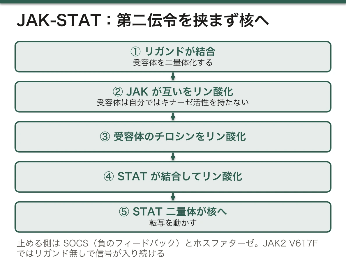
「経路が短い」ことが、そのまま薬の作りやすさになっている。JAK 阻害薬は、複数のサイトカインの信号をまとめて止められる(抗リウマチ薬の章)——「関節リウマチ・アトピー性皮膚炎・潰瘍性大腸炎・円形脱毛症に広がった」。
だから副作用も、止まる範囲の広さから予測できる(医療者を守る実務の章)——「帯状疱疹・結核の再燃・血球減少・脂質の上昇」。
JAK2 V617F 変異は、リガンドが無くても信号が入り続ける状態になる(骨髄増殖性腫瘍の章)——「真性多血症・本態性血小板血症・原発性骨髄線維症の診断に使われる」。
だからエリスロポエチンが低いのに赤血球が増える(エリスロポエチンの項)——「二次性多血症との鑑別に、エリスロポエチン値が役立つ理由になる」。
成長ホルモンもこの経路を使う(成長の異常の章)——「Laron 症候群では受容体側が壊れ、成長ホルモンは高いのに IGF-1 が低い」。
English version
Core
A cytokine receptor has no kinase activity of its own — instead a JAK (Janus kinase) is bound to it.
The route is short — ① the ligand dimerises the receptor → ② the JAKs phosphorylate one another → ③ they phosphorylate tyrosines of the receptor → ④ STAT binds and is phosphorylated → ⑤ STAT dimerises, enters the nucleus and drives transcription.
There is no second messenger in between — so it is fast and the amplification is small.
What stops it — SOCS (a negative feedback it induces itself), phosphatases.
What uses it — the interferons, many interleukins, erythropoietin, thrombopoietin, growth hormone, prolactin.
Going further
- That the pathway is short is directly what makes a drug easy to build. A JAK inhibitor can stop the signals of several cytokines together (the chapter on antirheumatic drugs) — it has spread to rheumatoid arthritis, atopic dermatitis, ulcerative colitis and alopecia areata.
- Hence the adverse effects can be predicted from the breadth of what is stopped (the chapter on the practicalities of protecting the practitioner) — herpes zoster, reactivation of tuberculosis, cytopenias, a rise in lipids.
- The JAK2 V617F mutation is a state in which the signal keeps coming without a ligand (the chapter on myeloproliferative neoplasms) — used in diagnosing polycythaemia vera, essential thrombocythaemia and primary myelofibrosis.
- Hence red cells increase while erythropoietin is low (the section on erythropoietin) — the reason the erythropoietin level helps in distinguishing secondary polycythaemia.
- Growth hormone uses this pathway too (the chapter on disorders of growth) — in Laron syndrome the receptor side is broken and growth hormone is high while IGF-1 is low.
カルシウム信号を薬で止める — カルシニューリン阻害薬
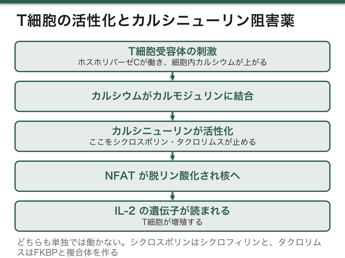
T 細胞が活性化する道筋は、カルシウム信号を通る。
- T 細胞受容体が刺激されると、ホスホリパーゼ C が働き 細胞内カルシウムが上がる
- カルシウムがカルモジュリンに結合する
- その複合体がカルシニューリン(脱リン酸化酵素)を活性化する
- カルシニューリンが NFAT を脱リン酸化し、核へ入れる
- NFAT が IL-2 の遺伝子を読ませ、T 細胞が増殖する
シクロスポリンとタクロリムスは、この 3 段目を止める。どちらも単独では働かず、細胞内の蛋白(イムノフィリン)と複合体を作ってカルシニューリンを阻害する——シクロスポリンはシクロフィリンと、タクロリムスは FKBP と結合する。
⚠️ 副作用の中心は腎毒性である。輸入細動脈を収縮させて腎血流を落とすため、移植した腎そのものを長期に痛めるという逆説が起きる。血中濃度の管理(トラフ値)が必須で、狭い治療域を外れると拒絶か中毒のどちらかになる。
| シクロスポリン | タクロリムス | |
| 特徴的な副作用 | 多毛・歯肉肥厚 | 耐糖能異常(移植後糖尿病)・振戦 |
| 共通 | 腎障害・高血圧・高カリウム血症・低マグネシウム血症・感染・二次発癌 | |
相互作用が非常に多い。どちらも CYP3A4 で代謝され、P 糖蛋白の基質でもある。
- 濃度が上がる——アゾール系抗真菌薬・マクロライド・カルシウム拮抗薬・グレープフルーツ果汁
- 濃度が下がる——リファンピシン・抗てんかん薬・セント・ジョーンズ・ワート(サプリメント)。拒絶反応を起こしうる
移植患者には「市販薬とサプリメントを自己判断で飲まない」と必ず伝える。
関連して、シロリムス(ラパマイシン)は FKBP に結合する点は同じだが、止める先が mTOR で、カルシニューリンではない。腎毒性が少ないかわりに、脂質異常・創傷治癒の遅れ・口内炎が問題になる。
免疫抑制薬を飲んでいる患者に新しい薬を出すときは、必ず相互作用を確認する。抗真菌薬1剤で中毒域に入る。
English version
Core
The route by which a T cell is activated passes through the calcium signal.
- The T-cell receptor is stimulated, phospholipase C works and intracellular calcium rises.
- Calcium binds calmodulin.
- That complex activates calcineurin (a phosphatase).
- Calcineurin dephosphorylates NFAT and lets it enter the nucleus.
- NFAT has the IL-2 gene read and the T cell proliferates.
Ciclosporin and tacrolimus stop the third step. Neither works alone: each forms a complex with an intracellular protein (an immunophilin) and inhibits calcineurin — ciclosporin with cyclophilin, tacrolimus with FKBP.
Going further
⚠️ The central adverse effect is nephrotoxicity. They constrict the afferent arteriole and reduce renal blood flow, giving the paradox that they damage the transplanted kidney itself over time. Monitoring of the blood level (the trough) is essential: outside the narrow therapeutic window lies either rejection or toxicity.
| Ciclosporin | Tacrolimus | |
|---|---|---|
| Characteristic adverse effects | Hirsutism, gingival hypertrophy | Impaired glucose tolerance (post-transplant diabetes), tremor |
| Shared | Renal injury, hypertension, hyperkalaemia, hypomagnesaemia, infection, secondary malignancy | |
The interactions are very many. Both are metabolised by CYP3A4 and are substrates of P-glycoprotein.
- The level rises — azole antifungals, macrolides, calcium antagonists, grapefruit juice.
- The level falls — rifampicin, antiepileptics, St John’s wort (a supplement). Rejection can follow.
Always tell a transplant patient not to take over-the-counter medicines or supplements on their own judgement.
Related: sirolimus (rapamycin) also binds FKBP, but what it stops is mTOR, not calcineurin. It has less nephrotoxicity, at the cost of dyslipidaemia, delayed wound healing and stomatitis.
In the clinic
When prescribing a new drug to a patient on immunosuppressants, always check the interactions. One antifungal takes them into the toxic range.
G タンパク質そのものが壊れると、病気になる
受容体の下流にある G タンパク質は、GTP を結合すると働き、自分で分解して止まる——内蔵のタイマーを持つスイッチである。このスイッチが入ったままになる/入らなくなることが、そのまま疾患になる。
| 働きが失われる | 偽性副甲状腺機能低下症(GNAS の変異) | PTH は高いのに効かない。低カルシウム・高リン血症。Albright 遺伝性骨異栄養症の身体的特徴を伴う |
| 働いたままになる | McCune-Albright 症候群(体細胞モザイクの GNAS 変異) | 多骨性線維性骨異形成・カフェオレ斑・思春期早発症。受精後に起きた変異なので、体の一部にだけ現れる |
| 腫瘍で働いたままになる | 下垂体腺腫の一部 | 成長ホルモンが出続ける(先端巨大症) |
同じ遺伝子の、機能喪失と機能獲得が、正反対の病気になる——GNAS はその代表例として繰り返し取り上げられる。
受容体の側の変異も、同じ形で整理できる。
- 常に活性化——LH 受容体の変異による家族性男性思春期早発症(LH が無くてもテストステロンが作られる)
- 働かない——V2 受容体の変異による腎性尿崩症(バソプレシンがあっても水を再吸収できない)
- 働かない——メラノコルチン 4 受容体(MC4R)の変異。単一遺伝子による肥満として最も頻度が高い
毒素は、この仕組みを外から乗っ取る。コレラ毒素は Gs の自己分解を止めて cAMP を作り続けさせ、百日咳毒素は Gi を働けなくして、やはり cAMP を上げる。止める仕組みを壊すことが、細菌にとっての攻撃になっている。
低分子量 G タンパク質(RAS)も同じ論理で癌になる。変異した RAS はGTP を分解できず、スイッチが入ったままになる。「働かせる変異」ではなく「止められなくする変異」だという点が要で、だから阻害薬の設計が長く困難だった。
⚠️ 体細胞モザイクという概念を、ここで押さえておく。McCune-Albright 症候群の GNAS 変異は全身にあれば致死的で、受精後に起きて体の一部にしか無いからこそ生まれてこられる。だから遺伝しないし、血液の検査では見つからない——病変部の組織で調べる必要がある。
思春期早発症+骨の変形+カフェオレ斑という組合せを見たら、1つの体細胞変異で説明できないかを考える。
English version
Core
The G protein downstream of a receptor works when it binds GTP and stops itself by hydrolysing it — a switch with a built-in timer. That this switch stays on, or will not go on, is directly a disease.
| Loss of function | Pseudohypoparathyroidism (a GNAS mutation) | PTH is high and does not work. Low calcium, high phosphate. With the physical features of Albright hereditary osteodystrophy |
| Stays on | McCune–Albright syndrome (a somatic mosaic GNAS mutation) | Polyostotic fibrous dysplasia, café-au-lait patches, precocious puberty. The mutation arose after fertilisation, so it appears only in part of the body |
| Stays on in a tumour | Some pituitary adenomas | Growth hormone keeps being released (acromegaly) |
Loss and gain of function in the same gene giving opposite diseases — GNAS is taken up again and again as the representative example.
Going further
Mutations on the receptor side can be arranged in the same form.
- Constitutively active — a mutation of the LH receptor gives familial male precocious puberty (testosterone is made without LH).
- Does not work — a mutation of the V2 receptor gives nephrogenic diabetes insipidus (water cannot be reabsorbed though vasopressin is present).
- Does not work — mutations of melanocortin 4 receptor (MC4R). The commonest monogenic obesity.
Toxins hijack this machinery from outside. Cholera toxin stops the self-hydrolysis of Gs and has cAMP made continuously, and pertussis toxin disables Gi, likewise raising cAMP. Breaking the mechanism that stops things is the bacterium’s attack.
The small G proteins (RAS) become cancers by the same logic. Mutant RAS cannot hydrolyse GTP and the switch stays on. The crux is that it is not a mutation that makes it work but a mutation that prevents it stopping, and this is why inhibitors were long difficult to design.
⚠️ Hold the concept of somatic mosaicism here. The GNAS mutation of McCune–Albright syndrome would be lethal if present throughout the body, and it is only because it arose after fertilisation and is present in part of the body that the child can be born. So it is not inherited and is not found in a blood test — the tissue of the lesion must be examined.
In the clinic
Seeing the combination of precocious puberty, bone deformity and café-au-lait patches, consider whether one somatic mutation explains it all.
まとめ
| 環状AMPと環状GMP — 増やす薬と、壊させない薬 | 環状AMP系:受容体 → Gs/Gi → アデニル酸シクラーゼ → 環状AMP → プロテインキナーゼAGsで増え、Giで減る分解するのはホスホジエステラーゼ環状GMP系:一酸化窒… |
|---|---|
| イノシトールリン脂質とカルシウム — 2つに分かれる信号 | Gq → ホスホリパーゼC → 膜のホスファチジルイノシトール二リン酸を2つに割るイノシトール三リン酸——水溶性。 |
| 細胞内カルシウムは、なぜ4桁も低く保たれているのか | 細胞外は約 1〜2 mmol/L、細胞質は約 100 nmol/L——1万倍の差を、絶えずエネルギーを使って維持している。 |
| JAK-STAT — サイトカインの信号が、最短で核へ届く | サイトカイン受容体は、自分ではキナーゼ活性を持たない——代わりに JAK(ヤヌスキナーゼ)が結合している。 |
| カルシウム信号を薬で止める — カルシニューリン阻害薬 | T 細胞が活性化する道筋は、カルシウム信号を通る。 |
| G タンパク質そのものが壊れると、病気になる | 受容体の下流にある G タンパク質は、GTP を結合すると働き、自分で分解して止まる——内蔵のタイマーを持つスイッチである。 |
このノートは医学生が授業の内容を自分の言葉でまとめたもので、診療の判断に使うものではありません。誤りに気づいたらお知らせください。