荷電した分子を電場の中で動かし、動く速さの差で分ける技術。原理・装置・測定量・医学での用途の4点を、それぞれ2〜3文で言えるようにしておく。
この実習の物理は電場中の荷電粒子にはたらく力と、媒質の粘性抵抗の釣り合いだけである。式は1本しかない。
- 電気泳動の原理 — 力の釣り合いで速さが決まる
- 電場と電圧 — 1点の「押す強さ」と2点間の「仕事」
- 紙の上でイオンを泳動する — Fe³⁺ と Cu²⁺ の移動度を比べる
- 医学での電気泳動 — 何を読んでいる検査か
- 1次元と2次元の電気泳動 — 性質を2つ使って重なりをほどく
- イオントフォレシス — 電流で薬を皮膚から送り込む
- 蛋白質の泳動の手順と、結果を歪めるもの
電気泳動の原理 — 力の釣り合いで速さが決まる
電場 E の中で電荷 q を持つ粒子は F = qE の力を受けて動く。同時に媒質から粘性抵抗 F = 6πηrv(Stokes の法則、η=粘度、r=半径、v=速度)を受ける。
2つの力が釣り合ったところで一定の速度になる。
qE = 6πηrv → v = qE / 6πηr
電場あたりの速度を電気泳動移動度 μ = v/E = q/6πηr という。移動度は電荷に比例し、大きさと粘度に反比例する——測定条件が同じなら物質固有の値になる。
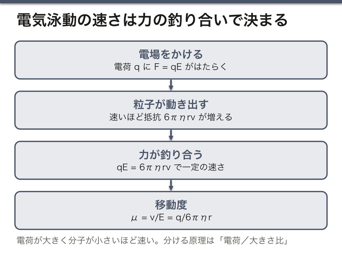
分ける原理は「電荷/大きさ比」である。電荷が大きく分子が小さいほど速い。だから同じ分子量でも荷電状態が違えば分かれるし、緩衝液の pH を変えれば分離のしかたが変わる——蛋白質は等電点より酸性側で正、塩基性側で負に帯電するからである。
SDS-PAGE はこの依存性をわざと壊す。SDS が蛋白質に一定の比率で結合して電荷を大きさに比例させるので、移動度が分子量だけで決まるようになる。「電荷で分ける」道具を「大きさで分ける」道具に作り替えたのが SDS-PAGE である。
ジュール熱が実験の限界を決める。電流が流れれば媒質が温まり、粘度 η が下がって移動度が上がる。温度勾配ができると帯(バンド)が歪む。だから電圧を上げれば速く終わるが、分離能は落ちる——冷却するか、定電力で流すかの選択になる。
English version
Core
A particle carrying charge q in an electric field E feels a force F = qE and moves. At the same time it feels a viscous drag from the medium, F = 6πηrv (Stokes’ law; η = viscosity, r = radius, v = velocity).
Where the two forces balance, the velocity becomes constant.
qE = 6πηrv → v = qE / 6πηr
The velocity per unit field is the electrophoretic mobility, μ = v/E = q/6πηr. The mobility is proportional to the charge and inversely proportional to the size and the viscosity — under the same conditions it is a value characteristic of the substance.
Going further
The principle of separation is the charge-to-size ratio. The greater the charge and the smaller the molecule, the faster. So molecules of the same molecular weight separate if their charge states differ, and changing the pH of the buffer changes the separation — because a protein is positively charged on the acid side of its isoelectric point and negatively on the basic side.
SDS-PAGE deliberately destroys this dependence. SDS binds protein in a fixed ratio and makes the charge proportional to the size, so the mobility comes to be set by molecular weight alone. SDS-PAGE is a tool for separating by charge remade into a tool for separating by size.
Joule heating sets the limit of the experiment. When current flows the medium warms, the viscosity η falls and the mobility rises. A temperature gradient distorts the bands. So raising the voltage finishes it sooner and costs resolution — the choice is between cooling and running at constant power.
電場と電圧 — 1点の「押す強さ」と2点間の「仕事」
電場 E(electric field)は、ある1点で単位電荷あたりにはたらく力である:E = F/q(単位 N/C)。向きを持つベクトルで、空間の点ごとに値がある。試験電荷を2倍にすれば受ける力も2倍になるが、E は変わらない——E は電荷を置く前からそこにある「場の性質」だからである。
電圧 V(電位差 potential difference)は、2点の間で電荷を運ぶのに要する、単位電荷あたりの仕事である:V = W/q(単位 J/C = V)。向きを持たないスカラーで、必ず「どこからどこまで」という2点について言う。
2つは仕事の定義 W = F·s でつながる。一様な電場なら、距離 d だけ運ぶ仕事は W = qE·d なので V = E·d、つまり E = V/d である。だから電場の単位は V/m とも書ける(N/C と同じ次元)。
電気泳動の装置で設定するのは電圧だが、イオンを押しているのは電場である。同じ 150 V でも電極間の距離が2倍なら電場は半分になり、イオンの速さも半分になる。移動度を電圧ではなく電場で定義するのはこのためである。
移動度 μ = v/E に、v = s/t(移動距離 ÷ 時間)と E = V/d を代入すると μ = s·d / (t·V) になる。測るのは移動距離 s・電極間距離 d・時間 t・電圧 V の4つだけである。単位は m·m/(s·V) = m²/(V·s)——移動度の「変わった単位」はこの代入から出てくる。
たとえるなら、電場は坂の傾き、電圧は坂の高低差である。同じ高低差でも、坂が短いほど傾きは急になる。
医療機器でも同じ区別が要る。神経や心筋を興奮させるのは電源の電圧そのものではなく、組織の中にできる電場と、それが流す電流である。除細動器のパッドが大きいのは、同じ電流を広い面積に分けて皮膚での電流の密度を下げ、熱傷を防ぐためでもある。
English version
Core
The electric field E is the force on a unit charge at one point: E = F/q (unit N/C). It is a vector with a direction, and it has a value at every point in space. If you double the test charge, the force doubles, but E does not change, because E is a property of the place, which exists before any charge is put there.
Voltage V (potential difference) is the work per unit charge needed to move a charge between two points: V = W/q (unit J/C = V). It is a scalar with no direction, and it always refers to two points: from where to where.
The two are linked by the definition of work, W = F·s. In a uniform field, the work to move a charge over a distance d is W = qE·d, so V = E·d, that is E = V/d. This is why the field can also be given in V/m (the same dimension as N/C).
Going further
On an electrophoresis apparatus you set the voltage, but what pushes the ions is the field. With the same 150 V, if the electrodes are twice as far apart, the field is half as strong and the ions move half as fast. This is why mobility is defined by the field, not by the voltage.
Put v = s/t (distance ÷ time) and E = V/d into μ = v/E and you get μ = s·d / (t·V). You only measure four things: the distance travelled s, the electrode distance d, the time t and the voltage V. The unit is m·m/(s·V) = m²/(V·s); the strange unit of mobility comes from this substitution.
As a picture: the field is the slope of a hill and the voltage is its height difference. For the same height, a shorter hill is steeper.
In the clinic
The same distinction matters for medical devices. What excites nerve and heart muscle is not the voltage of the source itself but the field inside the tissue and the current it drives. Defibrillator pads are large partly to spread the same current over a wide area, so that the current density at the skin is lower and burns are avoided.
紙の上でイオンを泳動する — Fe³⁺ と Cu²⁺ の移動度を比べる
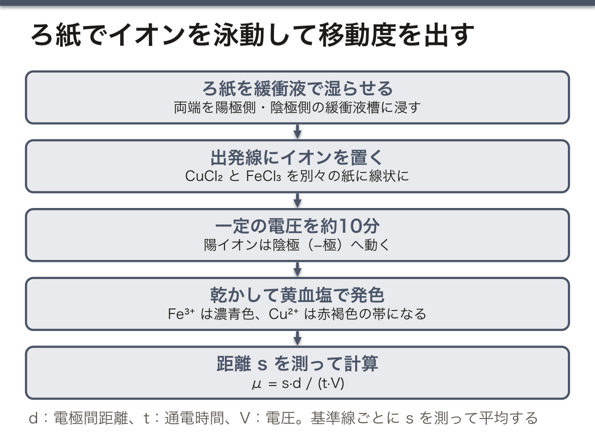
イオンの電気泳動は、緩衝液で湿らせたろ紙の上で行える。ろ紙の両端を陽極側と陰極側の緩衝液槽に浸し、中央の出発線に塩化銅(II) CuCl₂ と塩化鉄(III) FeCl₃ の溶液をそれぞれ別の紙に線状に置いて、百数十 V の一定電圧を10分ほどかける。
Cu²⁺ と Fe³⁺ は陽イオンなので陰極(−極)へ向かって動く(Cl⁻ は逆に陽極へ動くが、色が無いので見えない)。薄いイオンは紙の上でほとんど見えないので、泳動後に紙を乾かし、黄血塩(ヘキサシアニド鉄(II)酸カリウム K₄[Fe(CN)₆])の溶液で発色させる。Fe³⁺ は濃青色の沈殿(紺青、Prussian blue)、Cu²⁺ は赤褐色の沈殿をつくり、イオンが届いた所が色の帯として現れる。
出発線から帯の先端までの距離 s を、紙に引いた10本ほどの基準線ごとに測って平均し、μ = s·d/(t·V) で移動度を出す(d は電極間距離、t は通電時間)。
どちらが速いか——予想には2段階ある。電荷だけを見れば、3価の Fe³⁺ は2価の Cu²⁺ の1.5倍の力を受けるので速いはずである。しかし移動度は電荷と半径の比(μ = q/6πηr)で決まり、ここで効く半径は結晶中のイオン半径ではなく、まわりに水分子をまとった水和イオンの半径である。電荷密度の高い Fe³⁺ は水分子を強く引きつけて大きな水和殻を持つので、差は1.5倍まで開かない。無限希釈の水溶液での文献値はどちらも 10⁻⁸ m²/(V·s) の桁で、Fe³⁺ は Cu²⁺ の約1.3倍である。
実測値が文献値からずれる理由も物理で説明できる。①電気浸透——ろ紙のセルロースは表面が負に帯電しているので、紙の中の液そのものが陰極側へ流れ、陽イオンを余分に運ぶ。②電場が一様でない——電圧は緩衝液槽と濡れた紙とに抵抗の比で分かれるので、紙の上の電場は V/d と一致しない。③拡散と毛細管の流れ——帯の縁がにじみ、発熱で乾いた所へ液が吸い寄せられる。④Fe³⁺ は水中で加水分解して Fe(OH)²⁺ などに変わるので、実際に動いている粒子の電荷が +3 とは限らない。
Fe³⁺ と黄血塩の反応は、病理のベルリンブルー染色(鉄染色)と同じ化学である。組織や骨髄塗抹の中のヘモシデリン(Fe³⁺)が青く染まり、鉄欠乏と鉄過剰(ヘモクロマトーシス)の判断や、骨髄の環状鉄芽球(鉄芽球性貧血・骨髄異形成症候群)の確認に使う。
English version
Core
Ions can be separated by electrophoresis on filter paper wetted with buffer. The two ends of the paper dip into buffer tanks on the anode and cathode side. Solutions of copper(II) chloride CuCl₂ and iron(III) chloride FeCl₃ are put as a line on the start line of two separate strips, and a constant voltage of about 150 V is applied for about 10 minutes.
Cu²⁺ and Fe³⁺ are cations, so they move towards the cathode (− pole). (Cl⁻ moves the other way, to the anode, but it has no colour, so you cannot see it.) The dilute ions are almost invisible on paper, so after the run the paper is dried and developed with yellow prussiate (potassium hexacyanidoferrate(II), K₄[Fe(CN)₆]). Fe³⁺ forms a deep blue precipitate (Prussian blue) and Cu²⁺ a red-brown precipitate, so the place the ions reached appears as a coloured band.
The distance s from the start line to the front of the band is measured along about ten reference lines drawn on the paper and averaged. The mobility is then μ = s·d/(t·V), where d is the distance between the electrodes and t is the running time.
Going further
Which ion is faster? The prediction has two steps. Looking only at charge, Fe³⁺ (3+) feels 1.5 times the force on Cu²⁺ (2+), so it should be faster. But mobility depends on the ratio of charge to radius (μ = q/6πηr), and the radius that matters is not the ionic radius in a crystal but the radius of the hydrated ion, with its shell of water molecules. Fe³⁺ has a high charge density, holds water strongly and has a large hydration shell, so the difference does not reach 1.5 times. Literature values at infinite dilution in water are both of the order of 10⁻⁸ m²/(V·s), and Fe³⁺ is about 1.3 times Cu²⁺.
Why measured values differ from literature values can also be explained by physics. ① Electro-osmosis: the cellulose surface of the paper is negatively charged, so the liquid in the paper itself flows towards the cathode and carries the cations further. ② The field is not uniform: the voltage is shared between the buffer tanks and the wet paper according to their resistance, so the field on the paper is not equal to V/d. ③ Diffusion and capillary flow: the edges of the band spread, and liquid is pulled towards places that dry out from heating. ④ Fe³⁺ is hydrolysed in water to Fe(OH)²⁺ and similar ions, so the moving particle does not always carry +3.
In the clinic
The reaction of Fe³⁺ with yellow prussiate is the same chemistry as the Prussian blue (Perls) iron stain in pathology. Haemosiderin (Fe³⁺) in tissue or bone-marrow smears stains blue. It is used to judge iron deficiency and iron overload (haemochromatosis) and to find ring sideroblasts in the marrow (sideroblastic anaemia, myelodysplastic syndrome).
医学での電気泳動 — 何を読んでいる検査か
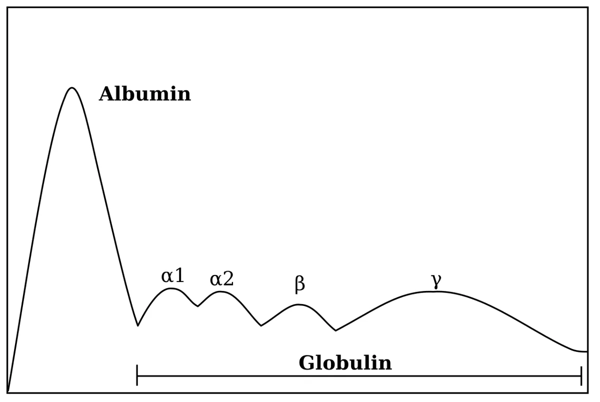
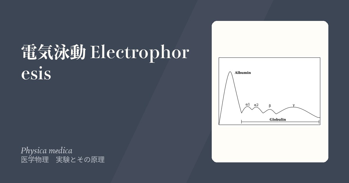
血清蛋白分画が代表。血清をセルロースアセテート膜またはアガロース上で泳動すると、陽極側からアルブミン → α1 → α2 → β → γ の5分画に分かれる。
| パターン | 所見 | 疾患 |
|---|---|---|
| M 蛋白 | γ 分画に鋭い単一のピーク | 多発性骨髄腫・MGUS |
| びまん性 γ 上昇 | 幅の広い γ の増加 | 慢性感染症・自己免疫疾患 |
| β-γ ブリッジング | β と γ の谷が埋まる | 肝硬変(IgA 増加) |
| アルブミン低下+α2 上昇 | — | ネフローゼ症候群 |
Hb 電気泳動は移動度の差が1アミノ酸の電荷の差から来る典型例である。HbS は β 鎖6番のグルタミン酸(負電荷)がバリン(電荷なし)に置換されているので、負電荷が1つ減り、陽極への移動が遅くなる。HbA → HbS → HbC の順に遅くなるのは、電荷が段階的に減るからである。「1個のアミノ酸の違いが、そのまま移動度の差として見える」——分子生物学と物理が直結する。
キャピラリー電気泳動と等電点電気泳動も原理の応用。等電点電気泳動は pH 勾配の中で泳動し、それぞれの蛋白が自分の等電点に達したところで電荷が 0 になって止まる——移動度の式で q = 0 になる点である。
免疫固定法(IFE)は泳動後に抗体を重ねて特定の免疫グロブリンだけを沈降させ、M 蛋白の種類(IgG κ など)を決める。電気泳動が「あるかどうか」、免疫固定が「何であるか」を答える。
English version
Core
Serum protein electrophoresis is the representative. Run serum on a cellulose acetate membrane or on agarose and it separates, from the anode, into five fractions: albumin → α1 → α2 → β → γ.
| Pattern | Finding | Disease |
|---|---|---|
| M protein | A sharp single peak in the γ fraction | Multiple myeloma, MGUS |
| Diffuse γ rise | A broad increase of γ | Chronic infection, autoimmune disease |
| β-γ bridging | The valley between β and γ fills in | Cirrhosis (increased IgA) |
| Low albumin with a raised α2 | — | Nephrotic syndrome |
Going further
Haemoglobin electrophoresis is the type case of a difference in mobility arising from the charge of a single amino acid. In HbS the glutamate (negative) at position 6 of the β chain is replaced by valine (uncharged), so one negative charge is lost and migration towards the anode is slower. HbA → HbS → HbC become slower in that order because the charge falls step by step. "A difference of one amino acid seen directly as a difference in mobility" — molecular biology and physics joined.
Capillary electrophoresis and isoelectric focusing are applications of the same principle. In isoelectric focusing the run is through a pH gradient, and each protein stops where it reaches its isoelectric point and its charge becomes zero — the point at which q = 0 in the mobility equation.
Immunofixation (IFE) overlays antibodies after the run and precipitates a particular immunoglobulin, identifying the class of the M protein (IgG κ and so on). Electrophoresis answers "is it there", immunofixation "what is it".
1次元と2次元の電気泳動 — 性質を2つ使って重なりをほどく
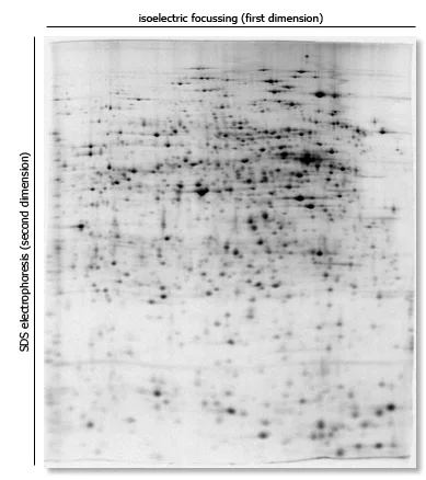
1次元電気泳動は、試料を1方向にだけ動かして分ける。分ける手がかりは実質1つ(電荷と大きさの比、SDS-PAGE なら分子量)なので、その値がたまたま近い蛋白質は同じバンドに重なる。血清蛋白分画の5本の帯も、1本1本は何十種類もの蛋白質の重なりである。
2次元電気泳動(two-dimensional electrophoresis)は、互いに独立な2つの性質で、直交する2方向に分ける。標準的な組み合わせは次の通り。
1次元目——等電点電気泳動(isoelectric focusing, IEF)。pH 勾配をつけたゲルの中で、各蛋白質は正味の電荷が 0 になる pH(等電点 pI)まで動いて止まる。
2次元目——そのゲルを横に寝かせ、直角の方向に SDS-PAGE で分子量の順に分ける。
結果は帯の列ではなく点(スポット)の地図になり、1枚のゲルで千を超える蛋白質を分けられる。
なぜ2つ目の性質は「独立」でなければならないのか。等電点と分子量はほとんど相関しないので、1次元目で重なった蛋白質も、2次元目では別々の位置に散らばる。分けられる数はおおまかに、1次元目で分けられる数と2次元目で分けられる数の積になる。同じ性質で2回分けても(たとえば SDS-PAGE を2回)点は対角線の上に並ぶだけで、何も増えない。
等電点電気泳動で蛋白質が「止まる」ことも移動度の式から出る。μ = q/6πηr の q が 0 になれば、電場があっても速度は 0 である。少しでも外へ拡散すると電荷を持って押し戻されるので、点は鋭く集束する(focusing という名前の由来)。
2次元電気泳動は、スポットを切り出して質量分析で同定するプロテオミクス(ある組織の蛋白質を網羅的に調べる研究)の基本の道具で、がんなどのバイオマーカー探しに使われてきた。日常の臨床検査で使うのは主に1次元の泳動——血清蛋白分画、M 蛋白の型を決める免疫固定法、確認検査のウエスタンブロット——である。
English version
Core
One-dimensional electrophoresis moves the sample in one direction only. It separates by essentially one property (the charge-to-size ratio, or molecular mass in SDS-PAGE), so proteins whose values happen to be close fall into the same band. Each of the five bands of serum protein electrophoresis is in fact a mixture of dozens of proteins.
Two-dimensional electrophoresis separates by two independent properties in two directions at right angles. The standard combination is:
First dimension: isoelectric focusing (IEF). In a gel with a pH gradient, each protein moves until it reaches the pH at which its net charge is zero (its isoelectric point, pI), and stops there.
Second dimension: the gel is laid sideways and the proteins are separated at right angles by SDS-PAGE, in order of molecular mass.
The result is not a row of bands but a map of spots, and more than a thousand proteins can be separated on one gel.
Going further
Why must the second property be independent? The isoelectric point and the mass of a protein are almost unrelated, so proteins that overlapped in the first dimension spread to different places in the second. Roughly, the number you can separate is the product of the numbers separated in each dimension. If you separate twice by the same property (for example SDS-PAGE twice), the spots only line up on a diagonal and nothing is gained.
The way a protein “stops” in isoelectric focusing also comes from the mobility equation. If q in μ = q/6πηr becomes 0, the speed is 0 even in a field. If the protein diffuses a little away, it gains a charge and is pushed back, so the spot is sharply focused; this is where the name comes from.
In the clinic
2D electrophoresis is the basic tool of proteomics (the study of all the proteins in a tissue): spots are cut out and identified by mass spectrometry, and the method has been used to search for biomarkers of cancer and other diseases. Routine clinical tests mainly use one-dimensional methods: serum protein electrophoresis, immunofixation to type an M-protein, and Western blotting as a confirmatory test.
イオントフォレシス — 電流で薬を皮膚から送り込む
イオントフォレシス(iontophoresis、イオン導入)は、弱い直流電流でイオンになった薬を皮膚から体内へ送り込む方法である。針を使わない、非侵襲的な経皮投与になる。
原理は電気泳動そのもので、同じ符号の電荷は反発することを使う。陽イオンの薬(例: リドカイン)は陽極の下に、陰イオンの薬(例: リン酸デキサメタゾン)は陰極の下に置く。電流を流すと薬のイオンは電極から押し出され、主に汗管や毛包を通って組織へ入る。もう1つの電極は離れた場所に貼り、回路を閉じる役をする。
送り込まれる量は、ほぼ電流 × 時間(電気量)に比例する。だから投与量は mA·min(ミリアンペア・分)で指定する。
薬を運ぶ仕組みは2つある。①電気的な反発(electromigration)——上に書いた、イオンが電場で押される効果。②電気浸透(electroosmosis)——皮膚は生理的な pH で負に帯電しているので、電流を流すと液そのものが陽極から陰極へ流れる。この流れは電荷を持たない分子も運ぶので、陽極側から入れる薬には追い風になる。
限界も物理から出てくる。運べるのは小さく、イオンになる薬で、量も少ない。電極では水の電気分解が起き、陽極に酸、陰極にアルカリができるので、電流の密度が高すぎたり時間が長すぎたりすると化学熱傷を起こす。緩衝剤を含んだ電極パッドを使うのはこのためである。
代表的な使い道は、足底腱膜炎(fasciitis plantaris。朝の歩き始めに踵が刺すように痛む)や滑液包炎(bursitis。肩・肘・股関節の近くの滑液包の炎症)への抗炎症薬(デキサメタゾン)の局所投与と、多汗症(hyperhidrosis)である。多汗症では薬を使わず、水道水に手足を浸して通電すると手掌・足底の発汗が減り、日本でも手掌多汗症の治療の選択肢として皮膚科で行われている。診断では、嚢胞性線維症の汗試験で、ピロカルピンをイオントフォレシスで皮膚に入れて汗を出させ、汗の Cl⁻ 濃度を測る。
English version
Core
Iontophoresis uses a weak direct current to drive an ionised drug through the skin into the body. It is a non-invasive way of giving a drug through the skin, with no needle.
The principle is electrophoresis itself: like charges repel. A cationic drug (for example lidocaine) is put under the anode, and an anionic drug (for example dexamethasone sodium phosphate) under the cathode. When current flows, the drug ions are pushed away from the electrode and enter the tissue, mainly through sweat ducts and hair follicles. The other electrode is placed somewhere else and only closes the circuit.
The amount delivered is roughly proportional to current × time (the charge), so the dose is given in mA·min (milliampere-minutes).
Going further
There are two ways the drug is carried. ① Electromigration: the ions are pushed by the field, as above. ② Electro-osmosis: skin is negatively charged at physiological pH, so when current flows, the liquid itself moves from the anode to the cathode. This flow carries even uncharged molecules, so it helps drugs given from the anode.
The limits also come from physics. Only small, ionisable drugs can be carried, and only in small amounts. Water is electrolysed at the electrodes, making acid at the anode and alkali at the cathode, so too high a current density or too long a time causes chemical burns. This is why buffered electrode pads are used.
In the clinic
Typical uses are local anti-inflammatory treatment (dexamethasone) of plantar fasciitis (fasciitis plantaris: stabbing heel pain with the first steps in the morning) and bursitis (inflammation of a bursa near the shoulder, elbow or hip), and the treatment of hyperhidrosis. For hyperhidrosis no drug is used: the hands or feet are put in tap water and current is passed, which reduces sweating of the palms and soles. In Japan, too, dermatologists use it as one of the options for palmar hyperhidrosis. In diagnosis, the sweat test for cystic fibrosis uses iontophoresis of pilocarpine into the skin to produce sweat, and the Cl⁻ concentration of the sweat is measured.
蛋白質の泳動の手順と、結果を歪めるもの
手順——①支持体(酢酸セルロース膜または寒天ゲル)を緩衝液に浸す ②試料を線状に置く ③一定電圧(または定電力)で泳動 ④固定・染色(蛋白なら Ponceau S やアミドブラック)⑤脱色 ⑥デンシトメーターで吸光度を走査し、各分画の面積比(%)を出す。
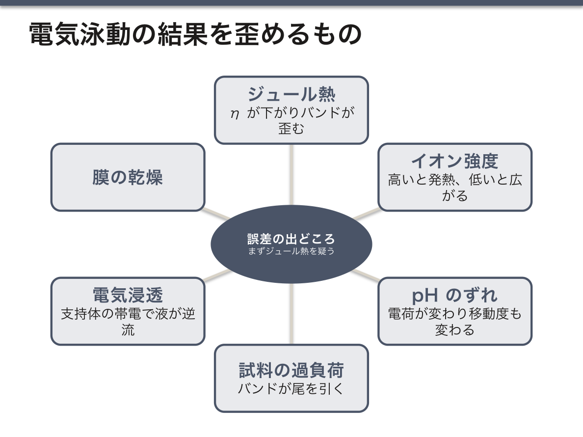
誤差の出どころ——①ジュール熱(バンドが歪む・端が速くなる)②緩衝液のイオン強度(高いと電流が増えて発熱、低いとバンドが広がる)③pH のずれ(電荷が変わり移動度が変わる)④試料の量が多すぎる(過負荷でバンドが尾を引く)⑤電気浸透(支持体自体が帯電し、緩衝液が逆流する)⑥膜の乾燥。
誤差を1つ挙げるなら、まずジュール熱——原理(v = qE/6πηr)から粘度 η の低下として説明できるため。
English version
Core
Procedure — ① the support (cellulose acetate membrane or agar gel) is soaked in buffer ② the sample is applied as a line ③ the run is at constant voltage (or constant power) ④ fixing and staining (Ponceau S or amido black for protein) ⑤ destaining ⑥ scanning the absorbance with a densitometer and obtaining the percentage area of each fraction.
Sources of error — ① Joule heating (bands distort; the edges run faster) ② the ionic strength of the buffer (too high and the current and heating rise; too low and the bands broaden) ③ a shift of pH (the charge changes and so does the mobility) ④ too much sample (overloading makes the bands tail) ⑤ electro-osmosis (the support itself is charged and the buffer flows back) ⑥ drying of the membrane.
To name one source of error, take Joule heating first — because it can be explained from the principle (v = qE/6πηr) as a fall in the viscosity η.
まとめ
| 電気泳動の原理 — 力の釣り合いで速さが決まる | 電場 E の中で電荷 q を持つ粒子は F = qE の力を受けて動く。 |
|---|---|
| 電場と電圧 — 1点の「押す強さ」と2点間の「仕事」 | 電場 E(electric field)は、ある1点で単位電荷あたりにはたらく力である:E = F/q(単位 N/C)。 |
| 紙の上でイオンを泳動する — Fe³⁺ と Cu²⁺ の移動度を比べる | イオンの電気泳動は、緩衝液で湿らせたろ紙の上で行える。 |
| 医学での電気泳動 — 何を読んでいる検査か | 血清蛋白分画が代表。血清をセルロースアセテート膜またはアガロース上で泳動すると、陽極側からアルブミン → α1 → α2 → β → γ の5分画に分かれる。 |
| 1次元と2次元の電気泳動 — 性質を2つ使って重なりをほどく | 1次元電気泳動は、試料を1方向にだけ動かして分ける。 |
| イオントフォレシス — 電流で薬を皮膚から送り込む | イオントフォレシス(iontophoresis、イオン導入)は、弱い直流電流でイオンになった薬を皮膚から体内へ送り込む方法である。 |
| 蛋白質の泳動の手順と、結果を歪めるもの | 手順——①支持体(酢酸セルロース膜または寒天ゲル)を緩衝液に浸す ②試料を線状に置く ③一定電圧(または定電力)で泳動 ④固定・染色(蛋白なら Ponceau S やアミドブラック… |
このノートは医学生が授業の内容を自分の言葉でまとめたもので、診療の判断に使うものではありません。誤りに気づいたらお知らせください。