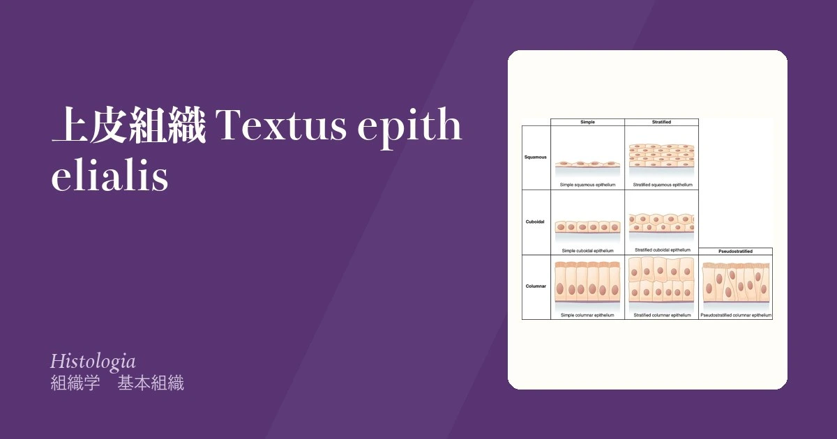
上皮の分類は2つの軸の掛け算でしかない。層の数(単層・重層・多列)×細胞の形(扁平・立方・円柱)。名前が長く見えるのは形容詞を並べているだけで、覚える対象は6種類ほどしかない。
大事なのは名前ではなくなぜそこにその上皮なのかである。物質を通す場所は薄い単層扁平、こすられる場所は厚い重層扁平、伸び縮みする場所は尿路上皮。形は要求される機能から決まっているので、部位と結びつけて覚えれば思い出す必要がなくなる。
そしてこの章は病理の入口でもある。上皮が本来と違う種類に置き換わる(化生)、異型を帯びる(異形成)、基底膜を破る(浸潤癌)——癌のほとんどは上皮から出るので、ここの理解がそのまま国試の腫瘍学に効く。
- 上皮
- 上皮の4つの型 — 被蓋上皮・腺上皮・筋上皮・感覚上皮
- 単層扁平上皮
- 単層立方上皮
- 単層円柱上皮
- 多列線毛円柱上皮(偽重層)
- 重層扁平上皮
- 尿路上皮(移行上皮)
- 密着結合(タイト結合)
- デスモソーム(接着斑)
- 微絨毛
- 杯細胞
- 外分泌腺
- 化生・異形成・上皮内癌 — 上皮が壊れていく順序
- 上皮Epithelium
- 上皮の4つの型 — 被蓋上皮・腺上皮・筋上皮・感覚上皮
- 単層扁平上皮Epithelium simplex squamosum
- 単層立方上皮Epithelium simplex cuboideum
- 単層円柱上皮Epithelium simplex columnare
- 多列線毛円柱上皮(偽重層)Epithelium pseudostratificatum columnare ciliatum
- 重層扁平上皮Epithelium stratificatum squamosum
- 尿路上皮(移行上皮)Epithelium transitionale
- 密着結合(タイト結合)Zonula occludens
- デスモソーム(接着斑)Macula adhaerens
- 微絨毛Microvilli
- 杯細胞Cellula caliciformis
- 外分泌腺Glandula exocrina
- 化生・異形成・上皮内癌 — 上皮が壊れていく順序
- まとめ
上皮Epithelium
体の表面と、管腔・体腔の内面を隙間なく覆う組織。特徴は5つ。
- 細胞が密に並び、細胞外基質がほとんど無い
- 基底膜の上に乗る
- 極性がある——管腔側(頂端面)と基底側で、膜も蛋白も働きも違う
- 血管が無い。栄養は下の結合組織から拡散で受ける
- 再生能が高い
「血管が無い」が効いてくる場面は多い。上皮内癌が転移しないのはこのためで、基底膜を破って初めて血管・リンパ管に手が届く。角膜が透明なのも血管が無いからで、血管が侵入すれば透明性を失う。上皮が厚くなりすぎると拡散が届かず表層から死ぬ——これが皮膚の角質層の正体である。
極性は上皮の定義そのものと言ってよい。小腸上皮では頂端面に微絨毛とSGLT1、基底側面にNa⁺/K⁺-ATPaseとGLUT2が配置され、この非対称があるから吸収という方向性のある仕事が成立する。極性が壊れることは上皮が上皮でなくなることを意味し、癌化の一段階(上皮間葉転換 EMT)でもある。
上皮に血管が無いことが、臨床の多くを決める。栄養は下の結合組織から拡散で届くので、上皮内癌(基底膜を越えていない)は転移しない——血管もリンパ管も上皮の中には無いからである。
角膜が無血管であることが角膜移植で拒絶反応が起きにくい理由になっている。
上皮は再生が速い。だから抗癌薬と放射線で最初に傷むのが粘膜(口内炎・下痢)であり、熱傷でも表皮が残っていれば(II 度浅層)瘢痕を残さず治る。再生の速さは、そのまま発癌の場でもある——ヒトの悪性腫瘍の大半は上皮から起こる。
極性を失うことが悪性化の一歩である。上皮間葉転換(EMT)で接着を捨てた細胞が浸潤・転移する。
English version
Core
The tissue that covers the body surface and lines cavities and lumina without a gap. Five characteristics:
- Cells are densely packed with almost no extracellular matrix.
- It rests on a basement membrane.
- It is polarised — the luminal (apical) and basal surfaces differ in membrane, in protein and in function.
- It has no blood vessels. Nutrition arrives by diffusion from the connective tissue beneath.
- It has a high capacity for regeneration.
Going further
"No blood vessels" matters in many places. Carcinoma in situ does not metastasise for exactly this reason: only by breaching the basement membrane does it reach blood and lymphatic vessels. The cornea is transparent because it is avascular, and it loses transparency if vessels invade. And if an epithelium becomes too thick, diffusion no longer reaches the surface and the superficial cells die — this is what the stratum corneum of the skin actually is.
Polarity may be called the definition of epithelium itself. In the small intestine, microvilli and SGLT1 are placed apically and Na⁺/K⁺-ATPase and GLUT2 basolaterally; it is this asymmetry that makes directional work such as absorption possible. To lose polarity is to stop being an epithelium, and it is one step in malignant transformation — epithelial–mesenchymal transition.
In the clinic
That epithelium has no vessels decides much of clinical practice. Nutrition arrives by diffusion from the connective tissue beneath, so carcinoma in situ, which has not crossed the basement membrane, cannot metastasise — there are neither blood nor lymphatic vessels within an epithelium. The avascularity of the cornea is also why corneal grafts are relatively free from rejection.
Epithelium regenerates rapidly. That is why mucosa is injured first by chemotherapy and radiation (stomatitis, diarrhoea), and why a burn heals without scarring if the epidermis survives (superficial partial thickness). The speed of renewal is also the setting for carcinogenesis — the great majority of human malignancies arise from epithelium.
Loss of polarity is a step towards malignancy. In epithelial–mesenchymal transition the cell discards its adhesions and becomes able to invade and metastasise.
上皮の4つの型 — 被蓋上皮・腺上皮・筋上皮・感覚上皮
ヨーロッパの組織学では、上皮を働きで4つの型に分けることが多い。
| 型 | 何をするか | 例 |
|---|---|---|
| 被蓋上皮(表面上皮) | 体の表面・管腔・体腔を覆う。層の数と細胞の形で細かく分ける | 表皮、腸の上皮、中皮 |
| 腺上皮 | 分泌に特化した細胞の集まり | 唾液腺、膵、内分泌腺 |
| 筋上皮 | 上皮細胞でありながら収縮する | 汗腺・乳腺・唾液腺の筋上皮細胞、瞳孔散大筋 |
| 感覚上皮(神経上皮) | 刺激を受け取る受容細胞を含む | 嗅上皮、味蕾、内耳の有毛細胞の上皮、(網膜) |
日本の教科書は、主に被蓋上皮を層と形で分類し、腺は別の章で、筋上皮と感覚上皮はそれぞれの器官の中で扱うことが多い。
発生では、上皮は三胚葉のすべてから生じる(外胚葉:表皮・角膜上皮、内胚葉:消化管・気道、中胚葉:腎の尿細管・中皮)。
働きは、保護(バリア)・吸収・分泌・排泄・物の輸送(線毛)・ガス交換・刺激の受容・収縮と多い。それぞれに合った構造——角化した層、微絨毛、分泌顆粒、線毛、薄い扁平な細胞、受容のための突起——を持つ。
感覚上皮がなぜ「上皮」なのか。受容細胞と支持細胞が基底膜の上に並び、細胞どうしが接着装置でつながり、頂上の面に受容のための構造(嗅細胞の線毛、味細胞の微絨毛、有毛細胞の不動毛)を出して刺激にさらしている。極性・接着・基底膜という上皮の条件をすべて満たしている。
感覚上皮の受容細胞は2種類ある。嗅細胞は受容細胞そのものがニューロンで、自分の軸索を中枢へ伸ばす(一次感覚細胞)。味細胞や有毛細胞は軸索を持たない上皮細胞で、感覚ニューロンの終末に信号を渡す(二次感覚細胞)。詳しくは視覚器の章の「感覚器の分類」で扱う。
嗅上皮は鼻腔の天井にあり、嗅細胞の軸索は篩骨の篩板の小さな穴を通って嗅球へ入る。頭部外傷で篩板が揺さぶられると、この細い線維が切れて嗅覚脱失が起こる。上皮の位置が、症状の起こり方を決めている。
English version
Core
European histology often divides epithelium into four types by function.
| Type | What it does | Examples |
|---|---|---|
| Covering (surface) epithelium | lines body surfaces, lumens and cavities; classified by number of layers and cell shape | epidermis, gut lining, mesothelium |
| Glandular epithelium | cells specialised for secretion | salivary glands, pancreas, endocrine glands |
| Myoepithelium | epithelial cells that contract | myoepithelial cells of sweat, mammary and salivary glands; dilator pupillae |
| Sensory epithelium (neuroepithelium) | contains receptor cells that receive stimuli | olfactory epithelium, taste buds, hair-cell epithelium of the inner ear, (retina) |
Japanese textbooks mostly classify covering epithelium by layers and shape, treat glands in a separate chapter, and discuss myoepithelium and sensory epithelium inside each organ.
In development, epithelium arises from all three germ layers (ectoderm: epidermis, corneal epithelium; endoderm: gut and airways; mesoderm: renal tubules, mesothelium).
Its functions are many: protection (barrier), absorption, secretion, excretion, transport (cilia), gas exchange, reception of stimuli and contraction. Each comes with a matching structure — keratinised layers, microvilli, secretory granules, cilia, thin flat cells, receptor processes.
Going further
Why is sensory epithelium called "epithelium"? Receptor and supporting cells stand on a basement membrane, are joined by junctions, and put receptor structures on their apical surface (cilia of olfactory cells, microvilli of taste cells, stereocilia of hair cells) to face the stimulus. It meets every condition of epithelium: polarity, junctions and basement membrane.
Receptor cells in sensory epithelium are of two kinds. The olfactory cell is itself a neuron and sends its own axon to the brain (primary sensory cell). Taste cells and hair cells are epithelial cells without axons and pass the signal to endings of sensory neurons (secondary sensory cells). The "classification of sense organs" in the chapter on the eye covers this further.
In the clinic
The olfactory epithelium is in the roof of the nasal cavity, and olfactory axons pass through small holes in the cribriform plate of the ethmoid to the olfactory bulb. If a head injury shakes the plate, these thin fibres tear and anosmia results. The position of the epithelium decides how the symptom arises.
単層扁平上皮Epithelium simplex squamosum
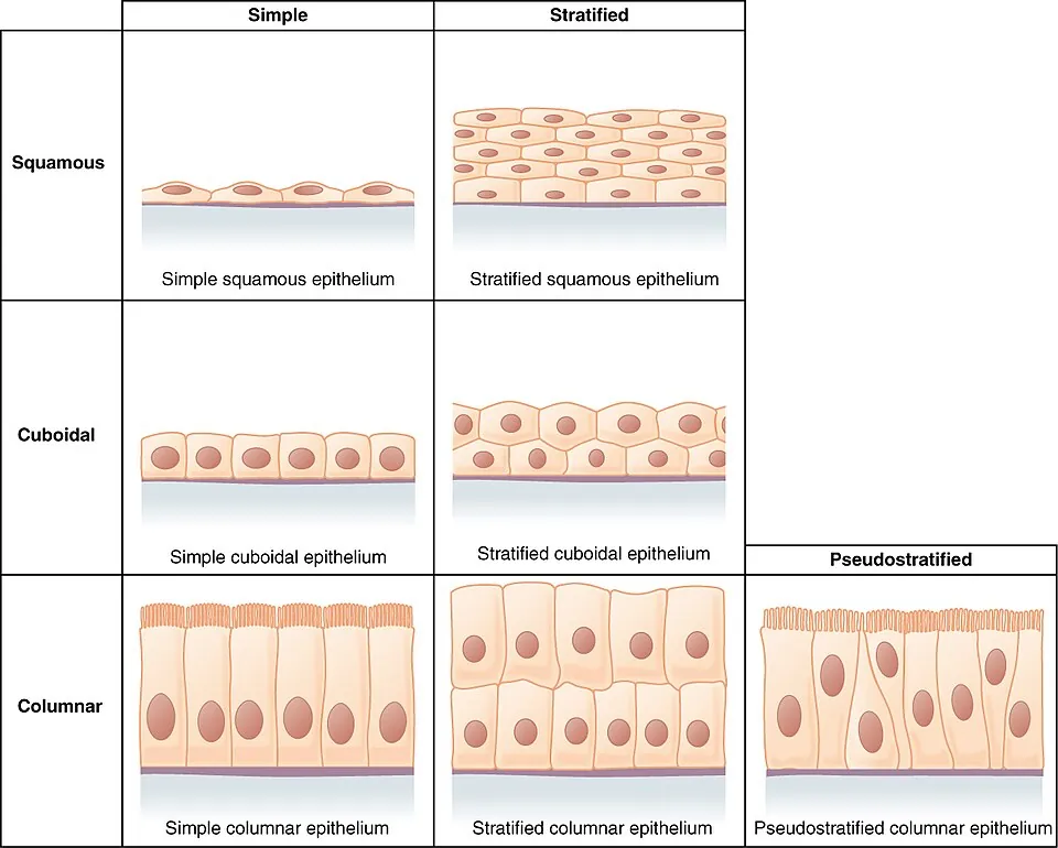
1層の、薄く平たい細胞。物質が通り抜ける場所に置かれる。
部位:肺胞(I型肺胞上皮)・血管の内皮・Bowman嚢・漿膜の中皮。
薄さが機能そのものなので、ここが厚くなれば即座に交換障害になる。
肺胞では呼吸膜の厚さが約0.2〜0.6μmで、I型肺胞上皮+融合した基底膜+毛細血管内皮の3層しかない。Fickの法則で拡散量は厚さに反比例するから、間質性肺炎・肺線維症・肺水腫でこの距離が伸びるだけで低酸素血症になる。
ここでCO₂ではなくO₂が先に落ちるのが重要。CO₂の拡散能はO₂の約20倍あるため、拡散障害では低酸素血症のみで PaCO₂ は正常か低下(代償性の頻呼吸による)という組み合わせになる。国試ではこの解離が問われる。
肺胞のI型上皮は障害に弱く再生しない。再生を担うのはII型肺胞上皮で、こちらが増殖してI型に分化する。だからびまん性肺胞傷害(ARDS)の回復期にはII型細胞の過形成が見られる。II型細胞は同時にサーファクタントを作っており、これが不足するのが新生児呼吸窮迫症候群である。
English version
Core
A single layer of thin, flattened cells, placed where substances must pass through.
Sites: pulmonary alveolus (type I pneumocyte), vascular endothelium, Bowman capsule, the mesothelium of serous membranes.
Thinness is the function itself, so any thickening here immediately impairs exchange.
Going further
In the alveolus the respiratory membrane is about 0.2–0.6 µm thick and consists of only three layers: the type I pneumocyte, the fused basal laminae, and the capillary endothelium. By Fick’s law diffusion is inversely proportional to thickness, so lengthening this distance in interstitial pneumonia, pulmonary fibrosis or oedema is by itself enough to cause hypoxaemia.
What matters is that oxygen falls before carbon dioxide. The diffusing capacity for CO₂ is about twenty times that for O₂, so a diffusion defect gives hypoxaemia with a normal or low PaCO₂, the latter from compensatory tachypnoea. This dissociation is asked about directly.
In the clinic
The type I pneumocyte is vulnerable to injury and does not regenerate. Regeneration is carried out by the type II pneumocyte, which proliferates and differentiates into type I. Hence type II hyperplasia is seen in the recovery phase of diffuse alveolar damage (ARDS).
The type II cell also makes surfactant, and its deficiency is neonatal respiratory distress syndrome.
単層立方上皮Epithelium simplex cuboideum
1層の、高さと幅がほぼ等しい細胞。分泌と吸収を担う。
部位:腎尿細管・甲状腺濾胞・腺の導管・卵巣表面。
腎の近位尿細管は単層立方上皮の典型で、頂端面に刷子縁(微絨毛)を持ち細胞質が濃い赤に染まる。ミトコンドリアが極端に多いためで、糸球体濾液の約65%をここで再吸収する能動輸送を支えている。
裏を返せば虚血に最も弱い部位でもある。急性尿細管壊死が近位尿細管とヘンレ係蹄上行脚(外髄質)に好発するのは、酸素需要が高いうえに直血管の末端で酸素分圧が低いという二重の不利による。
単層立方上皮は「輸送する上皮」である。腎尿細管がその代表で、細胞質が好酸性なのはミトコンドリアが多いから——能動輸送にエネルギーを使っていることが染色像に出ている。
だから虚血に最も弱い。急性尿細管壊死では近位尿細管の刷子縁が失われ、細胞が管腔へ剥落し、円柱を作る。尿沈渣の顆粒円柱・尿細管上皮円柱がその形態そのものである。
甲状腺濾胞上皮は機能状態で高さが変わる。刺激されると立方〜円柱になり、コロイドが減って辺縁に吸収空胞が出る(Basedow 病)。休止していると扁平になりコロイドが充満する。上皮の背丈が、ホルモンの需要を映している。
English version
Core
A single layer of cells whose height and width are about equal; secretion and absorption.
Sites: renal tubules, thyroid follicles, ducts of glands, the surface of the ovary.
Going further
The renal proximal tubule is the archetype: it has a brush border of microvilli apically and cytoplasm that stains deeply red, because it is packed with mitochondria, sustaining the active transport that reabsorbs about 65 per cent of the glomerular filtrate.
Which is exactly why it is the most vulnerable to ischaemia. Acute tubular necrosis has a predilection for the proximal tubule and the thick ascending limb in the outer medulla, from a double disadvantage: a high demand for oxygen together with a low oxygen tension at the far end of the vasa recta.
In the clinic
Simple cuboidal epithelium is transporting epithelium. The renal tubule is the representative, and its acidophilic cytoplasm reflects abundant mitochondria — the expenditure of energy on active transport appears in the staining.
Hence its vulnerability to ischaemia. In acute tubular necrosis the proximal brush border is lost, the cells desquamate into the lumen and form casts. The granular and tubular epithelial casts of the urinary sediment are that morphology itself.
The thyroid follicular epithelium changes height with its functional state. When stimulated it becomes cuboidal to columnar, the colloid diminishes and peripheral resorption vacuoles appear (Graves disease); when resting it becomes flattened and the follicle fills with colloid. The height of the epithelium mirrors the demand for hormone.
単層円柱上皮Epithelium simplex columnare
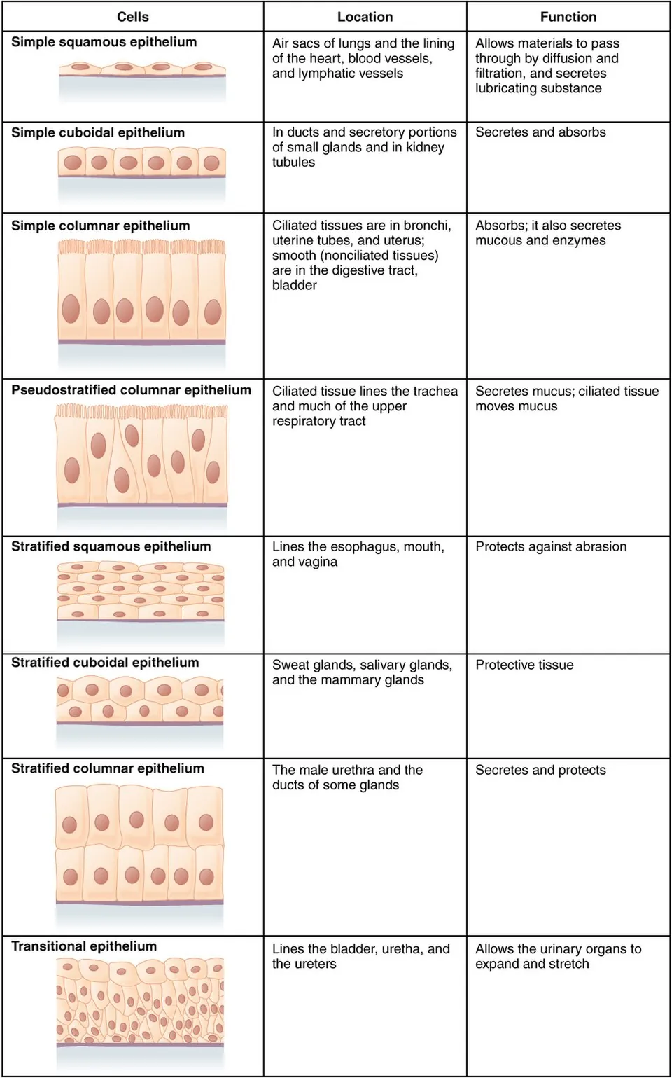
1層の、背の高い細胞。吸収と分泌に特化し、核は基底側に寄る。
部位:胃・小腸・大腸・胆嚢・子宮内膜・卵管。小腸では頂端面に微絨毛(刷子縁)、卵管・子宮では線毛を持つ。
小腸の表面積は3段階で拡張されている——輪状ヒダ(×3)→腸絨毛(×10)→微絨毛(×20)で、合計およそ600倍。この階層のどこが壊れるかで病態が変わる。
- セリアック病——絨毛が萎縮し陰窩が過形成。吸収面積そのものが失われる
- 乳糖不耐症——構造は正常だが微絨毛膜の酵素(ラクターゼ)が欠ける
前者は全般的な吸収不良(脂肪便・鉄欠乏・葉酸欠乏)、後者は乳糖だけの浸透圧性下痢。「構造の障害か、酵素の障害か」で症状の広がりが決まる。
吸収する上皮は、微絨毛を失うと吸収できなくなる。
セリアック病(グルテン過敏性腸症) — 絨毛の萎縮・陰窩の延長・上皮内リンパ球の増加。十二指腸生検で診断し、鉄と葉酸は近位で吸収されるので、まずこれらが欠乏する。
乳糖不耐症は微絨毛の刷子縁にあるラクターゼが足りない状態で、感染性腸炎の後に一過性に起こることがある。
杯細胞は粘液を出す。大腸で杯細胞が減るのは炎症の指標になる(潰瘍性大腸炎)。
腸上皮は3〜5日で入れ替わる。陰窩の幹細胞が供給源で、放射線と抗癌薬でここが傷むと粘膜が保てず下痢になる。回転の速さが、副作用の出やすさの理由である。
English version
Core
A single layer of tall cells, specialised for absorption and secretion, with the nucleus towards the base.
Sites: stomach, small and large intestine, gall bladder, endometrium, uterine tube. The small intestine has apical microvilli (brush border); the uterine tube and uterus have cilia.
Going further
The surface area of the small intestine is amplified in three stages — plicae circulares (×3) → villi (×10) → microvilli (×20), roughly 600-fold in total. Which stage fails determines the disease.
- Coeliac disease — the villi atrophy and the crypts become hyperplastic. The absorptive area itself is lost.
- Lactose intolerance — the structure is normal but the enzyme of the microvillous membrane (lactase) is missing.
The first gives generalised malabsorption (steatorrhoea, iron and folate deficiency); the second gives an osmotic diarrhoea confined to lactose. Whether the lesion is structural or enzymatic determines how wide the symptoms spread.
In the clinic
An absorbing epithelium that loses its microvilli cannot absorb.
Coeliac disease — villous atrophy, crypt elongation and increased intraepithelial lymphocytes. Diagnosed on duodenal biopsy; since iron and folate are absorbed proximally, these are the first deficiencies.
Lactose intolerance is a lack of lactase in the brush border and can occur transiently after infectious enteritis.
Goblet cells secrete mucus, and their depletion in the colon is an index of inflammation (ulcerative colitis).
The intestinal epithelium renews in three to five days from the crypt stem cells; when these are damaged by radiation or chemotherapy the mucosa cannot be maintained and diarrhoea follows. The speed of turnover is the reason for the toxicity.
多列線毛円柱上皮(偽重層)Epithelium pseudostratificatum columnare ciliatum
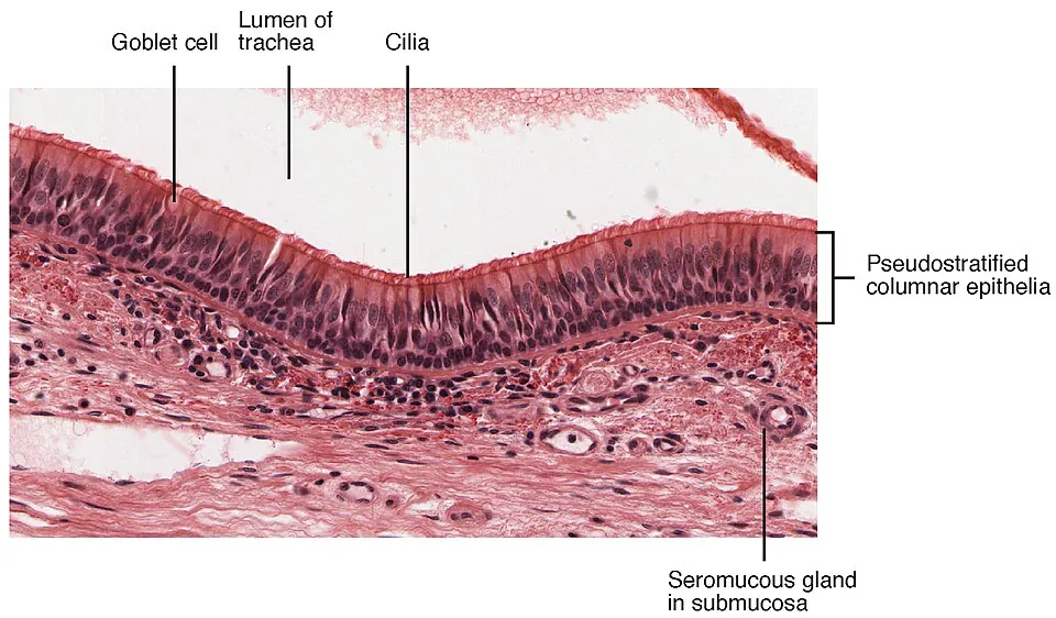
すべての細胞が基底膜に接しているのに、核の高さがそろわないため重層に見える。だから「偽」重層。
部位:気道(鼻腔〜気管支)・卵管・精巣上体・精管。
気道では線毛と杯細胞が組んで粘液線毛クリアランスを作り、粘液に絡めた異物を咽頭へ運び上げる。
この上皮は再生するときに元に戻れなくなることがある。慢性刺激(とくに喫煙)で線毛円柱上皮が重層扁平上皮に置き換わる=扁平上皮化生。物理的刺激には強くなるが、線毛を失うため粘液を運べなくなる。喫煙者の慢性咳嗽・易感染性・慢性気管支炎はここに始まる。
化生は可逆だが、刺激が続けば異形成→上皮内癌→浸潤癌へ進む。肺扁平上皮癌が中枢の太い気管支に、腺癌が末梢に生じるのは、化生が起きる場所の違いによる。
原発性線毛機能不全症(Kartagener 症候群)——ダイニンアーム欠損で線毛が動かない。気管支拡張症・慢性副鼻腔炎・内臓逆位の三徴。内臓逆位が加わるのは、胎生期のノード線毛が左右軸の決定に働いているためで、動かなければ左右がランダムに決まり約半数が逆位になる。精子鞭毛も同じ構造なので男性不妊を伴う。
English version
Core
Every cell touches the basement membrane, but the nuclei lie at different heights, so it appears stratified — hence pseudostratified.
Sites: airways (nasal cavity to bronchi), uterine tube, epididymis, ductus deferens.
In the airway, cilia and goblet cells together create mucociliary clearance, carrying trapped particles up to the pharynx.
Going further
This epithelium may fail to return to its original form when it regenerates. Under chronic irritation, especially smoking, ciliated columnar epithelium is replaced by stratified squamous — squamous metaplasia. It becomes more resistant to physical insult but, having lost its cilia, can no longer move mucus. The chronic cough, susceptibility to infection and chronic bronchitis of the smoker begin here.
Metaplasia is reversible, but if the irritation continues it proceeds to dysplasia → carcinoma in situ → invasive carcinoma. That squamous carcinoma of the lung arises in the large central bronchi while adenocarcinoma arises peripherally reflects where the metaplasia occurs.
In the clinic
Primary ciliary dyskinesia (Kartagener syndrome) — absent dynein arms, so the cilia do not move. The triad is bronchiectasis, chronic sinusitis and situs inversus.
Situs inversus is added because the nodal cilia of the embryo determine the left–right axis: if they do not move, laterality is decided at random and about half are inverted. Because the sperm flagellum has the same structure, male infertility accompanies it.
重層扁平上皮Epithelium stratificatum squamosum
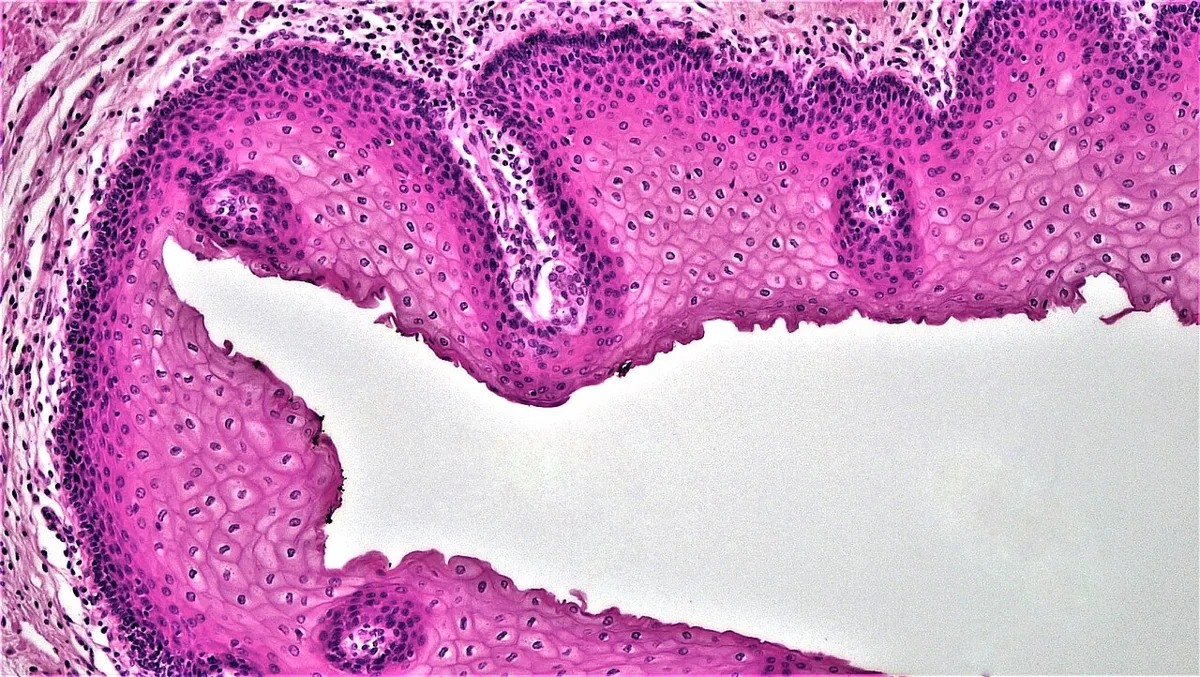
何層も積み重なり、表層ほど平たくなる。摩擦に耐えるための構造。基底層で分裂した細胞が押し上げられ、表層で脱落する。
- 角化型——表皮。表層の細胞は核を失いケラチンの塊になる(乾いた場所)
- 非角化型——口腔・食道・膣・肛門管下部・角膜(湿った場所。核が残る)
境目に注目すると病気の場所が見える。上皮の種類が切り替わる移行部は再生が盛んで、癌の好発部位になる。
- 食道胃接合部——重層扁平/単層円柱。逆流により円柱上皮化生(Barrett 食道)→腺癌
- 子宮頸部の扁平円柱上皮接合部(SCJ)——ここがHPV感染と頸癌の場
- 肛門管の歯状線——上が腺癌、下が扁平上皮癌
いずれも「2種類の上皮が出会う所」であり、細胞分裂が活発なところに発癌が起きるという原則の現れである。
Barrett 食道は逆流性食道炎の慢性化により、食道の重層扁平上皮が腸型の円柱上皮に置き換わる化生。食道腺癌の前癌病変であり、内視鏡で扁平上皮(白っぽい)と円柱上皮(赤い)の境界が口側へ移動して見える。日本の食道癌は扁平上皮癌が大半(飲酒・喫煙)だが、欧米では Barrett 由来の腺癌が多い——出身国で疫学が変わる点は国試でも問われる。
English version
Core
Many layers stacked up, becoming flatter towards the surface — a structure for withstanding friction. Cells divide in the basal layer, are pushed upward and are shed at the surface.
- Keratinised — the epidermis. Surface cells lose their nuclei and become masses of keratin (dry surfaces).
- Non-keratinised — oral cavity, oesophagus, vagina, lower anal canal, cornea (moist surfaces; the nuclei remain).
Going further
Look at the borders and the sites of disease appear. A junction where the type of epithelium changes is a site of active regeneration and a favoured site of carcinoma.
- Oesophagogastric junction — stratified squamous / simple columnar. Reflux causes columnar metaplasia (Barrett oesophagus) → adenocarcinoma.
- The squamocolumnar junction of the cervix — the site of HPV infection and of cervical carcinoma.
- The dentate line of the anal canal — adenocarcinoma above, squamous cell carcinoma below.
All are places where two kinds of epithelium meet, and all express the principle that carcinogenesis occurs where cell division is active.
In the clinic
Barrett oesophagus is metaplasia in which the stratified squamous epithelium of the oesophagus is replaced by intestinal-type columnar epithelium through chronic reflux oesophagitis. It is a premalignant condition for oesophageal adenocarcinoma, and endoscopically the border between squamous (pale) and columnar (red) epithelium is seen to have moved proximally.
Most oesophageal cancer in Japan is squamous cell carcinoma (alcohol and smoking), whereas in Western countries Barrett-derived adenocarcinoma predominates — that the epidemiology changes with the country is itself examined.
尿路上皮(移行上皮)Epithelium transitionale
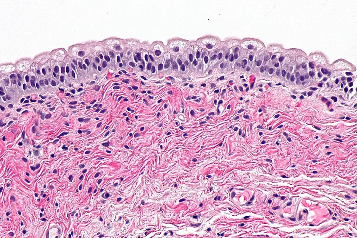
伸び縮みに特化した上皮。尿路(腎盂・尿管・膀胱・近位尿道)にのみ存在する。
空虚時は丸みを帯びた細胞が数層に重なり、表層に大きな傘細胞が並ぶ。充満すると細胞が滑って伸び、層が薄く平らに見える。
伸びるだけでなく尿を通さないことが本質的な仕事である。傘細胞の頂端膜はウロプラキンという蛋白の板で覆われ、さらに強固な密着結合で塞がれている。これが無ければ高浸透圧・酸性・尿素だらけの尿が組織側へ漏れる。
伸展時に余った膜は細胞内の円板状小胞として蓄えられ、必要に応じて頂端膜に組み込まれる——面積を増やす仕組みまで用意されている。
尿路上皮は腎盂から尿道まで一続きの同じ上皮であり、尿という同じ発癌物質にさらされる。そのため尿路上皮癌は多発・再発しやすい(field cancerization)。膀胱癌の治療後に腎盂・尿管まで含めた経過観察が要るのはこのためである。危険因子は喫煙と芳香族アミン(染料・ゴム工業)。無症候性肉眼的血尿が典型的な初発症状。
English version
Core
An epithelium specialised for distension, found only in the urinary tract — renal pelvis, ureter, bladder and proximal urethra.
When empty, rounded cells lie in several layers with large umbrella cells at the surface; when full, the cells slide over one another and the layer appears thin and flat.
Going further
Its essential work is not only to stretch but to be impermeable to urine. The apical membrane of the umbrella cell is covered by plaques of uroplakin and sealed by robust tight junctions. Without this, urine — hyperosmolar, acid and full of urea — would leak into the tissue.
The membrane left over when the epithelium is stretched is stored as discoid vesicles within the cell and inserted into the apical membrane when required: even the mechanism for increasing area is provided for.
In the clinic
The urothelium is one continuous epithelium from renal pelvis to urethra, exposed to the same carcinogens in the urine. Urothelial carcinoma is therefore multifocal and prone to recurrence — field cancerisation. This is why follow-up after treatment of bladder cancer must include the renal pelvis and ureter.
Risk factors are smoking and aromatic amines (dye and rubber industries). Painless macroscopic haematuria is the typical presenting symptom.
密着結合(タイト結合)Zonula occludens
隣り合う細胞の膜を帯状に密着させて隙間を塞ぐ結合。上皮の最も頂端側に、細胞をぐるりと取り巻いて位置する。
働きは2つ——細胞間の通り抜けを制限する(バリア)と、頂端側と基底側の膜蛋白が混ざらないよう仕切る(フェンス)。極性を保っているのはこの結合である。
構成蛋白はクローディンとオクルディン。クローディンの種類によって通す物とその量が決まるので、「密着結合=完全に閉じている」わけではない。腎尿細管ではクローディン16/19がMg²⁺・Ca²⁺の傍細胞路を作っており、この変異で家族性低Mg血症・高Ca尿症が起きる。
脳の血液脳関門も、内皮細胞どうしの密着結合が本体である。ここが緩む(炎症・腫瘍)と造影剤が漏れ、MRIで増強効果として見える。造影される=関門が壊れているという読み方ができる。
密着結合はバリアそのものである。
血液脳関門は脳の毛細血管内皮の密着結合で作られる。脂溶性の薬は通り、水溶性の薬は通らない——抗菌薬の選択(髄膜炎で第3世代セフェムを選ぶ)がここから導かれる。炎症では密着結合が緩むので、髄膜炎ではふだん通らない薬も届きやすくなる。
関門を欠く場所がある——脳室周囲器官(最後野・下垂体後葉・松果体)。最後野が化学受容器引金帯として働けるのは、血中の物質を直接感知できるからである。
血液精巣関門(Sertoli 細胞間)が破れると精子に対する自己抗体ができ、精管結紮術後や外傷後の不妊の一因になる。
コレラ毒素とロタウイルスは腸管上皮のバリアと輸送を壊し、水様性下痢を起こす。
English version
Core
A junction that seals the gap by fusing the membranes of adjacent cells in a belt. It lies at the most apical position, encircling the cell.
Two functions — it restricts passage between cells (a barrier), and it partitions apical from basolateral membrane proteins so they do not mix (a fence). It is this junction that maintains polarity.
Going further
The constituent proteins are claudins and occludin. The type of claudin determines what passes and how much, so a tight junction is not simply "completely closed". In the renal tubule, claudins 16 and 19 create a paracellular route for Mg²⁺ and Ca²⁺, and mutation causes familial hypomagnesaemia with hypercalciuria.
The blood–brain barrier is itself the tight junction between endothelial cells. When it loosens — in inflammation or around a tumour — contrast medium leaks and appears as enhancement on MRI. "It enhances" can be read as "the barrier is broken".
In the clinic
The tight junction is the barrier itself. The blood–brain barrier is formed by the tight junctions of cerebral capillary endothelium: lipid-soluble drugs cross, water-soluble drugs do not — from which the choice of antibiotic follows (a third-generation cephalosporin for meningitis). In inflammation the junctions loosen, so drugs that normally do not enter reach the CSF more easily.
Some places lack the barrier — the circumventricular organs (area postrema, neurohypophysis, pineal gland). The area postrema can act as the chemoreceptor trigger zone precisely because it can sample the blood directly.
If the blood–testis barrier (between Sertoli cells) is broken, antibodies against sperm develop — a cause of infertility after vasectomy or trauma. Cholera toxin and rotavirus destroy the barrier and transport of the intestinal epithelium and cause watery diarrhoea.
デスモソーム(接着斑)Macula adhaerens
細胞どうしを点で強く留める結合。カドヘリン(デスモグレイン・デスモコリン)が細胞間を橋渡しし、細胞内では中間径フィラメント(ケラチン)に繋がる。
引っ張りに耐える場所に多い——表皮・心筋・子宮頸部。
基底膜に対して細胞を留める片側版がヘミデスモソーム。
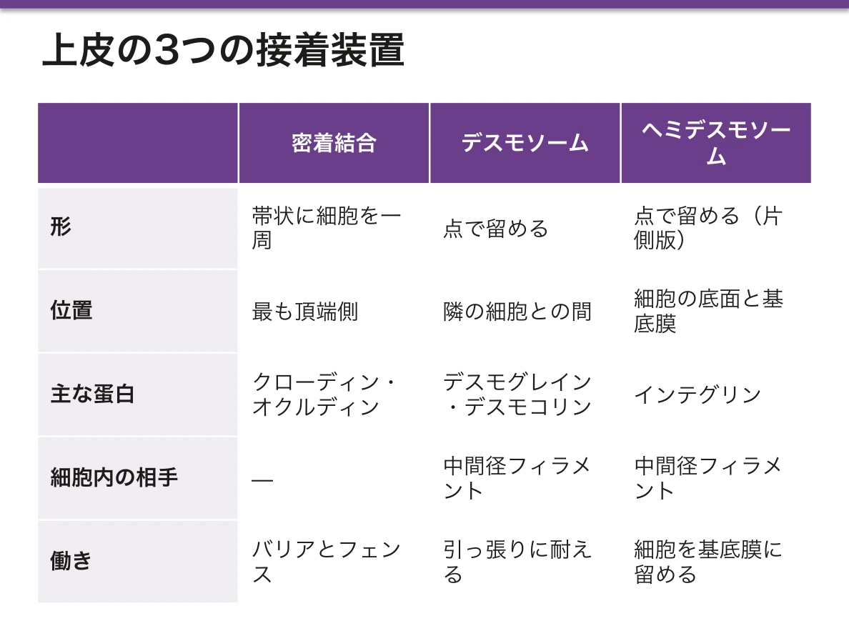
デスモソームは細胞の中まで力を伝える点が重要である。カドヘリン→デスモプラキン→ケラチン線維と繋がることで、上皮全体が1枚の布のように張力を分散する。
接着装置の階層を頂端側から並べると密着結合 → 接着帯(zonula adhaerens、アクチンに繋がる)→ デスモソームで、これを合わせて接着複合体と呼ぶ。ギャップ結合(nexus)はこれとは別で、接着ではなく細胞間の連絡を担う。
水疱の高さが、壊れた接着装置の高さで決まる。
- 尋常性天疱瘡——抗デスモグレイン3抗体。表皮内(基底層直上)で剥がれ、弛緩性水疱・Nikolsky 徴候陽性・口腔粘膜疹が先行
- 水疱性類天疱瘡——ヘミデスモソーム(BP180/BP230)に対する抗体。表皮下で剥がれるため緊満性水疱で、粘膜疹は乏しい
「デスモソーム=表皮内・弛緩性/ヘミデスモソーム=表皮下・緊満性」——接着の位置がそのまま臨床像になる。
English version
Core
A junction that fastens cells strongly at points. Cadherins (desmoglein, desmocollin) bridge the intercellular space and connect inside the cell to intermediate filaments (keratin).
Abundant where tension must be resisted — epidermis, cardiac muscle, cervix. The one-sided version that anchors the cell to the basement membrane is the hemidesmosome.
Going further
What matters is that the desmosome transmits force into the interior of the cell. Through cadherin → desmoplakin → keratin filament, the whole epithelium distributes tension like a single sheet of cloth.
Listed from the apical side, the adhesion apparatus is tight junction → zonula adherens (linked to actin) → desmosome, together the junctional complex. The gap junction (nexus) is separate: it provides communication, not adhesion.
In the clinic
The level of a blister is determined by the level of the adhesion that has failed.
- Pemphigus vulgaris — antibodies against desmoglein 3. Separation occurs within the epidermis, just above the basal layer: flaccid blisters, a positive Nikolsky sign, and oral lesions that come first.
- Bullous pemphigoid — antibodies against the hemidesmosome (BP180/BP230). Separation is subepidermal, so the blisters are tense and mucosal lesions are few.
Desmosome = intraepidermal and flaccid; hemidesmosome = subepidermal and tense — the position of the adhesion becomes the clinical picture.
微絨毛Microvilli
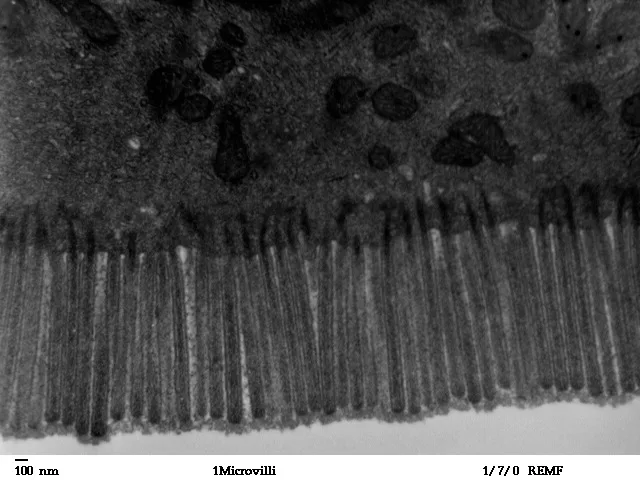
頂端面から出る細い突起。アクチンが芯で、動かない。目的は表面積を増やすこと。
密に並ぶと光学顕微鏡では刷子縁という帯として見える。部位は小腸・腎近位尿細管。
紛らわしい3つを区別する——微絨毛(アクチン・不動・吸収)/線毛(微小管9+2・動く・輸送)/不動毛(実は長い微絨毛。精巣上体・内耳)。
微絨毛膜には消化酵素そのものが埋め込まれている(マルターゼ・ラクターゼ・スクラーゼ・ペプチダーゼ)。これを膜消化と呼び、管腔内消化の最終段階を担う。だから微絨毛が壊れると、膵酵素が正常でも二糖類が分解できない。
線毛の9+2構造(周辺9対+中心2本の微小管、ダイニンアームで滑る)に対し、ノード線毛だけは9+0で回転運動をする。左右軸決定に関わるのはこちらである。
微絨毛封入体病は稀な先天性疾患だが、原理を示す。ミオシンVbの変異で微絨毛が頂端膜に出られず細胞内に取り込まれ、出生直後から難治性の水様下痢をきたす。吸収面が無ければ消化管は機能しないことがそのまま現れる。
English version
Core
Fine projections from the apical surface. The core is actin and they do not move. Their purpose is to increase surface area. When densely packed they appear by light microscopy as a band, the brush border. Sites: small intestine, renal proximal tubule.
Distinguish the three that are confused — microvillus (actin, immotile, absorption) / cilium (microtubules 9+2, motile, transport) / stereocilium (in fact a long microvillus; epididymis, inner ear).
Going further
Digestive enzymes themselves are embedded in the microvillous membrane — maltase, lactase, sucrase, peptidases. This is membrane digestion, the final stage after luminal digestion. So if the microvilli are destroyed, disaccharides cannot be broken down even when the pancreatic enzymes are normal.
Against the 9+2 of the motile cilium (nine peripheral doublets and a central pair, sliding by dynein arms), the nodal cilium alone is 9+0 and rotates. It is this one that determines the left–right axis.
In the clinic
Microvillus inclusion disease is rare but demonstrates the principle. A mutation in myosin Vb prevents the microvilli from reaching the apical membrane; they are internalised instead, and intractable watery diarrhoea begins immediately after birth. That the gut cannot function without an absorptive surface appears in its purest form.
杯細胞Cellula caliciformis
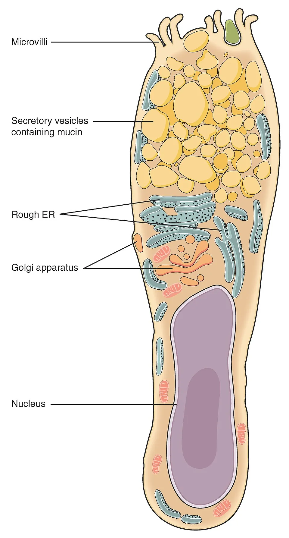
単細胞の外分泌腺。ムチンを分泌して粘液を作る。ムチン顆粒でふくらんだ頂端側と、押しつぶされた基底側の核でワイングラス状に見える。
部位:気道・小腸・大腸(大腸ほど多い)。HE では顆粒が抜けて白く見えるので、PAS 染色で確認する。
杯細胞の数はその場所が受けている刺激の記録になる。慢性気道炎症・喫煙では杯細胞が増生し(過形成)、粘液が過剰になる一方で線毛は減る。作る量が増えて運ぶ力が落ちるので、痰が出せず溜まる。慢性気管支炎の「3か月以上続く咳と痰を2年連続」という定義の実体はこれである。
逆に本来無いはずの場所に杯細胞が現れれば化生である。胃粘膜に杯細胞を含む腸型上皮が出るのが腸上皮化生で、H. pylori 感染 → 萎縮性胃炎 → 腸上皮化生 → 胃癌(腸型)という道筋の途中にある。
嚢胞性線維症——CFTR(Cl⁻チャネル)の異常で分泌液から水分が引けず、粘液が異常に粘稠になる。気道では喀出できず反復感染から気管支拡張症へ、膵では導管が詰まり膵外分泌不全(脂肪便・脂溶性ビタミン欠乏)、腸では胎便性イレウス。汗腺では逆にCl⁻を再吸収できず汗が塩辛い(汗中Cl⁻高値=発汗試験)。1つのチャネルの異常が臓器ごとに違う顔で出る。
English version
Core
A unicellular exocrine gland. It secretes mucin to form mucus. The apical end is distended with mucin granules and the basal nucleus is compressed, giving the shape of a wine glass.
Sites: airways, small intestine, large intestine (most numerous in the colon). In H&E the granules are washed out and appear white, so confirm with PAS.
Going further
The number of goblet cells is a record of the irritation that site has received. In chronic airway inflammation and smoking, goblet cells undergo hyperplasia and mucus becomes excessive while cilia diminish. Production rises as the capacity to transport falls, so sputum cannot be cleared and accumulates. This is the substance behind the definition of chronic bronchitis — cough and sputum for three months in two consecutive years.
Conversely, goblet cells appearing where they should not exist is metaplasia. Intestinal-type epithelium containing goblet cells in the gastric mucosa is intestinal metaplasia, part-way along the road H. pylori → atrophic gastritis → intestinal metaplasia → intestinal-type gastric carcinoma.
In the clinic
Cystic fibrosis — a defect of CFTR, a chloride channel, so water cannot be drawn into the secretions and the mucus becomes abnormally viscid. One channel defect presents with a different face in each organ:
- Airway — sputum cannot be expectorated; recurrent infection leads to bronchiectasis.
- Pancreas — the ducts obstruct, giving exocrine insufficiency (steatorrhoea, deficiency of fat-soluble vitamins).
- Intestine — meconium ileus.
- Sweat gland — conversely, chloride cannot be reabsorbed, so the sweat is salty; a high sweat chloride is the diagnostic test.
外分泌腺Glandula exocrina
上皮が下の結合組織へ落ち込んでできた腺で、導管を通じて体表・管腔へ分泌する。導管を失い血中へ出すのが内分泌腺。
分泌の様式で3つ——
- 漏出分泌(エクリン)——開口分泌のみ。細胞は無傷。大多数の腺・エクリン汗腺
- 離出分泌(アポクリン)——頂端の細胞質を一部ちぎって出す。乳腺・腋窩のアポクリン汗腺
- 全分泌(ホロクリン)——細胞まるごとが分泌物になる。脂腺
形での分類は導管が枝分かれするか(単一/複合)× 終末部の形(管状/腺房状/管状腺房状)の掛け算。名前は長いが2軸しかない。
臨床で効くのは全分泌=脂腺の理解である。脂腺は毛包に開口し、細胞が丸ごと壊れて皮脂になる。アンドロゲンで活性が上がるため思春期に増え、角化亢進で開口部が詰まれば面皰、そこにCutibacterium acnes が増えて炎症を起こすのが尋常性痤瘡である。「なぜ思春期に、なぜ顔と背中に」が腺の性質から説明できる。
分泌様式の違いが、疾患の場所を決める。
アポクリン汗腺(離出分泌)は腋窩・乳輪・外耳道・肛門周囲にしかない。だから化膿性汗腺炎はこの分布に一致して起こるし、乳房外 Paget 病も同じ場所に出る。分泌腺の分布を知っていれば、皮疹の部位から診断が絞れる。
アポクリン汗腺の分泌物そのものは無臭で、常在菌が分解して臭う——腋臭症の機序である。
脂腺(全分泌/ホロクリン)は細胞ごと崩れて分泌物になる。毛包脂腺系が詰まり、Cutibacterium acnes が増えるのが尋常性痤瘡で、アンドロゲンが脂腺を大きくするため思春期に増える。イソトレチノインが脂腺そのものを縮めるのは、この分泌様式を断つからである。
エクリン汗腺は全身にあり、体温調節を担う。嚢胞性線維症では汗の塩化物が高い——汗試験の根拠になる。
English version
Core
A gland formed by epithelium growing down into the connective tissue beneath, secreting onto a surface or into a lumen through a duct. A gland that loses its duct and secretes into the blood is an endocrine gland.
Three modes of secretion
- Merocrine (eccrine) — exocytosis only; the cell remains intact. Most glands, and the eccrine sweat gland.
- Apocrine — part of the apical cytoplasm is pinched off with the product. Mammary gland, axillary apocrine sweat gland.
- Holocrine — the whole cell becomes the secretion. Sebaceous gland.
Going further
Classification by form is the product of whether the duct branches (simple / compound) × the shape of the secretory portion (tubular / acinar / tubuloacinar). The names are long but there are only two axes.
What matters clinically is understanding holocrine = sebaceous gland. The sebaceous gland opens into the hair follicle, and the cell disintegrates entirely to become sebum. Androgens increase its activity, so it enlarges at puberty; if hyperkeratosis blocks the opening a comedone forms, and Cutibacterium acnes then proliferates and inflames it — acne vulgaris. Why at puberty, and why on the face and back, is explained by the nature of the gland.
In the clinic
The mode of secretion determines where disease occurs.
Apocrine sweat glands are found only in the axilla, areola, external auditory meatus and perianal region. Hidradenitis suppurativa therefore follows exactly that distribution, and extramammary Paget disease appears in the same places. Knowing the distribution of the glands narrows the diagnosis from the site of the rash. The apocrine secretion itself is odourless; the smell is produced by commensal bacteria breaking it down — the mechanism of body odour.
Sebaceous glands (holocrine) disintegrate entirely to form their secretion. Obstruction of the pilosebaceous unit with proliferation of C. acnes gives acne, which increases at puberty because androgens enlarge the gland. Isotretinoin works by shrinking the gland itself — it cuts off that mode of secretion.
Eccrine sweat glands are found all over the body and serve thermoregulation. In cystic fibrosis the sweat chloride is high — the basis of the sweat test.
化生・異形成・上皮内癌 — 上皮が壊れていく順序
慢性刺激に対して上皮が変化していく段階。どこまでが可逆かが要点。
- 化生 metaplasia——ある成熟上皮が別の成熟上皮に置き換わる。細胞自体は正常。可逆
- 異形成 dysplasia——核の腫大・極性の乱れ・分裂像の増加。前癌病変。軽度なら可逆
- 上皮内癌 carcinoma in situ——全層が癌細胞。ただし基底膜は破っていない。転移しない
- 浸潤癌——基底膜を破る。ここで転移が可能になる
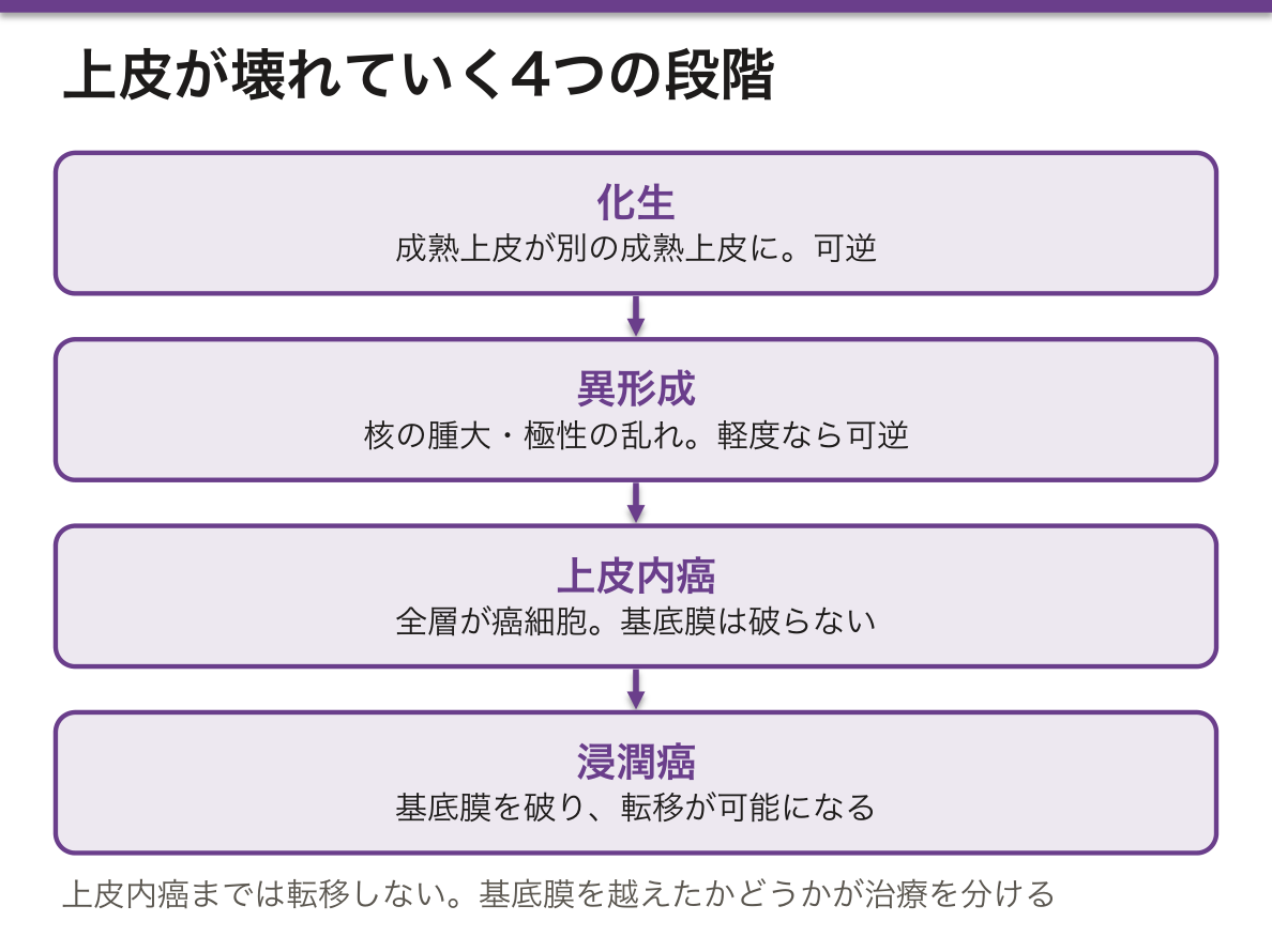
この4段階はそのまま診断と治療の分岐になる。上皮内癌までなら局所切除(内視鏡的切除・円錐切除)で根治しうるが、浸潤すればリンパ節郭清を含む手術と全身療法の対象になる。病期分類の最初の線が基底膜である理由がここにある。
代表的な経路を3つ押さえると応用が効く。
- 食道:逆流 → 円柱上皮化生(Barrett)→ 異形成 → 腺癌
- 胃:H. pylori → 萎縮性胃炎 → 腸上皮化生 → 異形成 → 腺癌
- 子宮頸部:HPV16/18 → CIN1〜3 → 上皮内癌 → 浸潤癌
いずれも原因の除去(PPI・除菌・ワクチン)が前段階で効くという共通点があり、予防医学の問題として問われる。
化生は「悪いもの」ではなく適応である点に注意。喫煙者の気道扁平上皮化生は刺激への合理的な応答であり、原因が除かれれば戻る。問題は適応の代償(線毛の喪失)と、刺激が続いた場合に次の段階へ進むことにある。国試では「化生は可逆・異形成は前癌・上皮内癌は基底膜を破らない」の3点が繰り返し問われる。
English version
Core
The stages through which epithelium changes under chronic irritation. The point is how far each is reversible.
- Metaplasia — one mature epithelium is replaced by another mature epithelium. The cells themselves are normal. Reversible.
- Dysplasia — nuclear enlargement, disordered polarity, increased mitoses. A premalignant lesion; reversible if mild.
- Carcinoma in situ — the full thickness is malignant, but the basement membrane is not breached. It does not metastasise.
- Invasive carcinoma — the basement membrane is breached. Metastasis becomes possible here.
Going further
These four stages are directly the branch points of diagnosis and treatment. Up to carcinoma in situ, local excision — endoscopic resection, conisation — can be curative; once invasion has occurred, the case belongs to surgery with lymph node dissection and systemic therapy. This is why the first line of staging is the basement membrane.
Three representative pathways cover most applications:
- Oesophagus: reflux → columnar metaplasia (Barrett) → dysplasia → adenocarcinoma.
- Stomach: H. pylori → atrophic gastritis → intestinal metaplasia → dysplasia → adenocarcinoma.
- Cervix: HPV 16/18 → CIN 1–3 → carcinoma in situ → invasive carcinoma.
All three share the feature that removing the cause works at the earlier stages — proton pump inhibitors, eradication, vaccination — and this is examined as a question of preventive medicine.
In the clinic
Note that metaplasia is not "something bad" but an adaptation. Squamous metaplasia in a smoker’s airway is a rational response to irritation and reverses when the cause is removed. The problem lies in the price of the adaptation — the loss of cilia — and in progression to the next stage if the irritation continues.
Three points are asked repeatedly: metaplasia is reversible, dysplasia is premalignant, and carcinoma in situ has not breached the basement membrane.
まとめ
| 上皮 | 体の表面と、管腔・体腔の内面を隙間なく覆う組織。 |
|---|---|
| 上皮の4つの型 — 被蓋上皮・腺上皮・筋上皮・感覚上皮 | ヨーロッパの組織学では、上皮を働きで4つの型に分けることが多い。 |
| 単層扁平上皮 | 1層の、薄く平たい細胞。物質が通り抜ける場所に置かれる。 |
| 単層立方上皮 | 1層の、高さと幅がほぼ等しい細胞。分泌と吸収を担う。 |
| 単層円柱上皮 | 1層の、背の高い細胞。吸収と分泌に特化し、核は基底側に寄る。 |
| 多列線毛円柱上皮(偽重層) | すべての細胞が基底膜に接しているのに、核の高さがそろわないため重層に見える。 |
| 重層扁平上皮 | 何層も積み重なり、表層ほど平たくなる。摩擦に耐えるための構造。 |
| 尿路上皮(移行上皮) | 伸び縮みに特化した上皮。尿路(腎盂・尿管・膀胱・近位尿道)にのみ存在する。 |
| 密着結合(タイト結合) | 隣り合う細胞の膜を帯状に密着させて隙間を塞ぐ結合。 |
| デスモソーム(接着斑) | 細胞どうしを点で強く留める結合。カドヘリン(デスモグレイン・デスモコリン)が細胞間を橋渡しし、細胞内では中間径フィラメント(ケラチン)に繋がる。 |
| 微絨毛 | 頂端面から出る細い突起。アクチンが芯で、動かない。 |
| 杯細胞 | 単細胞の外分泌腺。ムチンを分泌して粘液を作る。 |
| 外分泌腺 | 上皮が下の結合組織へ落ち込んでできた腺で、導管を通じて体表・管腔へ分泌する。 |
| 化生・異形成・上皮内癌 — 上皮が壊れていく順序 | 慢性刺激に対して上皮が変化していく段階。 |
このノートは医学生が授業の内容を自分の言葉でまとめたもので、診療の判断に使うものではありません。誤りに気づいたらお知らせください。