骨髄は「血を作る場所」であると同時に、B細胞が教育を受ける一次リンパ性器官でもある。
そして体液は、動脈から出て静脈に全部は戻らない。1日あたり2〜4 L が組織に残り、それをリンパ管が回収する——「戻せなくなったものが浮腫になる」。
- リンパ毛細管——盲端で始まる。基底膜が不完全で、弁が無く、係留線維で組織に引っ張られている
- だから組織圧が上がるほど、口が開いて吸い込む
この「基底膜が不完全」という一点が、リンパ行性転移の入口になる——「癌細胞が最も入りやすい管が、リンパ毛細管になる」。
骨髄の像は、造血の3系統と脂肪の比で読む。年齢と病気で、この比が動く。
- 骨髄 — 造血と脂肪の比で読む
- リンパ管 — 戻せなかったものを回収する
- 胸管 — 全身のリンパが、最後に1本へ集まる
- 乳び槽 — 脂だけが、血管を通らずに運ばれる
- 造血のニッチ — 幹細胞は、居場所によって振る舞いが変わる
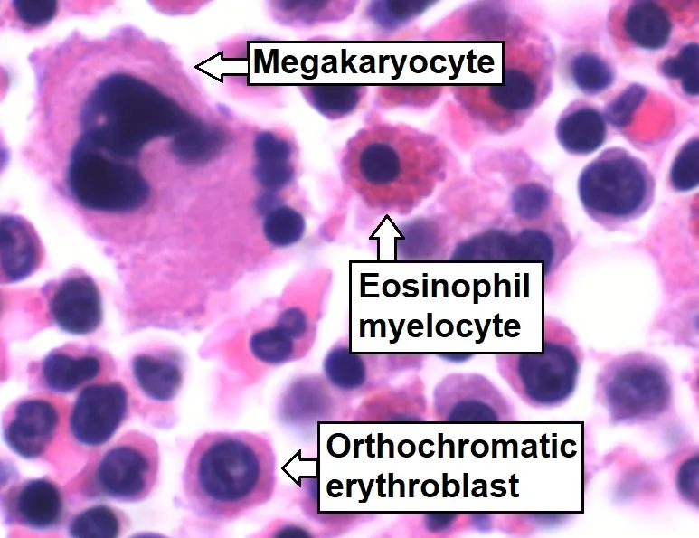
- 巨核球 — 類洞に腕を差し入れて、血小板をちぎる
- 血液骨髄関門 — 成熟した細胞だけが、血へ出られる
骨髄 — 造血と脂肪の比で読むMedulla ossium
赤色骨髄(造血あり)と黄色骨髄(脂肪)。加齢とともに黄色に置き換わる。
- 成人で造血が残る場所——椎体・胸骨・腸骨・肋骨・頭蓋・長管骨の近位端
- 細胞充実度の目安——およそ「100 − 年齢」%
構造——骨梁の間を洞様毛細血管が走り、造血細胞が間を埋める。血管周囲に間質細胞(ニッチ)。
成熟したものだけが血管へ出る——洞様毛細血管の内皮を、変形して通り抜ける。
- 赤芽球は核を捨ててから出る——だから末梢に有核赤血球があれば異常になる
- 巨核球は細胞質の断片だけを送り出す——巨核球そのものは骨髄に留まる
M/E比(顆粒球系/赤芽球系)——通常2〜4。どちらが増えたかを判断する。
「成熟したものだけが出る」という関門が、末梢血の異常所見の意味を決めている。幼若な細胞が末梢に出ていれば、関門が壊れているか、押し出されている(白血球の異常の章)——「出てはいけないものが出ていること自体が、所見になる」。
白赤芽球症(幼若顆粒球+有核赤血球)は、骨髄が置き換えられているときに出る。骨髄癌腫症・骨髄線維症(骨髄線維症の章)——「末梢血の1行が、骨髄が侵されていることを教える」。
そして涙滴赤血球が加われば、骨髄線維症をより強く疑う——「硬くなった骨髄を、無理に通ってきた形になる」。
造血の残る場所が、骨髄穿刺の部位を決めている。成人では後腸骨稜、乳児では脛骨も使える(骨髄検査の章)——「年齢によって、取れる場所が変わる」。
そして小児の骨髄は全身に広がっているので、白血病で骨痛が出やすい(小児白血病の章)——「歩きたがらないという訴えが、血液疾患の初発になる」。
骨髄のニッチが、造血幹細胞の維持に必須になる。移植した幹細胞が自分でここへ帰る(ホーミング)ので、点滴で投与できる(造血幹細胞移植の章)——「移植なのに手術をしない理由が、この帰巣性にある」。
細胞充実度は「100 から年齢を引く」を目安にする。年齢に対して低すぎる=低形成、高すぎる=過形成と読む。
骨髄穿刺は後腸骨稜で行う。成人で造血が残る扁平骨だからである。
吸えない(dry tap)ときは骨髄線維症・低形成・充実性の腫瘍浸潤を考え、生検(コア)で構造を見る。「穿刺で細胞を見る」と「生検で構造を見る」は別の検査である。
再生不良性貧血 — 汎血球減少+脂肪髄。骨髄異形成症候群 — 血球減少なのに骨髄は過形成(無効造血)。末梢血と骨髄の食い違いが診断の鍵になる。
骨髄は転移の場でもある。前立腺癌・乳癌・肺癌・神経芽腫。末梢血に幼若な血球が出る(白赤芽球症)のは骨髄が置き換えられているサインである。
English version
Core
Red marrow (haemopoietic) and yellow marrow (fat); with age it is replaced by yellow.
- Sites still haemopoietic in the adult — vertebrae, sternum, ilium, ribs, skull, and the proximal ends of the long bones.
- Rule of thumb for cellularity — about 100 minus the age per cent.
- Structure — sinusoidal capillaries run between the trabeculae with haemopoietic cells filling the spaces; stromal cells (the niche) lie around the vessels.
Only mature cells leave, deforming to pass through the sinusoidal endothelium.
- The erythroblast expels its nucleus before leaving — so nucleated red cells in the blood are abnormal.
- The megakaryocyte sends out only fragments of cytoplasm — the megakaryocyte itself stays in the marrow.
M:E ratio (granulocytic to erythroid) — normally 2–4; it tells you which lineage has increased.
Going further
The checkpoint that only mature cells leave gives meaning to abnormal findings in the blood. Immature cells in the blood mean the barrier is broken, or they are being pushed out — the very presence of what should not be there is the finding.
A leucoerythroblastic picture (immature granulocytes with nucleated red cells) appears when the marrow is being replaced — carcinomatosis of the marrow, myelofibrosis. One line of the blood count tells you the marrow is involved. If teardrop cells are added, myelofibrosis is more strongly suspected — the shape of a cell that has squeezed through a stiffened marrow.
Where haemopoiesis remains determines where marrow is aspirated: the posterior iliac crest in adults, and the tibia may be used in infants. And because the marrow of a child is spread throughout the skeleton, bone pain is common in leukaemia — a complaint of not wanting to walk can be the first presentation of a haematological disease.
The niche is essential to maintaining the stem cell. Transplanted stem cells find their own way back to it (homing), which is why they can be given by infusion — the reason a transplant requires no operation.
In the clinic
Take cellularity as 100 minus the age. Too low for the age is hypoplastic; too high is hyperplastic.
Marrow is aspirated from the posterior iliac crest, a flat bone still haemopoietic in the adult. A dry tap suggests myelofibrosis, hypoplasia or solid tumour infiltration, and a trephine biopsy is needed to see the architecture. Aspiration to see the cells and biopsy to see the structure are two different investigations.
Aplastic anaemia — pancytopenia with a fatty marrow. Myelodysplastic syndrome — cytopenia despite a hypercellular marrow (ineffective haemopoiesis). The discrepancy between blood and marrow is the key to the diagnosis.
The marrow is also a site of metastasis — prostate, breast, lung, neuroblastoma. Immature cells in the blood (a leucoerythroblastic picture) are the sign that the marrow is being replaced.
リンパ管 — 戻せなかったものを回収するVas lymphaticum
リンパ毛細管——盲端で始まる。内皮のみで、基底膜は不完全か欠く。周皮細胞が無い。
- 係留線維——内皮を周囲の膠原線維につなぐ。組織圧が上がると内皮が引っ張られて隙間が開く
- だから浮腫があるほど、よく吸い込む
集合リンパ管——弁が多い。弁と弁の間(リンパ管分節)が自律的に収縮して送る。
リンパ管が無い場所——中枢神経(古典的には)・軟骨・角膜・水晶体・上皮・骨髄。
最終的に胸管(左上半身と下半身全部)と右リンパ本幹が、静脈角へ注ぐ。
浮腫の機序は4つ——静水圧の上昇・膠質浸透圧の低下・血管透過性の亢進・リンパ還流の障害。
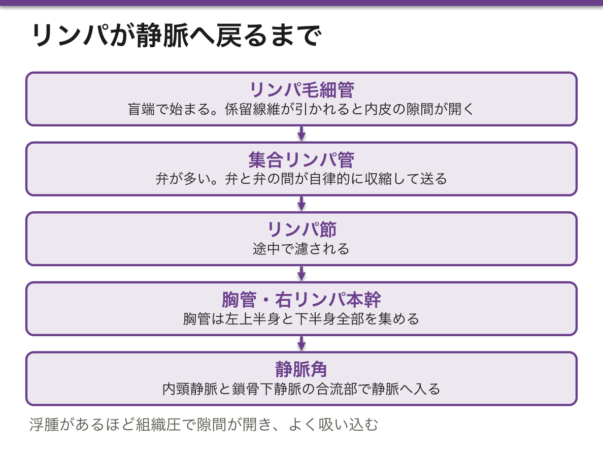
基底膜が不完全であることが、癌のリンパ行性転移の入口を作っている(転移の様式の章)——「血管より入りやすい管が、体の中を網の目に走っている」。
だから所属リンパ節の郭清が、外科手術の一部になる(リンパ節郭清の章)——「取るかどうかではなく、どこまで取るかが議論になる」。
センチネルリンパ節生検は、この流れの順番を利用している。最初に流れ着く1個を調べ、陰性なら郭清を省く(センチネルリンパ節の章)——「流れに順番があることが、手術を小さくした」。
そして郭清の後にリンパ浮腫が起きる。戻す道を切ったからになる(リンパ浮腫の章)——「治療そのものが、4つ目の機序を作っている」。
リンパ浮腫は圧痕を残しにくい。蛋白濃度が高く、線維化が進むからになる(浮腫の章)——「押して凹むかどうかが、機序を分ける手がかりになる」。
角膜にリンパ管も血管も無いことが、角膜移植の拒絶反応が少ない理由になる(角膜移植の章)——「免疫特権と呼ばれる状態が、脈管の欠如で説明される」。
リンパ管は「詰まると戻せなくなる」だけの道である。
リンパ浮腫 — 腋窩リンパ節郭清後の上肢、骨盤内リンパ節郭清後の下肢。蛋白に富む浮腫なので時間とともに線維化して硬くなり、非可逆になる。早期からの圧迫療法と用手的リンパドレナージが要る。患肢での採血・血圧測定・点滴を避けるのは蜂窩織炎を誘発すると一気に悪化するからである。
フィラリア症では成虫がリンパ管を閉塞し、象皮病になる。
係留線維の仕組み——組織圧が上がると内皮が引っ張られて隙間が開く。だから浮腫があるほどリンパは流れるという自己調節が働く。基底膜が不完全で周皮細胞が無いことが、癌細胞がリンパ管へ入りやすい理由でもある——癌腫がまずリンパ行性に転移するのはこの構造による。
English version
Core
Lymphatic capillary — begins blind. Endothelium only, with an incomplete or absent basal lamina and no pericytes.
- Anchoring filaments tie the endothelium to the surrounding collagen. When tissue pressure rises the endothelium is pulled and the gaps open — so the more oedema there is, the more it draws in.
- Collecting lymphatics — many valves. The segment between two valves (a lymphangion) contracts autonomously to propel the lymph.
Places with no lymphatics — the central nervous system (classically), cartilage, cornea, lens, epithelium, bone marrow.
Finally the thoracic duct (the left upper body and all of the lower body) and the right lymphatic duct drain into the venous angles.
Four mechanisms of oedema — raised hydrostatic pressure, reduced colloid osmotic pressure, increased permeability, and impaired lymphatic return.
Going further
The incomplete basal lamina creates the entrance for lymphatic metastasis — a network of vessels easier to enter than blood vessels runs throughout the body. This is why dissection of the regional nodes is part of cancer surgery — the debate is not whether to remove them but how far.
Sentinel node biopsy exploits the order of that flow: examine the first node the lymph reaches and, if it is negative, omit the dissection — the existence of an order to the flow made the operation smaller.
And after dissection, lymphoedema follows, because the route of return has been cut — the treatment itself creates the fourth mechanism.
Lymphoedema does not pit readily, because the protein concentration is high and fibrosis develops — whether it pits on pressure is a clue to the mechanism.
That the cornea has neither lymphatics nor blood vessels is why corneal grafts are rarely rejected — the state called immune privilege explained by the absence of vessels.
In the clinic
A lymphatic is simply a route that, once blocked, cannot return fluid.
Lymphoedema — of the arm after axillary dissection, of the leg after pelvic dissection. Being protein-rich, it fibroses and hardens with time and becomes irreversible. Compression and manual lymphatic drainage are needed from early on. Venepuncture, blood pressure measurement and infusion are avoided in the affected limb because cellulitis makes it deteriorate abruptly.
In filariasis the adult worms obstruct the lymphatics and cause elephantiasis.
The anchoring filament mechanism — rising tissue pressure pulls the endothelium and opens the gaps, so lymph flows more when there is oedema: a self-regulating arrangement.
The incomplete basal lamina and absent pericytes are also why cancer cells enter lymphatics readily — the structural reason carcinomas metastasise first by the lymphatic route.
胸管 — 全身のリンパが、最後に1本へ集まるDuctus thoracicus
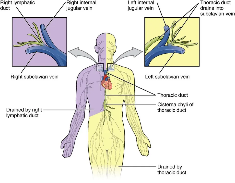
リンパは末端から中心へ一方通行で流れ、最後は2本の本幹から静脈角へ入る。
| 本幹 | 集める範囲 | 注ぐ場所 |
|---|---|---|
| 胸管 | 下半身の全部+左上半身(体の約3/4) | 左静脈角 |
| 右リンパ本幹 | 右上半身のみ | 右静脈角 |
胸管は第2腰椎の高さの乳び槽に始まり、大動脈裂孔を通って上行し、左静脈角(左内頸静脈と左鎖骨下静脈の合流部)へ注ぐ。
組織学的には静脈に似るが壁がさらに薄く、内膜・中膜・外膜の境が不明瞭で、弁が多い。
1日に約2〜4 L が戻る。
「左に偏っている」という一点が、臨床の多くを決めている。体の3/4のリンパが左静脈角へ集まる——「だから腹部・骨盤の癌の転移が、左鎖骨上窩に触れる(Virchow 転移)」(食道と胃の各論の章)。
逆に右鎖骨上窩のリンパ節腫大は、右上半身と胸部を示唆する。同じ鎖骨上窩でも、左右で探す場所が変わる——「触れた側が、探すべき臓器を教えている」。
壁が薄いことは、そのまま損傷のしやすさになる。食道癌の手術や左頸部の郭清で傷つくと乳び胸・乳び漏になり、脂肪と蛋白とリンパ球を失い続ける(胸腔と縦隔の章)——「漏れた液が白いのは、脂を運んでいるから」。
だから乳び漏の治療は、まず脂を減らすことになる。長鎖脂肪酸を止め、中鎖脂肪酸に置き換えると門脈へ直接吸収されるのでリンパを通らない(吸収の章)——「通る道を変えれば、漏れは止まる」。
左鎖骨上窩のリンパ節が腫れたら、腹部を探す。胸管が左静脈角に注ぐため、腹部・骨盤の悪性腫瘍が Virchow リンパ節に転移する。解剖を知っていれば、触診1つで検索範囲が決まる。
乳び胸 — 胸管の損傷。食道癌手術・外傷で起こる。白濁した胸水でトリグリセリドが高い。脂肪と蛋白とリンパ球を失い続けるので栄養障害と免疫低下を招く。脂肪を制限した食事(中鎖脂肪酸へ切り替える)が治療になる——中鎖脂肪酸は門脈へ直接入り、胸管を通らないからである。
右上半身だけが右リンパ本幹に集まる。体の 3/4 は胸管という非対称が、転移の広がり方の非対称を作っている。
English version
Core
Lymph flows one way from the periphery to the centre and finally enters the venous angles by two trunks.
| Trunk | Territory | Drains into |
|---|---|---|
| Thoracic duct | All of the lower body plus the left upper body — about three quarters of the body | Left venous angle |
| Right lymphatic duct | The right upper body only | Right venous angle |
The thoracic duct begins at the cisterna chyli at the level of L2, ascends through the aortic hiatus, and drains at the left venous angle (junction of the left internal jugular and subclavian veins).
Histologically it resembles a vein but with an even thinner wall, indistinct boundaries between the tunics, and numerous valves. About 2–4 litres return each day.
Going further
The single fact that it is left-sided decides much of the clinical picture. Three quarters of the body’s lymph gathers at the left venous angle — which is why metastasis from an abdominal or pelvic cancer is felt in the left supraclavicular fossa (Virchow node). Conversely, enlargement in the right supraclavicular fossa points to the right upper body and thorax. Even in the same fossa, the side tells you where to look.
The thin wall means it is easily injured. Damage during oesophageal surgery or left neck dissection causes chylothorax or a chyle leak, with continuing loss of fat, protein and lymphocytes — the fluid is white because it is carrying fat.
So treatment of a chyle leak begins by reducing the fat. Stopping long-chain fatty acids and substituting medium-chain ones, which are absorbed directly into the portal vein and do not pass through lymph — change the route and the leak stops.
In the clinic
If the left supraclavicular node is enlarged, search the abdomen. Because the thoracic duct drains to the left venous angle, abdominal and pelvic malignancies metastasise to the Virchow node. Knowing the anatomy, a single palpation defines the field of search.
Chylothorax — injury to the thoracic duct, from oesophageal surgery or trauma. Milky pleural fluid with a high triglyceride content. The continuing loss of fat, protein and lymphocytes causes malnutrition and immunosuppression. A fat-restricted diet, switching to medium-chain triglycerides, is the treatment — because medium-chain fatty acids enter the portal vein directly and do not pass through the thoracic duct.
Only the right upper body drains to the right lymphatic duct. The asymmetry, three quarters through the thoracic duct, creates the asymmetry in the spread of metastases.
乳び槽 — 脂だけが、血管を通らずに運ばれるCisterna chyli
小腸の絨毛の中心には、毛細血管ではなく1本の盲端のリンパ管がある(中心乳糜管)。
吸収された栄養は、そこで2つの道に分かれる。
| 吸収されるもの | 通る道 | 最初に着く場所 |
|---|---|---|
| 単糖・アミノ酸・短鎖/中鎖脂肪酸 | 毛細血管 → 門脈 | 肝 |
| 長鎖脂肪酸・コレステロール・脂溶性ビタミン | 中心乳糜管 → 胸管 | 左静脈角(=心臓) |
長鎖脂肪酸は腸上皮の中でトリグリセリドに再合成され、カイロミクロンとして放出される。大きすぎて毛細血管の壁を通れない。
食後のリンパは脂で白濁する。これを乳糜という。
脂だけがリンパを通ることが、薬物動態と毒性の両方に効いてくる。この経路は肝を経由しないので、初回通過効果を受けない(薬物動態の章)——「脂溶性の高い薬の一部が、思ったより強く効く理由になる」。
逆に脂溶性ビタミンの欠乏は、脂の吸収障害と必ず一緒に来る。胆汁が出ない・膵酵素が足りない・腸のリンパが詰まる、のどれでも A・D・E・K が同時に落ちる(脂溶性ビタミンの章)——「脂に溶けるものは、脂と運命を共にする」。
脂肪便を見たときの考え方も、この図から出てくる。①分解できない(膵) ②乳化できない(胆汁) ③取り込めない(粘膜) ④運び出せない(リンパ)(吸収不良の章)——「4段階のどこで止まったかを順に潰す」。
そして腸リンパ管拡張症は、④だけが壊れた病態になる。リンパが腸管腔へ漏れて蛋白とリンパ球を失い、低蛋白血症と浮腫とリンパ球減少が同時に出る——「浮腫とリンパ球減少が並んだら、腸から漏れていないかを考える」。
脂だけが門脈を通らない。この事実が薬理と栄養の両方に効いてくる。
長鎖脂肪酸はカイロミクロンになってリンパ管(胸管)へ入り、肝の初回通過を受けずに全身循環へ出る。中鎖脂肪酸は門脈へ直接入る。だから乳び胸・リンパ管拡張症では中鎖脂肪酸へ切り替える。
脂溶性ビタミン(A・D・E・K)も同じ道を通る。だから胆汁が出ない(閉塞性黄疸)・膵外分泌不全・回腸末端の切除では脂溶性ビタミンがまとめて欠乏する。ビタミン K 欠乏による出血傾向が閉塞性黄疸の術前に問題になる。
腸リンパ管拡張症ではリンパ管から蛋白が腸管腔へ漏れる(蛋白漏出性胃腸症)。低アルブミン血症とリンパ球減少が同時に起こる。
English version
Core
At the centre of an intestinal villus is not a capillary but a single blind-ended lymphatic, the lacteal. Absorbed nutrients divide there into two routes.
| What is absorbed | Route | First destination |
|---|---|---|
| Monosaccharides, amino acids, short- and medium-chain fatty acids | Capillary → portal vein | Liver |
| Long-chain fatty acids, cholesterol, fat-soluble vitamins | Lacteal → thoracic duct | Left venous angle — that is, the heart |
Long-chain fatty acids are re-esterified into triglyceride within the enterocyte and released as chylomicrons, which are too large to cross a capillary wall.
After a meal the lymph is white with fat — chyle.
Going further
That fat alone travels by lymph tells in both pharmacokinetics and toxicity. This route bypasses the liver, so there is no first-pass effect — the reason some highly lipid-soluble drugs act more strongly than expected.
Conversely, deficiency of the fat-soluble vitamins always comes together with a failure of fat absorption. Whether bile is absent, pancreatic enzymes are lacking, or the intestinal lymphatics are blocked, A, D, E and K fall together — what dissolves in fat shares the fate of fat.
The approach to steatorrhoea comes out of the same diagram: ① it cannot be broken down (pancreas) ② it cannot be emulsified (bile) ③ it cannot be taken up (mucosa) ④ it cannot be carried away (lymph) — work through the four in order.
Intestinal lymphangiectasia is the state in which only ④ is broken: lymph leaks into the lumen and protein and lymphocytes are lost, giving hypoproteinaemia, oedema and lymphopenia together — oedema alongside lymphopenia should raise the question of leakage from the gut.
In the clinic
Fat alone does not pass through the portal vein, and this bears on both pharmacology and nutrition.
Long-chain fatty acids become chylomicrons and enter the lymphatics (thoracic duct), reaching the systemic circulation without first-pass metabolism in the liver. Medium-chain fatty acids enter the portal vein directly. This is why medium-chain triglycerides are substituted in chylothorax and lymphangiectasia.
The fat-soluble vitamins (A, D, E, K) take the same route. So obstructive jaundice (no bile), pancreatic exocrine insufficiency and resection of the terminal ileum all cause them to be deficient together. Bleeding from vitamin K deficiency is a problem before surgery for obstructive jaundice.
In intestinal lymphangiectasia protein leaks from the lymphatics into the lumen (protein-losing enteropathy), giving hypoalbuminaemia and lymphopenia simultaneously.
造血のニッチ — 幹細胞は、居場所によって振る舞いが変わるNidus haemopoëticus
造血幹細胞は、骨髄のどこにいてもよいわけではない。特定の微小環境(ニッチ)に支えられて初めて自己複製できる。
- 骨内膜ニッチ——骨梁の表面。骨芽細胞が接する。静止した幹細胞を保つ
- 血管周囲ニッチ——類洞の周り。間葉系間質細胞と交感神経線維。分化と動員の場
ニッチが出す因子——幹細胞因子(SCF)・CXCL12(SDF-1)・トロンボポエチン・アンジオポエチン-1。
CXCL12 とその受容体 CXCR4 の結合が、幹細胞を骨髄に繋ぎ止めている。
骨髄の類洞は有窓性で、成熟した細胞だけが壁を通り抜けて血中へ出る。
「骨髄に留めているのは接着である」という理解が、そのまま移植の手技になる。CXCR4 を阻害する薬(プレリキサホル)や G-CSF を投与すると、幹細胞が繋ぎ目を離れて末梢血へ出てくる(造血を促す薬の章)——「腸骨を刺さずに、採血で幹細胞が採れる」。
だから末梢血幹細胞移植では、CD34 陽性細胞の数を数えて採取のタイミングを決める(造血幹細胞移植の章)——「動員という言葉が、分子の話としてそのまま成立する」。
ニッチの側が壊れて起きる血液疾患もある。骨髄線維症では間質が線維化してニッチが失われ、幹細胞が肝と脾へ移って髄外造血が起こる——「巨大な脾腫と、涙滴赤血球が並ぶ理由がここにある」。
白血病細胞は、正常な幹細胞のニッチを奪う。場所を取られることで正常造血が落ちるので、腫瘍量が少なくても汎血球減少が起こりうる(腫瘍総論の章)——「押し出されるのは、数ではなく場所の問題になる」。
ニッチの理解が、移植と動員の実務を作っている。
末梢血幹細胞移植 — G-CSF や CXCR4 拮抗薬(プレリキサホル)でニッチとの結合を外し、幹細胞を血中へ動員して採取する。骨髄を穿刺しなくても採れるようになった。
移植の前処置は患者の造血細胞を減らしてニッチを空けることが目的である。空き場所がなければ生着しない。
白血病幹細胞もニッチに守られる。抗癌薬が効きにくく、再発の源になると考えられている——ニッチから引き剥がして叩くという治療戦略の根拠になっている。
骨内膜ニッチ(静止)と血管ニッチ(増殖・分化)という2つの居場所の違いが、細胞の振る舞いを決める。
English version
Core
The haemopoietic stem cell cannot be anywhere in the marrow: it can self-renew only when supported by a particular microenvironment, a niche.
- Endosteal niche — the surface of the trabeculae, in contact with osteoblasts. Keeps the stem cell quiescent.
- Perivascular niche — around the sinusoids; mesenchymal stromal cells and sympathetic nerve fibres. The site of differentiation and mobilisation.
Factors released by the niche — stem cell factor (SCF), CXCL12 (SDF-1), thrombopoietin, angiopoietin-1. The binding of CXCL12 to its receptor CXCR4 tethers the stem cell in the marrow.
The marrow sinusoids are fenestrated, and only mature cells pass the wall into the blood.
Going further
Understanding that what holds the cells in the marrow is adhesion becomes directly the technique of transplantation. Giving a CXCR4 inhibitor (plerixafor) or G-CSF releases the stem cells from their tether and they appear in the peripheral blood — stem cells can be collected by venepuncture rather than by needling the ilium.
So in peripheral blood stem cell transplantation the CD34-positive cell count is used to decide when to harvest — "mobilisation" is a word that holds up literally at the molecular level.
There are haematological diseases that arise because the niche itself is destroyed. In myelofibrosis the stroma fibroses and the niche is lost, so stem cells move to liver and spleen and extramedullary haemopoiesis begins — here is the reason massive splenomegaly and teardrop cells appear together.
Leukaemic cells take over the niche of the normal stem cell. Normal haemopoiesis falls because the space has been taken, so pancytopenia can occur even with a small tumour burden — being crowded out is a question of place, not of number.
In the clinic
Understanding the niche has created the practice of transplantation and mobilisation.
Peripheral blood stem cell transplantation — G-CSF or a CXCR4 antagonist (plerixafor) breaks the bond with the niche, mobilising stem cells into the blood for collection. They can now be obtained without aspirating marrow.
The purpose of conditioning before transplantation is to reduce the patient’s own haemopoietic cells and empty the niche. Without vacant space there is no engraftment.
Leukaemic stem cells are also protected by the niche, are relatively resistant to chemotherapy and are thought to be the source of relapse — the rationale for strategies that prise them out of the niche before attacking them.
The difference between the two addresses — endosteal (quiescent) and vascular (proliferating and differentiating) — determines how the cell behaves.
巨核球 — 類洞に腕を差し入れて、血小板をちぎるMegacaryocytus
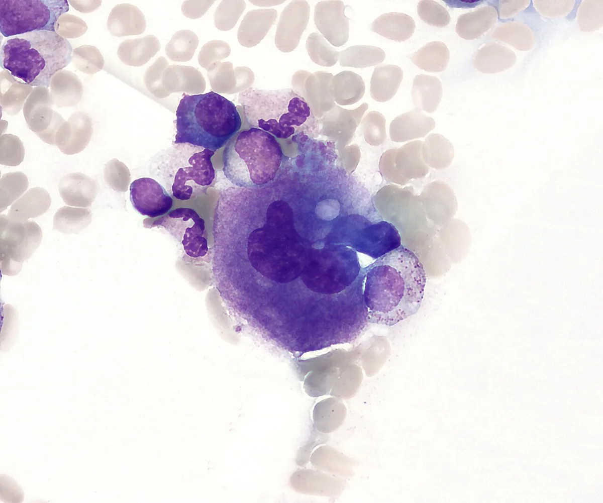
骨髄の中で最も大きい細胞(直径 50〜100 μm)で、核が分葉して1個の細胞に 16〜64 倍体の DNA が入っている。分裂せずに DNA だけ複製する(核内倍加)ため、核が巨大な塊に見える。標本で真っ先に目に入るので、骨髄と分かる目印になる。
血小板の作り方が独特で、細胞が割れて散るのではない。細胞質を細長い紐(前血小板)に伸ばし、それを類洞の壁の隙間から血流の中へ差し込む。流れる血が紐を引きちぎり、ちぎれた断片が血小板になる。1個の巨核球から 2,000〜5,000 個できる。
血小板に核が無い理由もここにある——もとから核の入っていない細胞質の切れ端だからである。
数の読み方。末梢の血小板が減っているとき、骨髄の巨核球が増えているか減っているかで原因が分かれる。
| 巨核球が増えている | 作ってはいるが末梢で壊されている/溜まっている | 免疫性血小板減少症(ITP)・脾機能亢進・DIC |
| 巨核球が減っている | 作る側が止まっている | 再生不良性貧血・化学療法後・白血病の浸潤 |
だから ITP では骨髄を見ると巨核球が多い。「血小板が減る病気なのに巨核球は増えている」という一見矛盾した像が、そのまま「末梢での破壊」という診断の根拠になる。
作られる量はトロンボポエチン(TPO、主に肝で産生)が決めている。血小板そのものが TPO を吸着して消費するので、血小板が減ると遊離 TPO が増えて産生が上がるという自動調節になっている。ITP に使う TPO 受容体作動薬は、この仕組みを外から押している薬である。
肝硬変で血小板が減る理由は2つ重なっている——脾腫による貯留と、肝で作る TPO の減少である。だから脾摘だけでは戻りきらないことがある。
English version
Core
The largest cell in the marrow (50–100 µm), with a lobulated nucleus containing 16–64 times the normal DNA in a single cell. It replicates its DNA without dividing (endomitosis), so the nucleus appears as a huge mass. It is the first thing the eye meets on a slide, and identifies the specimen as marrow.
The way platelets are made is unusual — the cell does not shatter. It extends its cytoplasm into long ribbons (proplatelets) and inserts them through the gaps in the sinusoid wall into the flowing blood. The flow tears the ribbons off, and the fragments are the platelets. One megakaryocyte yields 2,000–5,000.
Here too is the reason platelets have no nucleus — they are fragments of cytoplasm that never contained one.
Going further
How to read the number. When the peripheral platelet count is low, whether the marrow megakaryocytes are increased or decreased separates the causes.
| Megakaryocytes | Meaning | Examples |
|---|---|---|
| Increased | Production continues but they are destroyed or sequestered peripherally | Immune thrombocytopenia (ITP), hypersplenism, DIC |
| Decreased | Production has stopped | Aplastic anaemia, after chemotherapy, leukaemic infiltration |
So in ITP the marrow shows increased megakaryocytes. The apparently contradictory picture — a disease of low platelets in which megakaryocytes are raised — is itself the evidence for peripheral destruction.
The amount produced is set by thrombopoietin (made chiefly in the liver). Platelets themselves adsorb and consume TPO, so when platelets fall the free TPO rises and production increases — an automatic regulation. The TPO receptor agonists used in ITP push that mechanism from outside.
In the clinic
In cirrhosis the platelet count falls for two reasons together — sequestration in an enlarged spleen, and reduced hepatic production of thrombopoietin. This is why splenectomy alone may not fully correct it.
血液骨髄関門 — 成熟した細胞だけが、血へ出られる
骨髄の造血は類洞の外側(血管外)で行われている。できあがった血球は、類洞の内皮細胞を貫いて血管の中へ入る。この通り抜けが関門になっている。
- 変形できるものだけが通る——成熟した赤血球や白血球は核が小さい/柔らかいので通れる。有核赤血球や芽球は硬くて通れない
- 内皮に穴は開いていない——肝や脾の類洞と違い、骨髄の類洞は基底膜が乏しいものの内皮が連続している。細胞は隙間を抜けるのではなく、内皮細胞に一時的な孔を開けて通る
だから健常人の血には赤芽球も骨髄芽球も出てこない。末梢血に出ているかどうか自体が、関門の状態を語る。
関門が壊れる/迂回される3つの形。
| 白赤芽球症 | 幼若な赤血球と顆粒球が同時に末梢へ出る | 骨髄が線維や癌に置き換わっている(骨髄線維症・癌の骨髄転移) |
| 髄外造血 | 骨髄で作れず、肝・脾で作り始める | 脾腫・涙滴赤血球 |
| 幹細胞の動員 | 薬で意図的に関門を越えさせる | G-CSF や CXCR4 拮抗薬で末梢血幹細胞を採取する |
3番目が臨床で最も使われている。造血幹細胞は骨髄のニッチに CXCL12–CXCR4 で繋ぎ止められているので、その繋ぎを外せば血へ流れ出す。骨盤から骨髄を吸う代わりに、献血と同じ形で末梢血から幹細胞を集められるのはこの仕組みによる。
末梢血に赤芽球が出ていたら、まず骨髄の置き換わりを疑う。貧血の精査で「涙滴赤血球+白赤芽球症」が揃えば、骨髄線維症か癌の骨髄転移が上位に来る。
English version
Core
Haemopoiesis in the marrow takes place outside the sinusoids. The finished cells then pass through the sinusoidal endothelium into the vessel, and that passage is the barrier.
- Only what can deform passes — mature red and white cells have small or pliable nuclei and get through. Nucleated red cells and blasts are too rigid.
- There are no holes in the endothelium — unlike the sinusoids of liver and spleen, the marrow sinusoid has a continuous endothelium though little basal lamina. The cell does not slip through a gap but makes a temporary pore through an endothelial cell.
So neither erythroblasts nor myeloblasts appear in normal blood. Whether they are present is itself a statement about the barrier.
Going further
Three ways the barrier is broken or bypassed.
| Pattern | What is seen | Meaning |
|---|---|---|
| Leucoerythroblastic picture | Immature red and granulocytic cells together in the blood | The marrow is being replaced by fibrous tissue or tumour (myelofibrosis, marrow metastasis) |
| Extramedullary haemopoiesis | Production cannot occur in the marrow and begins in liver and spleen | Splenomegaly, teardrop cells |
| Stem cell mobilisation | The barrier is crossed deliberately with drugs | Peripheral blood stem cells harvested with G-CSF or a CXCR4 antagonist |
The third is the one most used clinically. The haemopoietic stem cell is tethered in the niche by CXCL12–CXCR4, so breaking that tether lets it flow into the blood. This is why stem cells can be collected in the same way as a blood donation, instead of aspirating marrow from the pelvis.
In the clinic
If nucleated red cells appear in the blood, suspect replacement of the marrow first.
In the investigation of anaemia, teardrop cells together with a leucoerythroblastic picture place myelofibrosis or marrow metastasis high on the list.
まとめ
| 骨髄 — 造血と脂肪の比で読む | 赤色骨髄(造血あり)と黄色骨髄(脂肪)。 |
|---|---|
| リンパ管 — 戻せなかったものを回収する | リンパ毛細管——盲端で始まる。内皮のみで、基底膜は不完全か欠く。 |
| 胸管 — 全身のリンパが、最後に1本へ集まる | リンパは末端から中心へ一方通行で流れ、最後は2本の本幹から静脈角へ入る。 |
| 乳び槽 — 脂だけが、血管を通らずに運ばれる | 小腸の絨毛の中心には、毛細血管ではなく1本の盲端のリンパ管がある(中心乳糜管)。 |
| 造血のニッチ — 幹細胞は、居場所によって振る舞いが変わる | 造血幹細胞は、骨髄のどこにいてもよいわけではない。 |
| 巨核球 — 類洞に腕を差し入れて、血小板をちぎる | 骨髄の中で最も大きい細胞(直径 50〜100 μm)で、核が分葉して1個の細胞に 16〜64 倍体の DNA が入っている。 |
| 血液骨髄関門 — 成熟した細胞だけが、血へ出られる | 骨髄の造血は類洞の外側(血管外)で行われている。 |
このノートは医学生が授業の内容を自分の言葉でまとめたもので、診療の判断に使うものではありません。誤りに気づいたらお知らせください。