筋は3種類しかない。骨格筋・心筋・平滑筋。区別する軸も3つだけ——横紋があるか/核はいくつでどこにあるか/随意か不随意か。この3点で標本を見れば必ず判別できる。
横紋の正体はアクチンとミオシンが規則正しく重なった縞である。だから横紋がある=サルコメアがある=長さが収縮力を決めるということになり、心不全の Frank-Starling 機構まで一本の線で繋がる。平滑筋に横紋が無いのは、同じ蛋白を使いながら配列が不規則だからにすぎない。
筋は興奮収縮連関という共通の仕組みで動く。細胞膜の脱分極 → Ca²⁺ の上昇 → アクチンとミオシンの滑り。この経路のどこが壊れたかで、重症筋無力症・悪性高熱・筋ジストロフィーが整理できる。
- 骨格筋
- 筋節(サルコメア)
- 興奮収縮連関 — T管と筋小胞体
- 心筋
- 平滑筋
- 筋疾患の読み方 — 神経原性か筋原性か
- 筋衛星細胞 — 骨格筋が、どこまで再生できるか
- 筋線維には型があり、染め分けられる
- 刺激伝導系の組織 — 特殊心筋
- 心臓の内分泌機能(ANP)と、心筋の再生能
- 平滑筋と心筋の発生 — どこから来たかで、再生の仕方が決まる
- 骨格筋Textus muscularis striatus
- 筋節(サルコメア)Sarcomerum
- 興奮収縮連関 — T管と筋小胞体
- 心筋Myocardium
- 平滑筋Textus muscularis levis
- 筋疾患の読み方 — 神経原性か筋原性か
- 筋衛星細胞 — 骨格筋が、どこまで再生できるかMyosatellitocytus
- 筋線維には型があり、染め分けられる
- 刺激伝導系の組織 — 特殊心筋Systema conducens cordis
- 心臓の内分泌機能(ANP)と、心筋の再生能
- 平滑筋と心筋の発生 — どこから来たかで、再生の仕方が決まるHistogenesis textus muscularis
- まとめ
骨格筋Textus muscularis striatus
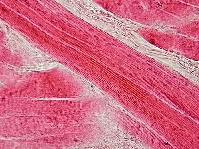
横紋あり・多核・核は辺縁・随意。1本の筋線維は多数の筋芽細胞が融合した合胞体で、核が細胞膜のすぐ内側に並ぶ。
被膜は3層——筋内膜(1本ずつ)→筋周膜(筋束)→筋外膜(筋全体)。血管と神経はこの層に沿って入る。
「核が辺縁にある」ことが標本上の決め手で、中心核が目立てば再生中か病的と読む。
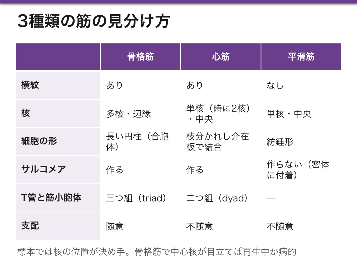
骨格筋線維は代謝で2型に分かれ、これが臨床の理解につながる。
- I型(遅筋・赤筋)——ミオグロビン・ミトコンドリア・毛細血管に富み、酸化的。疲れにくい。姿勢保持(ヒラメ筋)
- II型(速筋・白筋)——解糖に依存し、速いが疲れやすい。瞬発(大腿四頭筋の一部)
加齢・不動・ステロイドで先に萎縮するのは II型である。高齢者のサルコペニアで「立ち上がりが難しくなる」のが早い理由がここにあり、レジスタンストレーニングが推奨される根拠にもなる。
再生は筋線維の外、基底膜の内側にいる衛星細胞が担う。損傷時に活性化して融合し、新しい筋線維を作る。基底膜という管が残っているかどうかが再生の成否を決める点は、末梢神経の再生と同じ論理である。
合胞体だから、壊れると中身が血中へ大量に漏れる。横紋筋融解症ではCK の著明高値・ミオグロビン尿(赤褐色尿だが潜血陽性で赤血球は見えない)・高 K 血症・急性腎障害。スタチン・過度の運動・長時間の圧迫(挫滅症候群)が原因になる。大量輸液が治療の中心である。
Duchenne 型筋ジストロフィー — ジストロフィン(膜と細胞骨格をつなぐ)の欠損。X 連鎖潜性で男児に発症し、登はん性起立(Gowers 徴候)・腓腹筋の仮性肥大。筋生検では線維の大小不同・壊死と再生・脂肪と線維への置換。
筋の再生には限界がある。基底膜が保たれていれば衛星細胞が修復できるが、基底膜ごと壊れれば瘢痕になる——挫滅の程度が回復の見込みを決める。
English version
Core
Striated, multinucleate, nuclei peripheral, voluntary. A muscle fibre is a syncytium formed by the fusion of many myoblasts, with the nuclei lying just inside the sarcolemma.
Three sheaths — endomysium (each fibre) → perimysium (a fascicle) → epimysium (the whole muscle). Vessels and nerves enter along these layers.
That the nuclei are peripheral is the decisive feature on a slide; if central nuclei are prominent, read it as regenerating or diseased.
Going further
Skeletal muscle fibres divide into two metabolic types, and this leads to clinical understanding.
- Type I (slow, red) — rich in myoglobin, mitochondria and capillaries; oxidative and fatigue-resistant. Postural (soleus).
- Type II (fast, white) — glycolytic; fast but easily fatigued. Explosive movement.
It is type II that atrophies first with age, immobility and corticosteroids. This is why difficulty rising from a chair appears early in the sarcopenia of the elderly, and the basis for recommending resistance training.
Regeneration is carried out by the satellite cell, lying outside the fibre but inside the basal lamina. It is activated by injury, fuses and forms new fibres. Whether the tube of basal lamina survives decides the success of regeneration — the same logic as in peripheral nerve.
In the clinic
Being a syncytium, when it breaks its contents leak into the blood in quantity.
Rhabdomyolysis — markedly raised CK, myoglobinuria (red-brown urine, dipstick positive for blood but no red cells on microscopy), hyperkalaemia and acute kidney injury. Causes include statins, extreme exercise and prolonged compression (crush syndrome). Aggressive fluid therapy is the mainstay.
Duchenne muscular dystrophy — absence of dystrophin, which links membrane to cytoskeleton. X-linked recessive, affecting boys; Gowers sign and pseudohypertrophy of the calves. Biopsy shows variation in fibre size, necrosis and regeneration, and replacement by fat and fibrous tissue.
Regeneration has limits. If the basal lamina is preserved the satellite cells can repair; if it is destroyed as well, the result is scar — the degree of crushing determines the prospect of recovery.
筋節(サルコメア)Sarcomerum
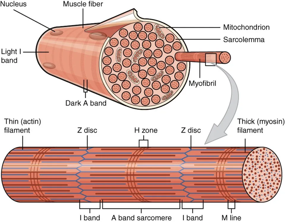
収縮の最小単位。Z帯からZ帯まで。
- A帯(暗い)——ミオシン(太いフィラメント)の長さ。収縮しても長さが変わらない
- I帯(明るい)——アクチン(細いフィラメント)だけの部分。収縮で短くなる
- H帯——A帯の中央、アクチンが届かない部分。収縮で短くなる
- Z帯——アクチンの係留点
収縮してもフィラメント自体は短くならない。互いに滑り込むだけ(滑り説)。
サルコメアの長さと発生張力の関係(長さ-張力曲線)が、そのまま心臓の生理につながる。アクチンとミオシンの重なりが最適なときに張力が最大になり、伸びすぎても縮みすぎても落ちる。
心筋では、拡張期に心室が伸ばされるほど収縮力が増す——これがFrank-Starling 機構である。前負荷が上がると心拍出量が増えるという循環生理の中核が、サルコメアの重なりという組織学的な事実から出ている。
タイチン(コネクチン)はZ帯からM線まで伸びる巨大な弾性蛋白で、受動的張力を生み、伸ばされた筋を元に戻す。心筋の拡張機能に関わり、その硬化がHFpEF(駆出率の保たれた心不全)に関与する。
サルコメアの構造がそのまま検査値になる。心筋トロポニン T・I は細いフィラメントの調節蛋白で、心筋にしかない型があるため心筋梗塞の特異的なマーカーになる。発症後3〜6時間で上昇し、1〜2週間続く——遅れて受診した患者でも診断できるのはこのためである。
横紋が消えるのは変性のサインで、心筋梗塞の初期像では核が消え、細胞質が好酸性になり、横紋が失われる(凝固壊死)。
強直(rigor mortis)はATP が枯渇してミオシン頭部がアクチンから外れなくなるために起こる。収縮ではなく「離れられない」状態である。
心不全の治療薬の一部はサルコメアの Ca 感受性やミオシンの働きに直接作用する。
English version
Core
The smallest unit of contraction, from Z line to Z line.
- A band (dark) — the length of the myosin (thick) filaments. Its length does not change with contraction.
- I band (light) — actin (thin) filaments only. Shortens on contraction.
- H zone — the centre of the A band, where actin does not reach. Shortens on contraction.
- Z line — the anchorage of actin.
The filaments themselves do not shorten on contraction; they simply slide past one another (the sliding filament theory).
Going further
The relation between sarcomere length and developed tension (the length–tension curve) leads directly into the physiology of the heart. Tension is maximal when the overlap of actin and myosin is optimal and falls when the sarcomere is either over-stretched or over-shortened.
In cardiac muscle, the more the ventricle is stretched in diastole the greater the force of contraction — the Frank–Starling mechanism. The central fact of circulatory physiology, that raising the preload raises the cardiac output, comes out of the histological fact of filament overlap.
Titin (connectin) is a giant elastic protein running from Z line to M line, generating passive tension and restoring a stretched muscle. It contributes to diastolic function in the heart, and its stiffening is implicated in HFpEF, heart failure with preserved ejection fraction.
In the clinic
The structure of the sarcomere becomes a laboratory value.
Cardiac troponin T and I are regulatory proteins of the thin filament, and because their cardiac isoforms exist nowhere else they are specific markers of myocardial infarction. They rise 3–6 hours after onset and remain for one to two weeks — which is why the diagnosis can still be made in a patient who presents late.
Loss of the striations is a sign of degeneration: in early infarction the nucleus disappears, the cytoplasm becomes eosinophilic and the striations are lost (coagulative necrosis).
Rigor mortis occurs because ATP is exhausted and the myosin head cannot detach from actin. It is not contraction but an inability to let go.
Some drugs for heart failure act directly on the calcium sensitivity of the sarcomere or on myosin itself.
興奮収縮連関 — T管と筋小胞体
電気信号を収縮に変える仕組み。
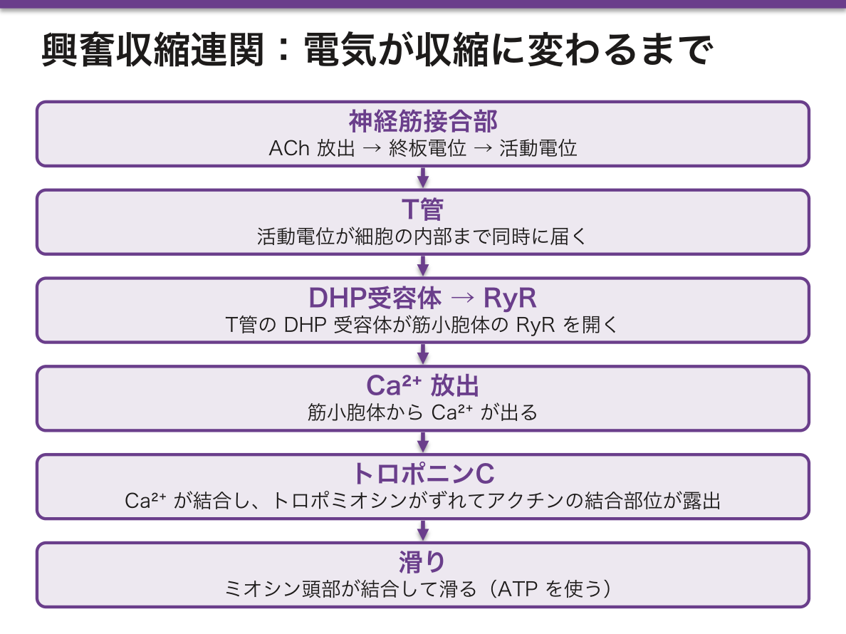
- 神経筋接合部でACh 放出 → 終板電位 → 活動電位
- 活動電位がT管(細胞膜が細胞内へ落ち込んだ管)を伝わり、内部まで同時に届く
- T管のDHP受容体が筋小胞体の RyR を開く
- 筋小胞体からCa²⁺ が放出される
- Ca²⁺ がトロポニンC に結合 → トロポミオシンがずれ → アクチンの結合部位が露出
- ミオシン頭部が結合して滑る(ATP を使う)
骨格筋ではT管1本+筋小胞体2つ=三つ組(triad)、心筋は二つ組(dyad)。
骨格筋と心筋で Ca²⁺ の出どころが違うのが決定的である。
- 骨格筋——DHP受容体とRyRが物理的に連結。細胞外Caをほとんど必要としない。だからCa拮抗薬が骨格筋の収縮力に影響しない
- 心筋——L型Caチャネルから入った少量の細胞外Caが引き金となり、筋小胞体からさらにCaが出る(Ca誘発性Ca放出)。細胞外Caに依存するため、Ca拮抗薬が心収縮力を落とす
この違いを知っていれば、薬理の暗記が要らなくなる。
硬直(死後硬直)も同じ経路で説明できる。ATP が枯渇するとミオシン頭部がアクチンから外れられず、結合したまま固まる。ATP は収縮ではなく「離れる」ために要るという点が要諦である。
悪性高熱症——RyR1 の変異。揮発性吸入麻酔薬やスキサメトニウムで筋小胞体からCa²⁺が制御不能に放出され、持続的な筋収縮・高体温・筋強直・横紋筋融解・高K血症をきたす。治療はダントロレン(RyR を直接ブロックする)。受容体1つの異常が、麻酔科の最重要救急になる。
English version
Core
The mechanism that converts an electrical signal into contraction.
- Acetylcholine is released at the neuromuscular junction → end-plate potential → action potential.
- The action potential travels along the T tubule (an invagination of the sarcolemma), reaching the interior of the fibre simultaneously.
- The DHP receptor of the T tubule opens the ryanodine receptor of the sarcoplasmic reticulum.
- Ca²⁺ is released from the sarcoplasmic reticulum.
- Ca²⁺ binds troponin C → tropomyosin shifts → the binding site on actin is exposed.
- The myosin head attaches and slides, using ATP.
Skeletal muscle has one T tubule with two cisternae (a triad); cardiac muscle has a dyad.
Going further
The decisive difference is where the calcium comes from.
- Skeletal muscle — the DHP receptor and the ryanodine receptor are physically coupled, and almost no extracellular calcium is needed. This is why calcium channel blockers do not affect the force of skeletal muscle.
- Cardiac muscle — a small amount of extracellular calcium entering through the L-type channel triggers further release from the sarcoplasmic reticulum (calcium-induced calcium release). Being dependent on extracellular calcium, the force of cardiac contraction is reduced by calcium channel blockers.
Know this difference and the pharmacology need not be memorised.
Rigor mortis is explained by the same pathway: when ATP is exhausted the myosin head cannot detach from actin and remains bound. The essential point is that ATP is required not for contraction but for letting go.
In the clinic
Malignant hyperthermia — a mutation of RyR1. Volatile anaesthetics and suxamethonium cause uncontrolled release of calcium from the sarcoplasmic reticulum, giving sustained contraction, hyperthermia, muscle rigidity, rhabdomyolysis and hyperkalaemia.
Treatment is dantrolene, which blocks the ryanodine receptor directly.
A defect in a single receptor becomes the most important emergency in anaesthesia.
心筋Myocardium
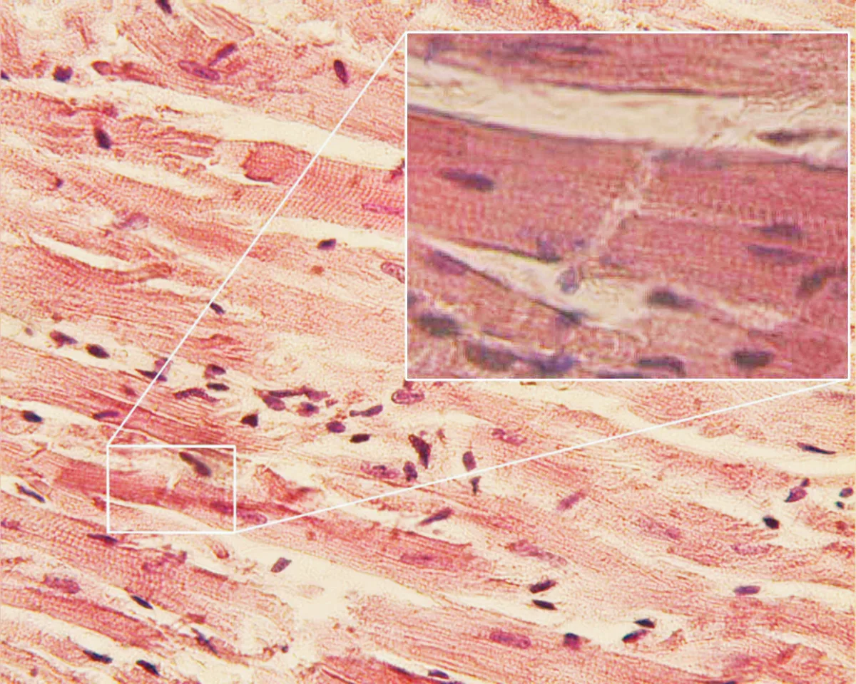
横紋あり・単核(時に2核)・核は中央・不随意。細胞は枝分かれし、端どうしが介在板 discus intercalatus で結合する。
介在板には3つの装置が入る。
- デスモソーム——引っ張りに耐える
- 接着帯——アクチンを繋ぐ
- ギャップ結合(nexus)——電気的に繋ぐ
ギャップ結合があるため、心筋は全体が機能的合胞体として同期して収縮する。
「機能的合胞体」であることが不整脈の理解の土台になる。細胞が電気的に繋がっているので、興奮は隣へ次々に伝わる。この性質が、リエントリー(旋回興奮)による頻脈の成立条件でもある。
心筋には再生能がほぼ無い。心筋梗塞で失われた心筋は線維性瘢痕に置き換わり、収縮しない領域が残る。これが心不全へ進む理由であり、「早期再灌流」が治療の中心である根拠そのものである。「時間は心筋なり」という標語は組織学的事実に基づく。
高齢の心筋細胞にはリポフスチン(消耗色素)が核周囲に蓄積し、褐色に見える(褐色萎縮)。ライソゾームで分解しきれなかった脂質の残骸で、分裂しない細胞(心筋・神経・肝)に溜まる点が共通する。
介在板の3つの装置が、それぞれ別の病気になる。
ギャップ結合は心筋を機能的合胞体にする。虚血でギャップ結合が壊れると伝導が乱れ、リエントリー性不整脈が起こる。梗塞後の心室頻拍の基盤である。
デスモソームの蛋白(プラコフィリン・デスモグレイン)の変異が不整脈原性右室心筋症を起こす。心筋が脂肪と線維に置き換わり、若年者の突然死の原因になる。接着装置の異常が、致死性不整脈になる。
心筋はほとんど再生しない。だから梗塞巣は線維性瘢痕に置き換わり、収縮に参加しない。心室瘤・心破裂・心不全はこの帰結である。
心房筋は ANP を分泌する——心筋は内分泌細胞でもある。BNP が心不全の指標になるのはこの性質による。
English version
Core
Striated, mononucleate (sometimes binucleate), nucleus central, involuntary. The cells branch and are joined end to end at the intercalated disc.
The intercalated disc contains three devices:
- Desmosome — resists tension.
- Adherens junction — links actin.
- Gap junction (nexus) — connects electrically.
Because of the gap junctions, the myocardium contracts synchronously as a functional syncytium.
Going further
Being a functional syncytium is the foundation for understanding arrhythmia. The cells are electrically connected, so excitation passes from one to the next — and this property is also the condition for re-entrant tachycardia.
Cardiac muscle has almost no regenerative capacity. Myocardium lost in infarction is replaced by fibrous scar, leaving a region that does not contract. This is why heart failure follows, and it is the very basis for making early reperfusion the centre of treatment. "Time is muscle" rests on a histological fact.
In the aged heart, lipofuscin accumulates around the nuclei and the muscle appears brown (brown atrophy). It is undigested lipid residue in lysosomes, and it is common to cells that do not divide — cardiac muscle, neurons and hepatocytes.
In the clinic
Each of the three devices in the intercalated disc becomes a separate disease.
Gap junctions make the myocardium a functional syncytium. When ischaemia damages them conduction becomes disordered and re-entrant arrhythmia follows — the substrate of ventricular tachycardia after infarction.
Mutations in desmosomal proteins (plakophilin, desmoglein) cause arrhythmogenic right ventricular cardiomyopathy, in which myocardium is replaced by fat and fibrous tissue and which causes sudden death in the young. A defect of an adhesion device becomes a lethal arrhythmia.
Cardiac muscle scarcely regenerates, so an infarct is replaced by fibrous scar that takes no part in contraction: ventricular aneurysm, cardiac rupture and heart failure follow.
Atrial muscle secretes ANP — the myocardium is also an endocrine tissue, and this is why BNP serves as an index of heart failure.
平滑筋Textus muscularis levis
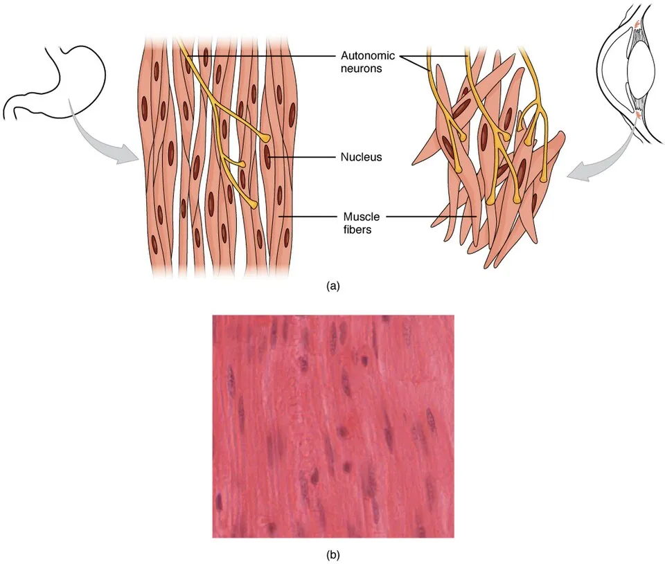
横紋なし・単核・核は中央(紡錘形の細胞の真ん中)・不随意。細胞は紡錘形で、収縮すると核がねじれてらせん状に見える。
場所:消化管・血管・気道・膀胱・子宮・立毛筋・虹彩。
アクチンとミオシンはあるがサルコメアを作らず、斜めに走って密体(Z帯に相当)に付着する。だから横紋が出ない。
収縮の制御が骨格筋と根本的に違う。平滑筋にはトロポニンが無い。Ca²⁺ はカルモジュリンに結合し、ミオシン軽鎖キナーゼ(MLCK)を活性化してミオシン側をリン酸化する。
骨格筋=アクチン側の制御(トロポニン)/平滑筋=ミオシン側の制御(MLCK)——この対比が薬理の理解を支える。
- NO・硝酸薬——cGMP↑ → MLC脱リン酸化を促進 → 血管拡張
- β2刺激薬——cAMP↑ → MLCK を抑制 → 気管支拡張
- Ca拮抗薬——細胞外Ca流入を止める → 血管平滑筋弛緩
平滑筋は収縮が遅く、持続し、消費エネルギーが少ない(latch state)。血管が一日中緊張を保てるのはこのためで、疲労する骨格筋との決定的な違いになる。
また平滑筋は分裂して増える能力を保っている。子宮平滑筋が妊娠で肥大・過形成するのも、動脈硬化で中膜の平滑筋が内膜へ遊走・増殖するのもこの性質による。
平滑筋は再生でき、増殖もする。これが骨格筋・心筋との決定的な違いで、そのまま病気の作られ方になる。
動脈硬化では中膜の平滑筋が内膜へ遊走して増殖し、線維性被膜を作る。ステント留置後の再狭窄も同じ現象で、薬剤溶出ステントは平滑筋の増殖を抑える薬を塗っている。
気管支喘息では気道平滑筋の肥厚がリモデリング(不可逆な狭窄)の主体になる。
子宮筋腫(平滑筋腫)は最も多い良性腫瘍の1つで、エストロゲン依存性なので閉経後に縮む。
平滑筋のマーカーはデスミンと α-SMAで、消化管間質腫瘍(GIST)との鑑別に使う(GIST は KIT/CD117 陽性で、由来は Cajal 介在細胞である)。
English version
Core
No striations, mononucleate, nucleus central (in the middle of a spindle-shaped cell), involuntary. On contraction the nucleus twists into a corkscrew.
Sites: gastrointestinal tract, vessels, airways, bladder, uterus, arrector pili, iris.
Actin and myosin are present but form no sarcomeres; they run obliquely and attach to dense bodies (equivalent to the Z line). Hence there are no striations.
Going further
The control of contraction is fundamentally different from skeletal muscle. Smooth muscle has no troponin. Ca²⁺ binds calmodulin, which activates myosin light chain kinase (MLCK) and phosphorylates the myosin side.
Skeletal muscle is regulated on the actin side (troponin); smooth muscle on the myosin side (MLCK) — this contrast supports the pharmacology.
- NO and nitrates — raise cGMP → promote dephosphorylation of myosin light chain → vasodilatation.
- β2 agonists — raise cAMP → inhibit MLCK → bronchodilatation.
- Calcium channel blockers — stop the entry of extracellular calcium → relax vascular smooth muscle.
Smooth muscle contracts slowly, sustains it and consumes little energy (the latch state). This is how a vessel can maintain tone all day — the decisive difference from fatigable skeletal muscle.
Smooth muscle also retains the ability to divide. Hypertrophy and hyperplasia of the uterus in pregnancy, and the migration and proliferation of medial smooth muscle into the intima in atherosclerosis, both follow from this property.
In the clinic
Smooth muscle can regenerate and can proliferate — the decisive difference from skeletal and cardiac muscle, and directly the way its diseases are built.
In atherosclerosis medial smooth muscle migrates into the intima, proliferates and forms the fibrous cap. Restenosis after stenting is the same phenomenon, and the drug-eluting stent is coated with an agent that suppresses smooth muscle proliferation.
In asthma thickening of the airway smooth muscle is the main element of remodelling (irreversible narrowing).
Uterine leiomyoma is one of the commonest benign tumours and, being oestrogen-dependent, shrinks after the menopause.
The markers of smooth muscle are desmin and α-SMA, used to distinguish it from gastrointestinal stromal tumour (KIT/CD117 positive, derived from the interstitial cell of Cajal).
筋疾患の読み方 — 神経原性か筋原性か
筋力低下を見たとき、まず筋そのものの病気か、支配神経の病気かを分ける。
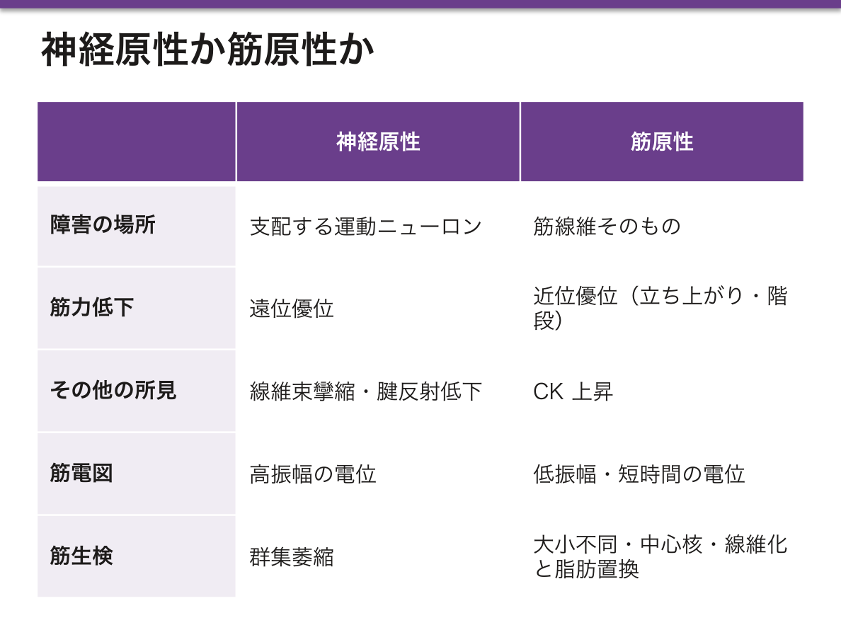
- 神経原性——支配する運動ニューロンの障害。遠位優位・線維束攣縮・腱反射低下・筋電図で高振幅の電位。生検で群集萎縮(同じ運動単位がまとめて萎縮)
- 筋原性——筋線維自体の障害。近位優位(立ち上がり・階段・腕を上げる動作が先に困る)・CK上昇・筋電図で低振幅短時間の電位。生検で大小不同・中心核・線維化と脂肪置換
Duchenne 型筋ジストロフィー——ジストロフィンの欠損(X連鎖潜性)。ジストロフィンは筋細胞の細胞骨格と基底膜を繋ぐ蛋白で、これが無いと収縮のたびに細胞膜が裂ける。
- CK著明高値(膜が壊れて漏れる)
- 登攀性起立(Gowers 徴候)——近位筋の脱力
- 腓腹筋の仮性肥大——筋が脂肪と線維組織に置き換わって太く見えるだけ
- 心筋も同じ蛋白を使うため拡張型心筋症を合併する
Becker 型は同じ遺伝子だが読み枠が保たれる変異で、量的・質的に不完全なジストロフィンが作られるため軽症。「フレームシフトか否か」で重症度が決まるという遺伝子と表現型の対応がきれいに出る例である。
神経筋接合部の病気はどちら側が壊れたかで症状が分かれる。重症筋無力症は後シナプスのACh受容体に対する自己抗体で、使うほど悪化(易疲労性)・眼瞼下垂と複視で発症・胸腺腫を合併。Lambert-Eaton 症候群は前シナプスのCaチャネル抗体で、使うと一時的に改善し、小細胞肺癌に伴うことが多い。「反復刺激で減るか増えるか」が検査上の決め手になる。
English version
Core
Faced with weakness, first separate disease of the muscle itself from disease of its nerve supply.
- Neurogenic — damage to the motor neuron. Distal predominance, fasciculation, reduced reflexes, high-amplitude potentials on EMG. Biopsy shows group atrophy (a whole motor unit atrophying together).
- Myopathic — damage to the fibre itself. Proximal predominance (rising from a chair, stairs, lifting the arms fail first), raised CK, low-amplitude short-duration potentials on EMG. Biopsy shows variation in fibre size, central nuclei, and replacement by fibrous tissue and fat.
Going further
Duchenne muscular dystrophy — absence of dystrophin (X-linked recessive). Dystrophin links the cytoskeleton of the muscle cell to the basal lamina, and without it the membrane tears with every contraction.
- Markedly raised CK — leaking through the broken membrane.
- Gowers sign — climbing up the body, from proximal weakness.
- Pseudohypertrophy of the calves — the muscle merely looks large because it has been replaced by fat and fibrous tissue.
- Cardiac muscle uses the same protein, so dilated cardiomyopathy accompanies it.
Becker type involves the same gene but with a mutation that preserves the reading frame, so an incomplete dystrophin is still made and the disease is milder. Severity determined by whether the mutation is a frameshift — an unusually clean correspondence between genotype and phenotype.
In the clinic
Diseases of the neuromuscular junction separate by which side is broken.
Myasthenia gravis — autoantibodies to the postsynaptic acetylcholine receptor. Worse with use (fatigability), presenting with ptosis and diplopia, and associated with thymoma.
Lambert–Eaton syndrome — antibodies to the presynaptic calcium channel. Temporarily improves with use, and is often associated with small cell lung cancer.
Whether the response decreases or increases on repetitive stimulation is the decisive test.
筋衛星細胞 — 骨格筋が、どこまで再生できるかMyosatellitocytus
筋線維の形質膜と基底膜のあいだにある、静止した幹細胞。核が1つで、細胞質に乏しい。
損傷すると活性化し、増殖して筋芽細胞となり、融合して既存の筋線維を修復するか、新しい筋管を作る。
再生した筋線維は、核が中心に並ぶ(中心核)——生検で「再生している」ことの指標になる。
| 筋 | 再生能 | 肥大の仕方 |
|---|---|---|
| 骨格筋 | ある(衛星細胞) | 肥大+核の追加 |
| 心筋 | ほとんど無い | 肥大のみ。壊れた所は線維化 |
| 平滑筋 | ある(自ら分裂できる) | 肥大+過形成 |
基底膜が残っていることが、再生の足場になる。
3種類の筋の再生能の差が、そのまま病気の治り方の差になる。心筋梗塞の後に瘢痕が残るのは、心筋が作り直せないから(循環器の病理の章)——「壊れた面積が、そのまま将来の心機能になる」。
子宮が妊娠で大きくなるのは、平滑筋が肥大と過形成の両方をするためになる(妊娠が体に起こす変化の章)——「骨格筋にはできない増え方をしている」。
衛星細胞が有限であることは、筋ジストロフィーの経過を説明する。壊れては再生を繰り返すうちに衛星細胞が枯れ、やがて脂肪と線維に置き換わる(筋疾患の章)——「生検で、壊死・再生・脂肪浸潤が同じ視野に並ぶ」。
基底膜が残るかどうかが、回復の質を分ける。基底膜まで壊れた挫滅では、足場が無いので瘢痕で埋まる(修復と創傷治癒の章)——「筋が『治る』と『固まる』の分かれ目が、この膜にある」。
筋力トレーニングで筋が太くなるときも、衛星細胞が核を供給している(運動の生化学の章)——「1本の線維が扱える細胞質の量に上限があるので、核を足す必要がある」。
基底膜が残っているかどうかが、再生できるかを決める。基底膜という「型」が残っていれば衛星細胞がその中で筋管を作れるが、型ごと壊れれば線維性瘢痕になる。挫滅と断裂で回復の見込みが違うのはこのためである。
筋ジストロフィーでは再生が追いつかない。壊死と再生を繰り返すうちに衛星細胞が枯渇し、最終的に脂肪と線維に置き換わる。
加齢によるサルコペニアも衛星細胞の減少が関わる。レジスタンス運動と十分な蛋白摂取が唯一のはっきりした対策である。
不動と廃用では筋線維が細くなる(萎縮)が数は減らないので、再び動かせば戻る——早期離床とリハビリテーションの根拠になっている。
English version
Core
A quiescent stem cell lying between the sarcolemma and the basal lamina of a muscle fibre; a single nucleus with scanty cytoplasm.
On injury it is activated, proliferates into myoblasts and either repairs the existing fibre or fuses to form new myotubes. Regenerated fibres have their nuclei in the centre — the index on biopsy that regeneration is occurring.
| Regenerative capacity | Mode of hypertrophy | |
|---|---|---|
| Skeletal | Yes (satellite cells) | Hypertrophy plus addition of nuclei |
| Cardiac | Almost none | Hypertrophy only; damage becomes fibrosis |
| Smooth | Yes (divides itself) | Hypertrophy plus hyperplasia |
The surviving basal lamina is the scaffold for regeneration.
Going further
The difference in regenerative capacity between the three muscles is directly the difference in how their diseases resolve.
A scar remains after myocardial infarction because cardiac muscle cannot be rebuilt — the area destroyed is directly the future cardiac function. The uterus enlarges in pregnancy because smooth muscle undergoes both hypertrophy and hyperplasia — a way of growing that skeletal muscle cannot manage.
That satellite cells are finite explains the course of muscular dystrophy. Through repeated destruction and regeneration the satellite cells are exhausted, and the muscle is eventually replaced by fat and fibrous tissue — necrosis, regeneration and fatty infiltration all appear in the same field on biopsy.
Whether the basal lamina survives separates recovery from repair. In a crush injury that destroys the basal lamina there is no scaffold, and the defect fills with scar — the difference between a muscle that heals and one that stiffens lies in this membrane.
Satellite cells also supply nuclei when muscle hypertrophies with training — there is a ceiling to the volume of cytoplasm one nucleus can serve, so nuclei must be added.
In the clinic
Whether the basal lamina survives decides whether regeneration is possible. If the "mould" remains, satellite cells can form myotubes within it; if the mould is destroyed, the result is fibrous scar. This is why the outlook differs between a crush and a clean tear.
In muscular dystrophy regeneration cannot keep pace. Repeated necrosis and regeneration exhaust the satellite cells and the muscle is finally replaced by fat and fibrous tissue.
Sarcopenia of ageing also involves a decline in satellite cells; resistance exercise with adequate protein intake is the only clearly effective measure.
In immobility and disuse the fibres become thinner (atrophy) but their number does not fall, so they return when movement resumes — the basis for early mobilisation and rehabilitation.
筋線維には型があり、染め分けられる
骨格筋は一様ではなく、性質の違う線維が混ざって並んでいる。ATPase 染色や免疫染色で染め分けると、市松模様(モザイク)に見える。
| I 型(遅筋・赤筋) | II 型(速筋・白筋) | |
| 代謝 | 酸化的。ミトコンドリアとミオグロビンが多い | 解糖系。グリコーゲンが多い |
| 収縮 | 遅く、疲れにくい | 速く、疲れやすい |
| 役割 | 姿勢の保持・持久 | 瞬発的な力 |
| 毛細血管 | 多い | 少ない |
型を決めているのは筋線維ではなく、支配している運動ニューロンである。1 個のニューロンが支配する線維(運動単位)は、すべて同じ型になる。神経を繋ぎ替えると、線維の型も変わる。
この配置が崩れる形で、疾患が読める。
| 群集萎縮/群化 | 同じ型の線維が塊で並ぶ | 神経原性。脱神経した線維を隣のニューロンが再支配し、その型に染め変えたため。モザイクが失われる |
| 大小不同・壊死と再生・脂肪への置換 | 型と無関係に散在する | 筋原性 |
| II 型線維の選択的萎縮 | 速筋だけが細くなる | 廃用・ステロイド・悪性腫瘍・加齢。最も頻度が高い所見の1つ |
| I 型線維の優位 | 遅筋ばかりになる | 先天性ミオパチーの一部 |
「モザイクが保たれているか」を最初に見る——これで神経原性か筋原性かの大枠が決まる。
加齢とサルコペニアは II 型から進む。だから高齢者はまず速い動き(立ち上がる・とっさに踏ん張る)が落ちる。転倒の予防に、歩行だけでなく素早い動きの訓練が要るのは、この選択的な萎縮のためである。
⚠️ 生検の部位を誤ると診断できない。進行しすぎた筋は脂肪に置き換わっていて何も分からないし、筋電図を刺した場所は人工産物が出る。中等度に侵された筋を、筋電図と反対側から採るのが原則である。
ステロイドを長く使う患者では、立ち上がりにくさを定期的に聞く。近位筋の II 型線維から落ちるので、症状の出方が決まっている。
English version
Core
Skeletal muscle is not uniform: fibres of different character are intermingled. Stained by ATPase or immunohistochemistry they appear as a chequerboard (mosaic).
| Type I (slow, red) | Type II (fast, white) | |
|---|---|---|
| Metabolism | Oxidative; rich in mitochondria and myoglobin | Glycolytic; rich in glycogen |
| Contraction | Slow, fatigue-resistant | Fast, easily fatigued |
| Role | Posture, endurance | Explosive force |
| Capillaries | Many | Few |
The type is determined not by the fibre but by the motor neuron that supplies it. All the fibres of one motor unit are of the same type, and cross-innervating a nerve changes the type of the fibre.
Going further
Disease is read as a breakdown of this arrangement.
| Finding | Appearance | Meaning |
|---|---|---|
| Group atrophy / type grouping | Fibres of one type in blocks | Neurogenic. Denervated fibres re-innervated by a neighbouring neuron and converted to its type. The mosaic is lost |
| Variation in size, necrosis and regeneration, fatty replacement | Scattered irrespective of type | Myopathic |
| Selective type II atrophy | Only fast fibres thin | Disuse, corticosteroids, malignancy, ageing. One of the commonest findings |
| Type I predominance | Almost all slow fibres | Some congenital myopathies |
Look first at whether the mosaic is preserved — that settles the broad division into neurogenic and myopathic.
Ageing and sarcopenia proceed from type II, so what the elderly lose first is quick movement — standing up, or catching oneself. The need to train quick movements as well as walking, for the prevention of falls, follows from this selective atrophy.
⚠️ Sampling the wrong site makes diagnosis impossible. A muscle too far advanced has been replaced by fat and shows nothing, and a muscle in which EMG needles have been placed shows artefact. The rule is to take a moderately affected muscle, on the side opposite to the EMG.
In the clinic
In patients on long-term corticosteroids, ask regularly about difficulty rising from a chair. Because the type II fibres of the proximal muscles fail first, the way the symptoms appear is predetermined.
刺激伝導系の組織 — 特殊心筋Systema conducens cordis
神経ではなく、変化した心筋細胞でできている。共通の特徴は収縮装置(筋原線維)が乏しく、グリコーゲンに富み、明るく大きく見えること。
洞房結節——上大静脈と右心房の境(分界溝の上端)。最も小さい「P 細胞(歩調取り細胞)」が中心動脈を取り巻く。固有の自動能が最も速い(毎分60〜100)ので、ここが歩調取りになる。
房室結節——心房中隔の下部、Koch 三角の中。細胞が細く、伝導が遅い——この遅れが、心房が収縮し終えてから心室が収縮するための「間」を作る。
房室束(His 束)——線維骨格を貫く唯一の筋の連絡。ここが切れると完全房室ブロック。
Purkinje 線維——最も太く、最も明るく、筋原線維が周辺に押しやられている。心内膜下を走り、伝導が最も速い(毎秒約4m)。標本で見分けるのはこれ。
自動能は洞房結節 60〜100 > 房室結節 40〜60 > Purkinje 20〜40 と下るほど遅い。上位が止まると下位が肩代わりする(補充調律)。
English version
Core
Made of modified cardiac muscle, not of nerve. The common features are scanty myofibrils, abundant glycogen, and a pale, large appearance.
- Sinoatrial node — at the junction of the superior vena cava and right atrium. The smallest cells, the P (pacemaker) cells, surround a central artery. Its intrinsic rate is the fastest (60–100 per minute), so it is the pacemaker.
- Atrioventricular node — in the lower interatrial septum, within the triangle of Koch. The cells are slender and conduction is slow — and that delay creates the interval that allows the atria to finish contracting before the ventricles begin.
- Bundle of His — the only muscular connection through the fibrous skeleton. If it is interrupted, complete heart block.
- Purkinje fibres — the largest and palest, with myofibrils pushed to the periphery. They run subendocardially and conduct fastest (about 4 m/s). These are what to look for on a slide.
Going further
Intrinsic rates fall down the system — sinoatrial node 60–100 > atrioventricular node 40–60 > Purkinje 20–40. If a higher pacemaker fails, a lower one takes over (an escape rhythm).
心臓の内分泌機能(ANP)と、心筋の再生能
心臓は内分泌器官でもある。心房筋細胞(特に右心房)の細胞質には、電子密度の高い分泌顆粒がある——これが組織学的な証拠。
ANP(心房性ナトリウム利尿ペプチド)は心房壁が伸展されると放出される(=循環血液量が増えたとき)。働きは①腎でのナトリウム排泄と利尿 ②血管拡張 ③レニン・アルドステロン・バソプレシンの抑制——要するにレニン-アンジオテンシン系の逆。心室からは BNP が出て、心不全の指標に使われる。
再生能——長く「心筋は再生しない」とされてきたが、炭素14 を用いた研究で、ヒトの心筋細胞は年に約1%(加齢で0.3%程度に低下)入れ替わることが示された。ただし梗塞巣を埋めるには全く足りない。
だから梗塞後の修復は再生ではなく線維性瘢痕——収縮しない組織に置き換わるため、心不全と不整脈の基盤になる。「わずかに再生するが、実質的には瘢痕治癒」と答えるのが正確。
心筋細胞は多くが二核で、多倍体化している——これが分裂しにくい理由の一つ。再生医療では iPS 由来心筋細胞の移植が試みられている。
English version
Core
The heart is also an endocrine organ. The cytoplasm of atrial myocytes, especially in the right atrium, contains electron-dense secretory granules — the histological evidence.
ANP is released when the atrial wall is stretched, that is, when the circulating volume rises. Its actions are ① natriuresis and diuresis in the kidney ② vasodilatation ③ inhibition of renin, aldosterone and vasopressin — in short, the opposite of the renin–angiotensin system. The ventricles release BNP, used as an index of heart failure.
Regenerative capacity — it was long held that cardiac muscle does not regenerate, but carbon-14 studies have shown that human cardiomyocytes turn over at about 1 per cent a year, falling to around 0.3 per cent with age. This is nowhere near enough to fill an infarct. So repair after infarction is not regeneration but fibrous scar — replacement by tissue that does not contract, and the basis of heart failure and arrhythmia.
The accurate answer is: it regenerates slightly, but in practice it heals by scarring.
Going further
Many cardiomyocytes are binucleate and polyploid — one of the reasons they divide poorly. In regenerative medicine, transplantation of iPS-derived cardiomyocytes is being attempted.
平滑筋と心筋の発生 — どこから来たかで、再生の仕方が決まるHistogenesis textus muscularis
| 由来 | でき方 | 生後の再生 | |
|---|---|---|---|
| 骨格筋 | 沿軸中胚葉 → 体節 → 皮筋節 → 筋節(四肢と舌へは移動する前駆細胞) | 筋芽細胞が融合して筋管 → 多核の筋線維 | 筋衛星細胞から再生できる |
| 心筋 | 側板中胚葉の臓側葉(心臓を作る領域) | 心内膜の管を包む心筋外套(心筋心外膜板)から。細胞は融合せず、介在板でつながる | ほとんど再生しない |
| 平滑筋 | 多くは臓側中胚葉の間葉(消化管・気道・血管)。大動脈弓や頭頸部の血管では神経堤、生殖器などではその場の間葉 | 間葉細胞が細長くなり、平滑筋アクチンなどを作る | 分裂して増えられる |
例外として、瞳孔括約筋と瞳孔散大筋は神経外胚葉(眼杯)から、筋上皮細胞は上皮から生じる。
心筋の前駆細胞では NKX2-5・GATA4・TBX5 などの転写因子が、平滑筋では SRF と心筋ミオシン関連因子(myocardin)が分化を進める。刺激伝導系(Purkinje 線維を含む)は神経ではなく、心筋細胞が特殊化したものである。
心筋細胞は胎児期には盛んに分裂するが、生後まもなく2核になったり核が倍数化したりして、細胞周期から外れる。その後の心臓は細胞の数ではなく1個ずつが大きくなる(肥大)ことで成長する。だから心筋が死んだあとは、同じ細胞で埋め戻せず線維で埋まる(瘢痕)。
平滑筋細胞は分裂能を保ち、刺激によって収縮型から、増殖して基質を作る合成型へ性質を変えられる。妊娠した子宮が、細胞が大きくなる(肥大)だけでなく数も増える(過形成)のは、この能力による。
Holt-Oram 症候群(TBX5 の変異)では、心房中隔欠損と上肢(親指・前腕)の形成異常が組み合わさる。TBX5 が心臓を作る領域と前肢の芽の両方で働くからである。NKX2-5 の変異は心房中隔欠損に房室伝導障害を伴いやすい——刺激伝導系が心筋から作られることと合う。
心臓の流出路の中隔は神経堤の細胞が作るので、神経堤の異常(22q11.2 欠失など)では円錐動脈幹の奇形(Fallot 四徴症、総動脈幹症など)が起こる。
血管の平滑筋が合成型へ変わって内膜で増えることが、粥状硬化や、ステントを入れたあとの再狭窄の一因になる。
English version
Core
| Origin | How it forms | Regeneration after birth | |
|---|---|---|---|
| Skeletal | paraxial mesoderm → somite → dermomyotome → myotome (migrating precursors for limbs and tongue) | myoblasts fuse into myotubes → multinucleated fibres | from satellite cells |
| Cardiac | splanchnic layer of lateral plate mesoderm (heart-forming field) | from the myoepicardial mantle around the endocardial tube; cells do not fuse and are joined by intercalated discs | almost none |
| Smooth | mostly mesenchyme of splanchnic mesoderm (gut, airways, vessels); neural crest for aortic-arch and head–neck vessels; local mesenchyme in the genital tract | mesenchymal cells elongate and make smooth-muscle actin and other proteins | can divide and increase |
Exceptions: the sphincter and dilator pupillae come from neuroectoderm (optic cup), and myoepithelial cells come from epithelium.
Transcription factors such as NKX2-5, GATA4 and TBX5 drive cardiac progenitors; SRF and myocardin drive smooth muscle. The conducting system (including Purkinje fibres) is not nervous tissue but specialised cardiomyocytes.
Going further
Cardiomyocytes divide actively in the fetus, but soon after birth they become binucleate or polyploid and leave the cell cycle. After that, the heart grows by enlarging each cell (hypertrophy), not by adding cells. So dead heart muscle cannot be replaced by the same cells and is filled with fibres (scar).
Smooth muscle cells keep the ability to divide and can switch from a contractile to a synthetic state that multiplies and makes matrix. This is why the pregnant uterus grows by both larger cells (hypertrophy) and more cells (hyperplasia).
In the clinic
In Holt-Oram syndrome (TBX5 mutation), atrial septal defect and upper-limb (thumb, forearm) malformation occur together, because TBX5 works both in the heart-forming field and in the forelimb bud. NKX2-5 mutations often give atrial septal defect with atrioventricular conduction block — consistent with the conducting system being made from cardiomyocytes.
Neural crest cells build the septum of the cardiac outflow tract, so neural crest defects (such as 22q11.2 deletion) cause conotruncal anomalies (tetralogy of Fallot, truncus arteriosus and others).
Vascular smooth muscle switching to the synthetic state and multiplying in the intima contributes to atherosclerosis and to restenosis after stenting.
まとめ
| 骨格筋 | 横紋あり・多核・核は辺縁・随意。1本の筋線維は多数の筋芽細胞が融合した合胞体で、核が細胞膜のすぐ内側に並ぶ。 |
|---|---|
| 筋節(サルコメア) | 収縮の最小単位。Z帯からZ帯まで。 |
| 興奮収縮連関 — T管と筋小胞体 | 電気信号を収縮に変える仕組み。神経筋接合部でACh 放出 → 終板電位 → 活動電位活動電位がT管(細胞膜が細胞内へ落ち込んだ管)を伝わり、内部まで同時に届くT管のDHP受容体が筋… |
| 心筋 | 横紋あり・単核(時に2核)・核は中央・不随意。 |
| 平滑筋 | 横紋なし・単核・核は中央(紡錘形の細胞の真ん中)・不随意。 |
| 筋疾患の読み方 — 神経原性か筋原性か | 筋力低下を見たとき、まず筋そのものの病気か、支配神経の病気かを分ける。 |
| 筋衛星細胞 — 骨格筋が、どこまで再生できるか | 筋線維の形質膜と基底膜のあいだにある、静止した幹細胞。 |
| 筋線維には型があり、染め分けられる | 骨格筋は一様ではなく、性質の違う線維が混ざって並んでいる。 |
| 刺激伝導系の組織 — 特殊心筋 | 神経ではなく、変化した心筋細胞でできている。 |
| 心臓の内分泌機能(ANP)と、心筋の再生能 | 心臓は内分泌器官でもある。心房筋細胞(特に右心房)の細胞質には、電子密度の高い分泌顆粒がある——これが組織学的な証拠。 |
| 平滑筋と心筋の発生 — どこから来たかで、再生の仕方が決まる | 由来でき方生後の再生骨格筋沿軸中胚葉 → 体節 → 皮筋節 → 筋節(四肢と舌へは移動する前駆細胞)筋芽細胞が融合して筋管 → 多核の筋線維筋衛星細胞から再生できる心筋側板中胚葉の… |
このノートは医学生が授業の内容を自分の言葉でまとめたもので、診療の判断に使うものではありません。誤りに気づいたらお知らせください。