神経は、両端で形を変える。受け取る側(感覚終末)と渡す側(運動終板・自律神経終末)で、構造も臨床の意味も違う。
- 被膜のある受容器——触覚・圧覚・振動。層状の被膜が「変化」だけを拾う
- 自由神経終末——痛覚・温度覚。被膜が無い。最も多い
- 固有受容器——筋紡錘とGolgi腱器官。長さと張力を測る
そして神経細胞体は、末梢では神経節に集まる——脊髄神経節(感覚・偽単極性)と自律神経節(多極性)。この2つは標本で見分けられる。
神経筋接合部は、最も薬と病気が集中する場所になる——「1か所の構造の理解で、重症筋無力症・筋弛緩薬・有機リン中毒・ボツリヌスが全部つながる」。
神経組織の基本は神経組織の章、膜電位は膜電位の章、末梢神経障害は末梢神経の章にある。
- 感覚終末 — 何を測っているかで形が違う
- 神経筋接合部と神経節 — 薬と病気が集まる場所
- 自律神経節 — 乗り換える場所が、薬の効き方を決める
- 末梢神経の変性と再生 — 切れた先で何が起きるか
- 神経線維の太さ — 速さを決め、そして薬の効き方を決める
- 神経周膜 — 神経の中にも関門があり、圧が上がる
- 神経生検 — どこを、いつ採るか
- 上皮内の神経終末 — 上皮の中で終わる神経
- 筋紡錘と運動終板 — 測る装置と、渡す装置
感覚終末 — 何を測っているかで形が違うCorpusculum lamellosum
| 受容器 | 場所 | 測るもの | 順応 |
|---|---|---|---|
| Meissner 小体 | 真皮乳頭(無毛部) | 触覚・低周波の振動 | 速い |
| Pacini 小体(層板小体) | 皮下・骨膜・腸間膜 | 高周波の振動・圧 | 最も速い |
| Merkel 盤 | 表皮基底層 | 持続する触圧・形 | 遅い |
| Ruffini 小体 | 真皮・関節包 | 伸展・ずれ | 遅い |
| 自由神経終末 | ほぼ全身 | 痛覚・温度覚・粗大触覚 | 遅い |
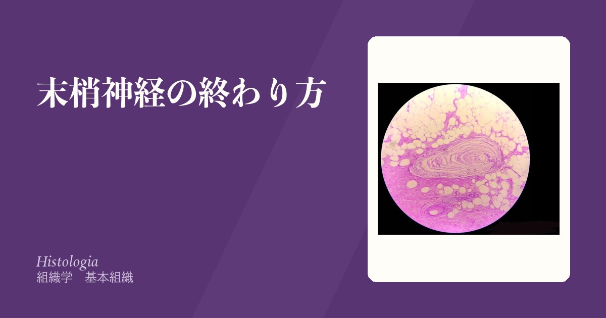
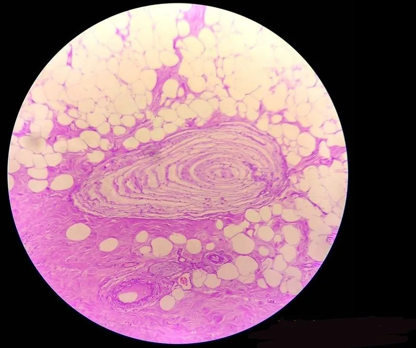
Pacini 小体は、玉ねぎ状の同心円の層板が特徴で、標本で最も見つけやすい。
筋紡錘——錘内筋線維を被膜が包む。Ia線維が長さの変化を、γ運動線維が感度を調節する。
Golgi 腱器官——腱にあり、張力を測る。Ib線維。
「速い順応」が何を意味するかが、この表を使える知識に変える。速い受容器は変化しか拾わないので、服を着ていることを忘れられる——「感じ続けないことが、機能になっている」。
Pacini 小体の層板が、この順応の仕組みそのものになる。層が圧の持続を吸収し、変化だけを軸索に届ける——「構造が、そのまま濾波器になっている」。
筋紡錘とγ運動線維が、深部腱反射の土台になる(伸張反射の章)——「打腱器で叩いているのは腱ではなく、筋を急に伸ばしている」。
だから反射の亢進と減弱で、病変の高さが分かる。亢進は上位運動ニューロン、減弱は反射弓のどこか(神経診察の章)——「1つの反射に、感覚・脊髄・運動の3つが乗っている」。
Golgi 腱器官は張力が過大になると抑制をかける。これが痙縮の治療や、ストレッチの理屈に使われる(痙縮の管理の章)——「持続した伸張が、筋を緩める」。
糖尿病性神経障害では、細い線維(自由神経終末)から先に傷む。だから痛覚と温度覚の低下が先行し、振動覚は後になる(糖尿病性神経障害の章)——「足の傷に気づけないことが、最初の危険になる」。
何を測る受容器かが分かると、神経障害の診かたが決まる。
振動覚は太い有髄線維(Aβ)が運ぶので、128Hz 音叉での振動覚検査は、太い線維の障害を最も早く捉える。糖尿病性ニューロパチーのスクリーニングで足趾から調べるのは、長さ依存性に遠位から侵されるからである。
モノフィラメント(10g)で触圧覚を調べるのは糖尿病足病変の予防——痛みを感じない足が潰瘍を作る。
Pacini 小体は最も順応が速いので「変化」しか感じない。服を着ている感覚がすぐ消えるのはこのためである。
Merkel 盤は順応が遅く、持続的な圧と形を感じる。Merkel 細胞癌は稀だが悪性度が高く、高齢者の露光部に生じる。
English version
Core
| Receptor | Site | Measures | Adaptation |
|---|---|---|---|
| Meissner corpuscle | Dermal papillae (hairless skin) | Touch, low-frequency vibration | Fast |
| Pacinian (lamellated) corpuscle | Hypodermis, periosteum, mesentery | High-frequency vibration, pressure | Fastest |
| Merkel disc | Basal layer of epidermis | Sustained touch and pressure, form | Slow |
| Ruffini corpuscle | Dermis, joint capsule | Stretch, shear | Slow |
| Free nerve ending | Almost everywhere | Pain, temperature, crude touch | Slow |
The Pacinian corpuscle, with its onion-like concentric lamellae, is the easiest to find on a slide.
Muscle spindle — intrafusal fibres within a capsule; Ia fibres report change in length and γ motor fibres adjust the sensitivity. Golgi tendon organ — in the tendon, measuring tension; Ib fibres.
Going further
Understanding what "fast adaptation" means turns this table into usable knowledge. A fast receptor reports only change, which is why one can forget that one is wearing clothes — not continuing to feel is itself the function.
The lamellae of the Pacinian corpuscle are that mechanism: they absorb sustained pressure and deliver only the change to the axon — the structure is itself a filter.
The muscle spindle and the γ fibre are the basis of the deep tendon reflex — the tendon hammer is not striking the tendon but suddenly stretching the muscle. So an increased or decreased reflex localises the lesion: increased means an upper motor neuron lesion, decreased means somewhere in the reflex arc — one reflex carries sensory, spinal and motor components together.
The Golgi tendon organ inhibits when tension becomes excessive, which is used in the management of spasticity and is the rationale of stretching — sustained stretch relaxes a muscle.
In diabetic neuropathy the small fibres (free nerve endings) are damaged first, so pain and temperature are lost before vibration — the first danger is being unable to notice a wound on the foot.
In the clinic
Knowing what a receptor measures determines how neuropathy is examined.
Vibration is carried by large myelinated (Aβ) fibres, so testing with a 128 Hz tuning fork detects large-fibre damage earliest. Screening for diabetic neuropathy begins at the toes because the involvement is length-dependent and distal.
A 10 g monofilament tests protective touch and pressure for the prevention of diabetic foot disease — a foot that cannot feel pain ulcerates.
The Pacinian corpuscle adapts fastest and senses only change — hence the sensation of clothing disappears at once.
The Merkel disc adapts slowly and senses sustained pressure and form. Merkel cell carcinoma is rare but highly malignant, arising on sun-exposed skin in the elderly.
神経筋接合部と神経節 — 薬と病気が集まる場所Junctio neuromuscularis
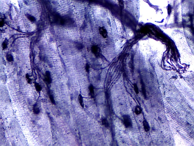
神経筋接合部(運動終板)——1本の運動ニューロンと筋線維の接点。
- シナプス前——アセチルコリンを含む小胞。電位依存性Caチャネル(P/Q型)
- シナプス間隙——基底膜にアセチルコリンエステラーゼが埋まる
- シナプス後——ひだ(junctional fold)にニコチン性アセチルコリン受容体が密集
ここを狙うもの。
| 作用点 | 病気/薬 |
|---|---|
| Caチャネル(前) | Lambert-Eaton 症候群 |
| 放出そのもの(前) | ボツリヌス毒素 |
| 受容体(後) | 重症筋無力症・非脱分極性筋弛緩薬 |
| エステラーゼ | コリンエステラーゼ阻害薬・有機リン |
神経節——脊髄神経節は偽単極性で、細胞体が丸く整列し衛星細胞が囲む。自律神経節は多極性で、細胞が不規則に散る。
作用点が前か後かで、症状の出方が逆になる。Lambert-Eaton 症候群では反復刺激で筋力が増し、重症筋無力症では減る(神経筋接合部疾患の章)——「前が壊れれば溜まって増え、後が壊れれば減り続ける」。
だから電気生理検査で、高頻度反復刺激の反応を見る——「同じ筋力低下という訴えを、波形の増減で分ける」。
Lambert-Eaton 症候群は、小細胞肺癌に伴うことが多い。腫瘍随伴症候群として、癌より先に現れることがある(腫瘍随伴症候群の章)——「神経の症状から、肺の検索が始まる」。
有機リン中毒は、エステラーゼを止めてアセチルコリンを過剰にする。ムスカリン症状(縮瞳・分泌過多・徐脈)とニコチン症状(線維束攣縮・筋力低下)が同時に出る(中毒の章)——「1つの酵素が止まると、2種類の受容体が同時に暴れる」。
脊髄神経節が偽単極性であることが、帯状疱疹の分布を説明する。ウイルスが1つの神経節に潜み、その皮膚分節だけに出る(帯状疱疹の章)——「細胞体が集まっている場所が、そのまま潜伏場所になる」。
自律神経節が腸管に無い部分が、Hirschsprung 病になる(Hirschsprung病の章)——「神経節細胞が『無い』ことを、生検で証明する病気になる」。
神経筋接合部は、前・間・後のどこが壊れるかで病気が分かれる。
シナプス後(アセチルコリン受容体に対する自己抗体) — 重症筋無力症。日内変動(夕方に悪化)・易疲労性・眼瞼下垂と複視で発症。反復刺激で振幅が減る(waning)、アイスパック試験・テンシロン試験、胸腺腫の合併を必ず調べる。コリンエステラーゼ阻害薬が効く。
シナプス前(P/Q 型 Ca チャネル抗体) — Lambert–Eaton 症候群。waxing、小細胞肺癌。
間隙(アセチルコリンエステラーゼ) — 有機リン中毒で酵素が阻害され、ムスカリン症状(縮瞳・流涎・気道分泌過多・徐脈)。アトロピンと PAMで治療する。
筋弛緩薬もここに働く——非脱分極性(ロクロニウム)は競合的で、スガマデクスで拮抗できる。
English version
Core
Neuromuscular junction (motor end plate) — the contact between one motor neuron and a muscle fibre.
- Presynaptic — vesicles of acetylcholine; voltage-gated P/Q-type calcium channels.
- Synaptic cleft — acetylcholinesterase embedded in the basal lamina.
- Postsynaptic — junctional folds with nicotinic acetylcholine receptors densely packed at their crests.
| Site of action | Disease / drug |
|---|---|
| Calcium channel (pre) | Lambert–Eaton syndrome |
| Release itself (pre) | Botulinum toxin |
| Receptor (post) | Myasthenia gravis, non-depolarising muscle relaxants |
| Esterase | Cholinesterase inhibitors, organophosphates |
Ganglia — the spinal ganglion is pseudounipolar, its round cell bodies arranged regularly and surrounded by satellite cells; the autonomic ganglion is multipolar with cells scattered irregularly.
Going further
Whether the lesion is pre- or postsynaptic reverses the behaviour of the weakness. In Lambert–Eaton syndrome strength increases with repetitive stimulation; in myasthenia gravis it decreases — a presynaptic fault allows transmitter to accumulate, a postsynaptic one only ever loses. So electrophysiology looks at the response to high-frequency repetitive stimulation — the same complaint of weakness is separated by whether the waveform grows or shrinks.
Lambert–Eaton syndrome commonly accompanies small cell lung cancer and, as a paraneoplastic syndrome, may appear before the tumour — a neurological symptom starts the search of the chest.
Organophosphate poisoning stops the esterase and floods the junction with acetylcholine, producing muscarinic (miosis, hypersecretion, bradycardia) and nicotinic (fasciculation, weakness) features together — one enzyme stopped, and two kinds of receptor run wild at once.
That the spinal ganglion is pseudounipolar explains the distribution of shingles: the virus lies latent in a single ganglion and erupts in that dermatome alone — where the cell bodies gather is where the virus hides. And the absence of autonomic ganglia in a segment of bowel is Hirschsprung disease — a disease diagnosed by proving on biopsy that ganglion cells are absent.
In the clinic
Diseases of the junction divide by whether the fault is pre-, intra- or postsynaptic.
Postsynaptic (autoantibodies to the acetylcholine receptor) — myasthenia gravis. Diurnal variation (worse in the evening), fatigability, presenting with ptosis and diplopia. Decrement (waning) on repetitive stimulation, ice-pack and edrophonium tests, and always look for a thymoma. Cholinesterase inhibitors help.
Presynaptic (antibodies to P/Q-type calcium channels) — Lambert–Eaton syndrome: waxing, and small cell lung cancer.
The cleft — organophosphates inhibit acetylcholinesterase, giving muscarinic features (miosis, salivation, bronchial secretion, bradycardia). Treat with atropine and pralidoxime.
Muscle relaxants also act here: non-depolarising agents (rocuronium) are competitive and can be reversed with sugammadex.
自律神経節 — 乗り換える場所が、薬の効き方を決めるGanglion autonomicum
自律神経は必ず2つのニューロンを直列につなぐ。その乗り換え地点が神経節になる。
| 交感神経 | 副交感神経 | |
|---|---|---|
| 節前線維の出る場所 | 胸髄・上部腰髄(T1〜L2) | 脳幹(III・VII・IX・X)と仙髄(S2〜S4) |
| 神経節の位置 | 中枢の近く(交感神経幹・椎前神経節) | 効果器の近く/壁の中 |
| 節前線維 | 短い | 長い |
| 節後線維 | 長い | 短い |
| 節前の伝達物質 | どちらもアセチルコリン(ニコチン性受容体) | |
| 節後の伝達物質 | ノルアドレナリン(汗腺だけアセチルコリン) | アセチルコリン(ムスカリン性受容体) |
組織像——大型で核小体の明瞭な神経細胞体が散在し、その周りを衛星細胞が囲む。
副腎髄質は例外——節前線維が直接クロム親和性細胞に終わる。細胞そのものが「節後ニューロンの代わり」になる。
「節前は両方ともアセチルコリン」という点が、薬理の骨格になる。神経節を遮断する薬は交感も副交感も同時に切ってしまうので、副作用が全身に及ぶ(自律神経作用薬の章)——「だから節遮断薬は、ほとんど使われなくなった」。
逆に節後で伝達物質が分かれることが、選択的な薬を可能にしている——「β1 だけ、M3 だけ、という狙い撃ちが成立する場所は、節より先になる」。
副腎髄質が「変形した神経節」であることは、その腫瘍の性質を説明する。褐色細胞腫は神経堤由来で、傍神経節腫(パラガングリオーマ)と連続した病気になる(副腎の章)——「副腎の外にできることがあるのは、同じ由来の細胞が節に沿って分布しているから」。
壁在神経節が失われる病気も、ここに繋がる。Hirschsprung 病は神経堤細胞の移動が途中で止まり、遠位の腸に神経節細胞が無い(無神経節部)(子どもの腹の章)——「拡張しているのは正常側で、狭いほうが病気の側になる」。
だから直腸生検で「神経節細胞が無いこと」を証明する——「あるものを見つけるのではなく、無いことを確かめる診断になる」。
乗り換えの場所が、副作用の出方を決める。
節前線維はどちらもアセチルコリン(ニコチン受容体)。節後は交感がノルアドレナリン、副交感がアセチルコリン(ムスカリン受容体)。例外が汗腺で、交感神経支配なのにアセチルコリンを使う——抗コリン薬で発汗が止まるのはこのためである。
抗コリン薬の副作用は口渇・便秘・尿閉・散瞳と閉塞隅角緑内障の誘発・頻脈・せん妄——ムスカリン受容体がある場所を数えれば全部出てくる。高齢者では認知機能低下と転倒のリスクになる。
交感神経節は脊柱の傍らにあり、節前が短く節後が長い。副交感神経節は標的臓器のすぐそばにあるので作用が限局的である。交感神経は全身一斉に、副交感神経は臓器ごとに働く——解剖がそのまま生理になっている。
English version
Core
The autonomic system always places two neurons in series, and the ganglion is where they change over.
| Sympathetic | Parasympathetic | |
|---|---|---|
| Origin of preganglionic fibres | Thoracic and upper lumbar cord (T1–L2) | Brainstem (III, VII, IX, X) and sacral cord (S2–S4) |
| Position of ganglion | Near the centre (sympathetic trunk, prevertebral ganglia) | Near or within the wall of the target organ |
| Preganglionic fibre | Short | Long |
| Postganglionic fibre | Long | Short |
| Preganglionic transmitter | Acetylcholine in both (nicotinic receptors) | |
| Postganglionic transmitter | Noradrenaline (acetylcholine at sweat glands alone) | Acetylcholine (muscarinic receptors) |
Histology — large cell bodies with prominent nucleoli, scattered, each surrounded by satellite cells.
The adrenal medulla is the exception: preganglionic fibres end directly on the chromaffin cells. The cells themselves stand in place of the postganglionic neuron.
Going further
That both preganglionic transmitters are acetylcholine is the skeleton of the pharmacology. A ganglion-blocking drug cuts sympathetic and parasympathetic at the same time, so its effects are systemic — which is why ganglion blockers are scarcely used any more. Conversely, the divergence of transmitter beyond the ganglion is what makes selective drugs possible — "β1 only" or "M3 only" is achievable only distal to the ganglion.
That the adrenal medulla is a modified ganglion explains the nature of its tumours: phaeochromocytoma is of neural crest origin and is continuous with paraganglioma — it can arise outside the adrenal because the same cells are distributed along the ganglia.
Loss of the intramural ganglia belongs here too. In Hirschsprung disease the migration of neural crest cells stops part-way and the distal bowel has no ganglion cells — the dilated segment is the normal one and the narrow segment is the diseased one. So rectal biopsy is used to prove the absence of ganglion cells — a diagnosis made by confirming that something is not there.
In the clinic
Where the changeover happens determines the pattern of side effects.
Preganglionic fibres are cholinergic (nicotinic) in both divisions. Postganglionic fibres are noradrenergic in the sympathetic and cholinergic (muscarinic) in the parasympathetic. The exception is the sweat gland, which is sympathetically innervated but uses acetylcholine — which is why anticholinergics stop sweating.
The side effects of anticholinergic drugs — dry mouth, constipation, urinary retention, mydriasis with precipitation of angle-closure glaucoma, tachycardia, delirium — are obtained simply by listing where muscarinic receptors are. In the elderly they are a cause of cognitive decline and falls.
The sympathetic ganglia lie beside the vertebral column with short preganglionic and long postganglionic fibres; the parasympathetic ganglia sit next to the target organ, so their action is localised. The sympathetic acts throughout the body at once, the parasympathetic organ by organ — the anatomy is directly the physiology.
末梢神経の変性と再生 — 切れた先で何が起きるか
軸索が切れると、細胞体から切り離された遠位部が全長にわたって崩れる(Waller 変性)。
- 1〜2日——軸索が断片化し、髄鞘が崩れる
- 数日〜数週——Schwann 細胞とマクロファージが残骸を貪食する
- Schwann 細胞が増殖し、基底膜の筒の中に列(Büngner 帯)を作る
- 近位断端から成長円錐が伸び、この筒をたどって再生する(1日あたり約1 mm)
細胞体側でも変化が起きる——核が辺縁へ寄り、Nissl 小体が溶ける(中心性染色質融解)。
損傷の程度で3つに分ける。
| 分類 | 壊れているもの | 回復 |
|---|---|---|
| 一過性伝導障害〈neurapraxia〉 | 髄鞘のみ(軸索は連続) | 数日〜数週で完全に戻る |
| 軸索断裂〈axonotmesis〉 | 軸索が断裂、筒(基底膜)は残る | ゆっくり戻る |
| 神経断裂〈neurotmesis〉 | 神経幹ごと断裂 | 手術しないと戻らない |
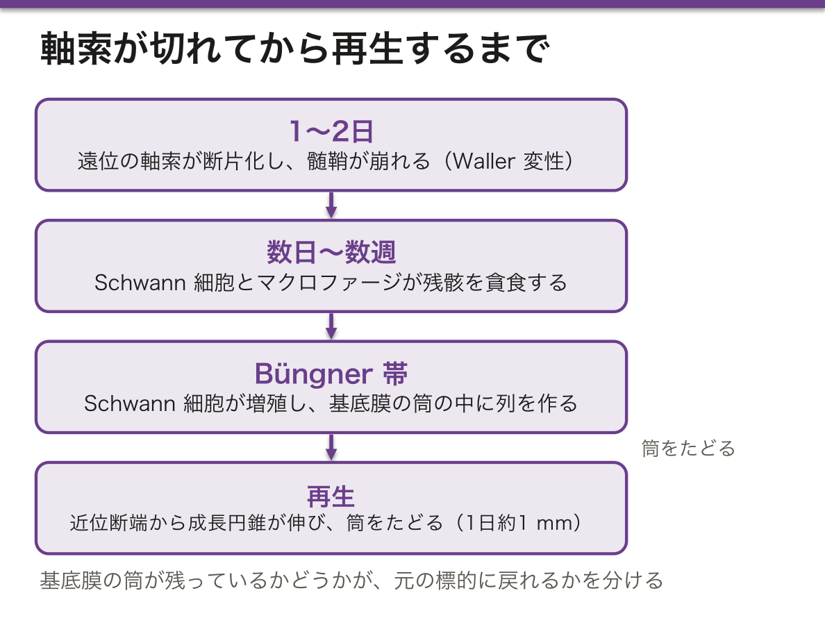
「筒が残っているか」の一点が、治るか治らないかを分けている。基底膜の筒が道案内になるので、それが残れば軸索は元の標的へ戻れる——「案内が失われれば、伸びた軸索は行き先を間違える」。
だから神経腫(切断端で軸索が渦を巻いた塊)ができる。行き場を失った軸索が、瘢痕の中で無秩序に増える。触ると激痛が走る(装具・義肢の章)——「断端が痛いことの実体が、これになる」。
1日1 mm という速さは、そのまま予後の計算に使える。肘で尺骨神経が損傷すれば手内在筋まで約30 cm で、およそ10か月かかる(神経伝導検査の章)——「待ってよい期間と、手術を決める期限が数字で出る」。
中枢神経では、この再生が起こらない。オリゴデンドロサイトは Büngner 帯を作らず、逆に軸索の伸長を抑える分子を出す(グリアの章)——「末梢は治り、中枢は治らないという差が、支持細胞の側にある」。
神経伝導検査の時期にも影響する。Waller 変性が完成する前(受傷後数日以内)は、遠位を刺激すると反応が出てしまう——「早すぎる検査は、切れていないと誤読させる」。
末梢神経は再生する。ただし条件と速さがある。
1日約 1mm(1か月で約 3cm)という速さから、回復までの期間を患者に説明できる。肘の尺骨神経損傷で手内在筋が戻るまで1年以上——その前に筋が線維化すれば、神経が届いても動かない。時間との競争になる。
Tinel 徴候 — 再生の先端を叩くと放散痛が出る。叩いて痛む位置が末梢へ移動していけば、再生が進んでいる——外来で経過を追える指標になる。
Schwann 細胞と基底膜の管(Büngner 帯)が道しるべになる。管が失われると軸索が迷走し、神経腫を作る。
中枢では再生しない——稀突起膠細胞が抑制因子を出し、グリア瘢痕が壁になる。末梢と中枢で予後がまったく違うのはこの環境の差による。
English version
Core
When an axon is cut, the part separated from the cell body breaks down along its whole length — Wallerian degeneration.
- 1–2 days — the axon fragments and the myelin breaks up.
- Days to weeks — Schwann cells and macrophages phagocytose the debris.
- Schwann cells proliferate and form columns within the tube of basal lamina — the bands of Büngner.
- A growth cone extends from the proximal stump and regenerates along the tube at about 1 mm a day.
The cell body changes too — the nucleus moves to the rim and the Nissl substance disperses (central chromatolysis).
| Classification | What is broken | Recovery |
|---|---|---|
| Neurapraxia | Myelin only (axon continuous) | Complete, in days to weeks |
| Axonotmesis | Axon severed, tube preserved | Slow but complete |
| Neurotmesis | The whole nerve trunk severed | Not without surgery |
Going further
Whether the tube remains is the single thing that separates recovery from failure. The basal lamina tube acts as a guide, so if it survives the axon reaches its original target — lose the guide and the growing axon goes to the wrong place.
Hence the neuroma: axons with nowhere to go proliferate chaotically within scar, forming a whorled mass at the stump that is exquisitely painful to touch — this is the substance of a painful stump.
The figure of 1 mm a day can be used directly for prognosis: an ulnar nerve injury at the elbow is about 30 cm from the intrinsic muscles of the hand, so roughly ten months — the period one may wait, and the deadline for deciding on surgery, come out as numbers.
In the CNS this regeneration does not occur: oligodendrocytes make no bands of Büngner and instead release inhibitors of axonal growth — the difference between peripheral recovery and central failure lies on the side of the supporting cell.
It also affects the timing of nerve conduction studies: before Wallerian degeneration is complete (within a few days of injury) stimulation distal to the lesion still produces a response — a study done too early will be misread as an intact nerve.
In the clinic
Peripheral nerve regenerates — but with conditions and at a set speed. From about 1 mm a day (3 cm a month) the period to recovery can be explained to the patient. An ulnar nerve injury at the elbow needs more than a year for the intrinsic muscles of the hand — and if the muscle has fibrosed before then, it will not move even when the nerve arrives. It is a race against time.
Tinel sign — tapping over the advancing front produces radiating paraesthesia. If the point that tingles moves distally, regeneration is progressing — an indicator that can be followed in clinic.
The Schwann cells and the tube of basal lamina (bands of Büngner) are the guide. If the tube is lost the axons wander and form a neuroma.
In the CNS there is no regeneration — oligodendrocytes release inhibitors and the glial scar forms a wall. The wholly different prognosis of peripheral and central injury comes from this difference of environment.
神経線維の太さ — 速さを決め、そして薬の効き方を決めるFibra nervosa
| 分類 | 髄鞘 | 速さ | 運ぶもの |
|---|---|---|---|
| Aα(Ia・Ib) | 厚い | 70〜120 m/秒 | 運動・筋紡錘と腱器官からの固有感覚 |
| Aβ(II) | あり | 30〜70 | 触覚・圧覚・振動覚 |
| Aγ | あり | 15〜30 | 錘内筋線維(感度の調節) |
| Aδ(III) | 薄い | 5〜30 | 速い痛み(鋭い)・冷覚 |
| B | 薄い | 3〜15 | 交感神経の節前線維 |
| C(IV) | 無い | 0.5〜2 | 遅い痛み(鈍い)・温覚・かゆみ・節後の自律神経 |
速さは直径と髄鞘で決まる——太いほど速く、髄鞘があれば跳躍伝導でさらに速い。
「痛みが二度来る」という現象が、この表から直接出てくる。Aδ による鋭い痛みが先に届き、C による鈍く続く痛みが後から来る(感覚の章)——「速さの違う2本の線が、1つの刺激を2回伝えている」。
局所麻酔薬は、細い線維から先に効く(区域麻酔の章)——「痛覚 → 温度覚 → 触覚 → 運動、の順に消える。だから『触られているのは分かるが痛くない』状態が作れる」。
圧迫では逆に、太い線維から先に障害される(末梢神経障害の章)——「正座でしびれるとき、まず運動と触覚が落ちて痛覚が残るのは、この向きの違いによる」。
ゲートコントロール説も、太さの違いで説明される(慢性の痛みの章)——「Aβ を刺激すると C 線維の入力が抑えられる。さすると痛みが和らぐことの説明になる」。
神経伝導検査が測っているのは、最も速い線維になる(神経の電気を測る章)——「小径線維だけが障害される病態(小径線維ニューロパチー)では、検査が正常に出る」。
太さの違いが、麻酔と虚血の効き方の順番を決める。
局所麻酔薬は細い線維から先に効く。自律神経(B)→ 痛覚・温度覚(Aδ・C)→ 触覚(Aβ)→ 運動(Aα)の順で、硬膜外麻酔で「痛みは取れているが足は動く」状態が作れるのはこのためである。回復は逆順になる。
圧迫と虚血は太い線維から先に落ちる。正座のしびれで触覚と深部感覚が先に消え、痛覚が残るのはこの順序による。麻酔と圧迫で順番が逆という点が試験でよく問われる。
Aδ と C の違いが痛みの二相性を作る。Aδ=鋭く速い一次痛、C=鈍く遅い二次痛。ぶつけた直後の鋭い痛みと、後から来るじんじんする痛みである。
神経伝導検査は太い有髄線維しか測れないので、小径線維ニューロパチーでは正常に出る——「検査が正常でも痛みは本物」という重要な限界になる。
English version
Core
| Class | Myelin | Speed | Carries |
|---|---|---|---|
| Aα (Ia, Ib) | Thick | 70–120 m/s | Motor; proprioception from spindle and tendon organ |
| Aβ (II) | Yes | 30–70 | Touch, pressure, vibration |
| Aγ | Yes | 15–30 | Intrafusal fibres (adjusting sensitivity) |
| Aδ (III) | Thin | 5–30 | Fast (sharp) pain, cold |
| B | Thin | 3–15 | Preganglionic sympathetic |
| C (IV) | None | 0.5–2 | Slow (dull) pain, warmth, itch, postganglionic autonomic |
Speed is set by diameter and myelination — thicker is faster, and myelin adds saltatory conduction.
Going further
The phenomenon that pain arrives twice comes straight out of this table. The sharp pain carried by Aδ arrives first and the dull, persisting pain carried by C follows — two lines of different speed transmitting one stimulus twice.
Local anaesthetics affect thin fibres first — pain → temperature → touch → motor, which is why a state can be produced in which the patient can feel being touched but has no pain.
Compression works in the opposite order, affecting thick fibres first — which is why, when a leg goes numb after kneeling, motor function and touch fade first while pain remains.
The gate control theory is also explained by calibre: stimulating Aβ suppresses C fibre input — the explanation for why rubbing a painful place helps.
A nerve conduction study measures only the fastest fibres — so in conditions affecting small fibres alone (small fibre neuropathy) the study is normal.
In the clinic
The difference in calibre sets the order in which anaesthesia and ischaemia take effect.
Local anaesthetic affects thin fibres first: autonomic (B) → pain and temperature (Aδ, C) → touch (Aβ) → motor (Aα). This is why epidural anaesthesia can abolish pain while the legs still move, and recovery occurs in the reverse order.
Compression and ischaemia affect thick fibres first. When a leg goes numb after kneeling, touch and deep sensation disappear first while pain remains. That the order is opposite for anaesthesia and for compression is frequently examined.
The difference between Aδ and C gives the two phases of pain: Aδ = sharp, fast, first pain; C = dull, slow, second pain — the sharp pain on striking oneself and the throbbing that follows.
Nerve conduction studies measure only large myelinated fibres, so they are normal in small fibre neuropathy — an important limitation: a normal study does not mean the pain is not real.
神経周膜 — 神経の中にも関門があり、圧が上がるPerineurium
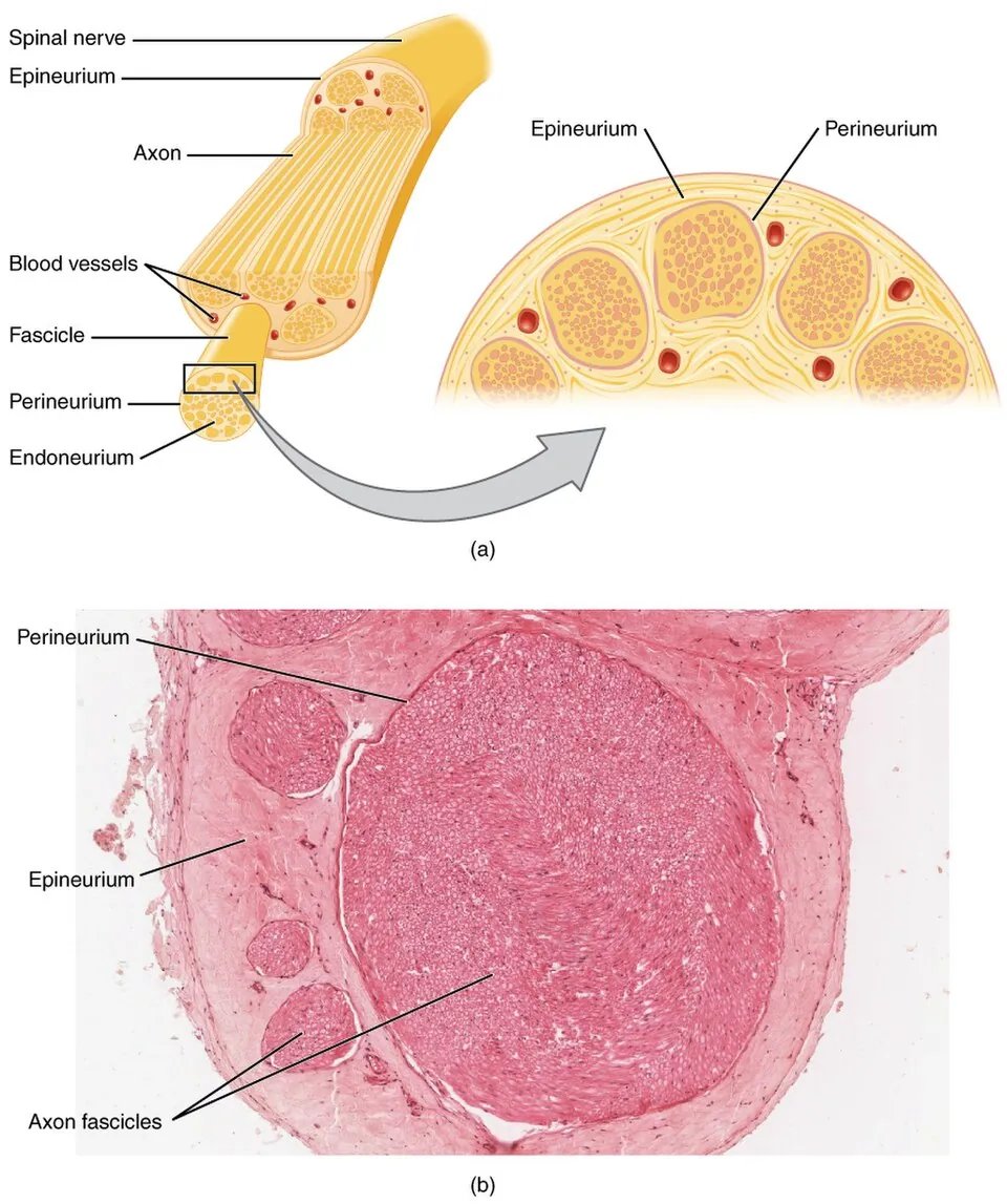
末梢神経は3層の膜に包まれる——神経内膜・神経周膜・神経外膜。
| 層 | 包むもの | 特徴 |
|---|---|---|
| 神経内膜 | 個々の神経線維 | 細い膠原線維と、内膜内の毛細血管 |
| 神経周膜 | 神経束 | 扁平な細胞が同心円状に並び、密着結合を持つ。血液神経関門を作る |
| 神経外膜 | 神経幹全体 | 疎性結合組織・脂肪・栄養血管 |
神経周膜が閉じた区画を作るので、内部の圧(神経内圧)が上がりうる。
内膜内の毛細血管は密着結合を持ち、リンパ管は無い——だから一度入った浮腫が引きにくい。
「閉じた区画である」ことが、絞扼性神経障害の本体になる。外から圧されると内圧が上がり、内膜内の毛細血管が虚血になる(手の神経の章)——「手根管症候群は、狭い場所を通ることの帰結になる」。
だから症状が夜間と早朝に強い——「臥位で組織圧が上がり、手首が屈曲位を取るからになる」。
関門があることは、薬と免疫の到達を制限している(末梢神経障害の章)——「関門が弱い場所(神経根・後根神経節・自律神経節)が、Guillain-Barré 症候群や糖尿病で先に侵される」。
神経ブロックでは、この膜が薬の広がり方を決めている(神経ブロックの章)——「神経束の中へ針を進めると、圧が上がって神経を傷める。だから束の外に置く」。
神経縫合では、外膜どうしを合わせるか、束ごとに合わせるかを選ぶ(縫う技術の章)——「束の並びを合わせられれば、運動と感覚が混線しにくい」。
神経周膜は関門であり、同時に閉じた区画を作る。
血液神経関門を作るので、薬も抗体も入りにくい。後根神経節と神経終末は関門が弱いため、そこが自己免疫と中毒の標的になりやすい。
閉じた区画なので、内圧が上がると虚血になる。これが絞扼性ニューロパチーの本体である。手根管症候群(正中神経)、肘部管症候群(尺骨神経)、腓骨神経麻痺(腓骨頭部)。いずれも「トンネルの中で圧が上がる」という同じ機序である。
浮腫を起こす状態で発症しやすい——妊娠・甲状腺機能低下症・関節リウマチ・透析(アミロイド)。手根管症候群の背景疾患を探す根拠になる。
神経ブロックでは周膜の外(神経上膜内)に薬を置く。神経束の中へ直接注入すると神経を損傷するので、超音波で層を確認しながら行う。
English version
Core
A peripheral nerve is invested by three layers.
| Layer | Invests | Features |
|---|---|---|
| Endoneurium | Each individual fibre | Fine collagen fibres and intrafascicular capillaries |
| Perineurium | A fascicle | Flattened cells in concentric layers with tight junctions; forms the blood–nerve barrier |
| Epineurium | The whole nerve trunk | Loose connective tissue, fat, nutrient vessels |
Because the perineurium creates a closed compartment, the pressure inside (the endoneurial pressure) can rise. The intrafascicular capillaries have tight junctions and there are no lymphatics — so once oedema has entered, it does not readily leave.
Going further
Being a closed compartment is the substance of entrapment neuropathy. External compression raises the internal pressure and renders the intrafascicular capillaries ischaemic — carpal tunnel syndrome is the consequence of passing through a narrow place. Hence symptoms are worst at night and in the early morning — tissue pressure rises when lying down and the wrist adopts a flexed position.
The existence of a barrier limits the access of drugs and of immunity — the places where the barrier is weak (the nerve root, the dorsal root ganglion, the autonomic ganglion) are attacked first in Guillain–Barré syndrome and in diabetes.
In nerve block, these membranes determine how the drug spreads — advancing the needle into a fascicle raises the pressure and injures the nerve, so the drug is placed outside the fascicle.
In nerve repair, one chooses between apposing the epineurium and apposing individual fascicles — if the fascicles can be matched, motor and sensory fibres are less likely to be crossed.
In the clinic
The perineurium is both a barrier and the wall of a closed compartment.
As the blood–nerve barrier it excludes drugs and antibodies. The dorsal root ganglion and the nerve terminals have a weak barrier, and are therefore the targets of autoimmunity and toxins.
As a closed compartment, a rise in internal pressure causes ischaemia — the substance of entrapment neuropathy: carpal tunnel syndrome (median nerve), cubital tunnel syndrome (ulnar nerve), peroneal palsy at the fibular head. All share one mechanism: pressure rising inside a tunnel.
They occur readily in conditions causing oedema — pregnancy, hypothyroidism, rheumatoid arthritis, dialysis (amyloid) — which is the reason to look for an underlying cause in carpal tunnel syndrome.
In nerve block the drug is placed outside the perineurium, within the epineurium; injecting directly into a fascicle damages the nerve, so ultrasound is used to identify the layers.
神経生検 — どこを、いつ採るか
末梢神経障害の多くは、病歴・診察・神経伝導検査で診断できる。生検はそれでも分からないときにだけ行う——採った場所に永久の感覚障害が残るからである。
採るのはほぼ腓腹神経(外果の後方)。純粋な感覚神経で、運動が失われないためである。足の外側に痺れが残ることを、必ず事前に説明する。
生検でしか分からないことがある場面に限る。
- 血管炎——治療が免疫抑制になるので、証明の価値が大きい
- アミロイドーシス——Congo red と免疫染色
- ハンセン病・サルコイドーシス・悪性腫瘍の浸潤
- 遺伝性ニューロパチー——ただし遺伝子検査が優先されるようになった
見る所見は「何が失われたか」と「分布が均一か」の2つである。
| 軸索性 | 有髄線維の密度が減る。太い線維から先に減る | 最も多い型(糖尿病・アルコール・薬剤) |
| 脱髄性 | 髄鞘だけが失われ、玉ねぎ様肥厚ができる | 脱髄と再髄鞘化を繰り返した跡。慢性炎症性脱髄性多発根神経炎・Charcot-Marie-Tooth 病 |
| 束のあいだでばらつく | ある束は保たれ、隣の束は脱落 | 血管炎を強く示唆する——栄養血管が詰まった領域だけが傷むため |
「ばらつき」を見るために、束を横断して観察できる採り方が要る。
⚠️ 小径線維は、通常の検査では見えない。痛みと温度を伝える細い線維の障害(小径線維ニューロパチー)は神経伝導検査が正常で、腓腹神経生検でも捉えにくい。皮膚生検で表皮内の神経線維の密度を測る方法が使われる——3 mm のパンチで採れるので侵襲が小さい。糖尿病の早期の神経障害を捉える方法として注目されている。
「検査が正常なのに痛い」患者を、心因性と決めつけない——見ていない線維があるという可能性を先に考える。
生検を勧める前に、結果で治療が変わるかを自問する。変わらないなら、感覚障害を残してまで採る理由が無い。
English version
Core
Most peripheral neuropathy can be diagnosed from the history, examination and nerve conduction studies. Biopsy is done only when those still leave the question open — because a permanent sensory deficit remains where the nerve was taken.
The nerve taken is almost always the sural nerve (behind the lateral malleolus), because it is purely sensory and no motor function is lost. Always explain beforehand that numbness on the lateral foot will remain.
Restrict it to situations that only biopsy can settle:
- Vasculitis — the value of proof is high because treatment is immunosuppression.
- Amyloidosis — Congo red and immunostaining.
- Leprosy, sarcoidosis, infiltration by malignancy.
- Hereditary neuropathy — though genetic testing now takes precedence.
Going further
Two things are looked for: what has been lost, and whether the distribution is uniform.
| Pattern | Appearance | Meaning |
|---|---|---|
| Axonal | Reduced density of myelinated fibres, large fibres first | The commonest type (diabetes, alcohol, drugs) |
| Demyelinating | Myelin alone lost, with onion-bulb formations | The trace of repeated demyelination and remyelination: CIDP, Charcot–Marie–Tooth disease |
| Variation between fascicles | One fascicle spared, the next depleted | Strongly suggests vasculitis — only the territory of an occluded vasa nervorum is damaged |
To see that variation, the specimen must be taken so that fascicles can be compared in cross-section.
⚠️ Small fibres are invisible to the usual tests. Damage confined to the thin fibres carrying pain and temperature (small fibre neuropathy) gives normal conduction studies and is hard to capture even on sural nerve biopsy. Skin biopsy measuring intraepidermal nerve fibre density is used instead — a 3 mm punch, so the procedure is minor. It is of interest for detecting early diabetic neuropathy.
Do not conclude that a patient whose tests are normal but who is in pain has a psychogenic problem — consider first that there are fibres you have not looked at.
In the clinic
Before recommending a biopsy, ask yourself whether the result will change the treatment. If it will not, there is no reason to take a nerve and leave a sensory deficit.
上皮内の神経終末 — 上皮の中で終わる神経
上皮には血管が入らないが、神経は入る。基底膜を貫いて上皮細胞の間を上る。
自由神経終末——最も多い。無髄の終末が上皮細胞の間に分岐して終わる。痛覚・温度覚・粗い触覚。角膜上皮は特に密で、これが角膜の強い痛みの理由。
Merkel 細胞と神経終末の複合体——表皮基底層にあるMerkel 細胞が、円板状の神経終末とシナプス様の接触をつくる。順応の遅い触覚受容器で、形と縁の識別を担う。指腹・口唇・毛包周囲に多い。
味蕾と嗅上皮も、上皮内に感覚装置を持つ例。味蕾は上皮細胞が受容器になり、神経が下で受け取る(二次感覚細胞)のに対し、嗅細胞は神経細胞そのものが上皮に並ぶ(一次感覚細胞)——この違いが最頻出。
Merkel 細胞は神経堤由来か表皮由来か長く論争があり、現在は表皮の角化細胞系由来とされる。Merkel 細胞癌は皮膚の高悪性度腫瘍。
English version
Core
Vessels do not enter an epithelium, but nerves do: they pierce the basal lamina and ascend between the epithelial cells.
- Free nerve endings — the most numerous. Unmyelinated terminals branch and end between the epithelial cells. Pain, temperature and crude touch. The corneal epithelium is especially densely supplied, which is why the cornea hurts so much.
- Merkel cell–neurite complex — the Merkel cell of the basal epidermis makes a synapse-like contact with a disc-shaped ending. A slowly adapting touch receptor responsible for discriminating form and edges; abundant on the finger pads, lips and around hair follicles.
The taste bud and the olfactory epithelium are further examples of sensory apparatus within epithelium. In the taste bud an epithelial cell is the receptor and the nerve receives from beneath (a secondary sensory cell); in the olfactory epithelium the neuron itself lies within the epithelium (a primary sensory cell) — this distinction is the one most often asked.
Going further
Whether the Merkel cell is of neural crest or epidermal origin was long disputed; it is now held to derive from the keratinocyte lineage of the epidermis. Merkel cell carcinoma is a high-grade cutaneous malignancy.
筋紡錘と運動終板 — 測る装置と、渡す装置
筋紡錘 fusus neuromuscularis——筋の中にある紡錘形の被膜に包まれた装置で、筋の長さと、その変化の速さを測る。
中身は錘内筋線維(核袋線維と核鎖線維)。周りの錘外筋線維より細く、中央部に収縮能が無い。感覚支配はIa 群(中央に環状らせん終末。速さに応答)とII 群(花瓶状終末。長さに応答)。運動支配は γ 運動ニューロンで、両端だけを縮めて筋が短くなっても感度を保つ(α-γ 連関)。
Ia 群 → 同名筋の α 運動ニューロンへ単シナプス性に戻るのが伸張反射(腱反射)。
Golgi 腱器官は腱にあり張力を測る(Ib 群、抑制性)。「紡錘=長さ・並列、腱器官=張力・直列」で対にする。
運動終板——軸索終末が筋線維の表面のくぼみに収まる。シナプス後膜はひだ状に折り畳まれ、その頂上に ACh 受容体が密集する。間隙にアセチルコリンエステラーゼ。
臨床——重症筋無力症は受容体への自己抗体、Lambert-Eaton 症候群は前膜の Ca チャネルへの抗体、ボツリヌス毒素は ACh の放出を止める。どの部位が壊れるかで病名が決まる。
English version
Core
Muscle spindle — a fusiform, encapsulated device within the muscle that measures the length of the muscle and the rate at which it changes. It contains intrafusal fibres (nuclear bag and nuclear chain), thinner than the extrafusal fibres and non-contractile in their central portion.
- Sensory supply — group Ia (annulospiral endings centrally, responding to rate) and group II (flower-spray endings, responding to length).
- Motor supply — γ motor neurons, contracting only the ends so that sensitivity is preserved as the muscle shortens (α–γ coactivation).
Group Ia returns monosynaptically to the α motor neuron of the same muscle — the stretch (tendon) reflex. The Golgi tendon organ lies in the tendon and measures tension (group Ib, inhibitory).
Pair them as: spindle = length, in parallel; tendon organ = tension, in series.
Motor end plate — the axon terminal sits in a depression on the muscle fibre. The postsynaptic membrane is thrown into folds with acetylcholine receptors concentrated at their crests, and acetylcholinesterase lies in the cleft.
Going further
Clinically — myasthenia gravis is an autoantibody against the receptor, Lambert–Eaton syndrome an antibody against the presynaptic calcium channel, and botulinum toxin stops the release of acetylcholine. Which part is broken decides the name of the disease.
まとめ
| 感覚終末 — 何を測っているかで形が違う | 受容器場所測るもの順応Meissner 小体真皮乳頭(無毛部)触覚・低周波の振動速いPacini 小体(層板小体)皮下・骨膜・腸間膜高周波の振動・圧最も速いMerkel 盤表皮基底… |
|---|---|
| 神経筋接合部と神経節 — 薬と病気が集まる場所 | 神経筋接合部(運動終板)——1本の運動ニューロンと筋線維の接点。 |
| 自律神経節 — 乗り換える場所が、薬の効き方を決める | 自律神経は必ず2つのニューロンを直列につなぐ。 |
| 末梢神経の変性と再生 — 切れた先で何が起きるか | 軸索が切れると、細胞体から切り離された遠位部が全長にわたって崩れる(Waller 変性)。 |
| 神経線維の太さ — 速さを決め、そして薬の効き方を決める | 分類髄鞘速さ運ぶものAα(Ia・Ib)厚い70〜120 m/秒運動・筋紡錘と腱器官からの固有感覚Aβ(II)あり30〜70触覚・圧覚・振動覚Aγあり15〜30錘内筋線維(感度の調節… |
| 神経周膜 — 神経の中にも関門があり、圧が上がる | 末梢神経は3層の膜に包まれる——神経内膜・神経周膜・神経外膜。 |
| 神経生検 — どこを、いつ採るか | 末梢神経障害の多くは、病歴・診察・神経伝導検査で診断できる。 |
| 上皮内の神経終末 — 上皮の中で終わる神経 | 上皮には血管が入らないが、神経は入る。基底膜を貫いて上皮細胞の間を上る。 |
| 筋紡錘と運動終板 — 測る装置と、渡す装置 | 筋紡錘 fusus neuromuscularis——筋の中にある紡錘形の被膜に包まれた装置で、筋の長さと、その変化の速さを測る。 |
このノートは医学生が授業の内容を自分の言葉でまとめたもので、診療の判断に使うものではありません。誤りに気づいたらお知らせください。