耳は1つの器官に見えて、音を集めて運ぶ部分(外耳・中耳)と感じる部分(内耳)からなる。内耳はさらに、聴覚(蝸牛)と平衡覚(前庭・半規管)を、つながった1つの袋の中で受け持つ。ほとんど全部が側頭骨の中に彫り込まれているので、骨の地図(錐体部・鼓室部・乳様突起)と重ねて覚えるのが近道である。
臨床で効くのは「隣に何があるか」である。中耳の小部屋は6つの壁に囲まれ、上は中頭蓋窩、下は内頸静脈、前は内頸動脈、後ろは乳突蜂巣、内側は内耳と顔面神経に接している。中耳炎の合併症も、手術で気をつける場所も、この壁の地図から出てくる。有毛細胞や内リンパの組織は聴覚器・平衡覚器(組織学)、側頭骨そのものは頭蓋の骨、蝸牛神経核・前庭神経核は延髄・橋・菱形窩、下丘と内側膝状体は中脳・間脳にある。
- 外耳と耳介 — 形の名前と、皮膚の神経
- 外耳道と鼓膜 — 曲がった管と、傾いた膜
- 鼓室 — 6つの壁と、その向こう側
- 耳小骨と鼓室の筋 — 音を渡す鎖と、そのブレーキ
- 耳管と乳突蜂巣 — 中耳の換気口と、奥の空洞
- 骨迷路 — 前庭・半規管・蝸牛
- 膜迷路 — 内リンパの閉じた袋と、3種類の受容器
- 聴覚路と平衡覚路 — 両側へ上る音、4方向へ散る平衡
- 外耳と耳介 — 形の名前と、皮膚の神経Auricula
- 外耳道と鼓膜 — 曲がった管と、傾いた膜Meatus acusticus externus et membrana tympanica
- 鼓室 — 6つの壁と、その向こう側Cavitas tympani
- 耳小骨と鼓室の筋 — 音を渡す鎖と、そのブレーキOssicula auditus
- 耳管と乳突蜂巣 — 中耳の換気口と、奥の空洞Tuba auditiva
- 骨迷路 — 前庭・半規管・蝸牛Labyrinthus osseus
- 膜迷路 — 内リンパの閉じた袋と、3種類の受容器Labyrinthus membranaceus
- 聴覚路と平衡覚路 — 両側へ上る音、4方向へ散る平衡Via acustica et via vestibularis
- まとめ
外耳と耳介 — 形の名前と、皮膚の神経Auricula
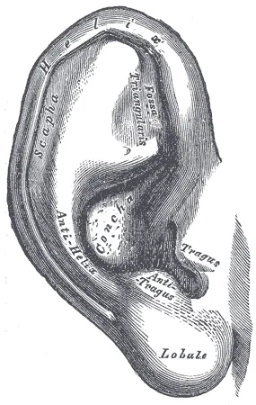
耳は外耳 auris externa・中耳 auris media・内耳 auris internaの3部に分かれる。外耳は耳介 auriculaと外耳道 meatus acusticus externusからなり、鼓膜は外耳と中耳の境として外耳の側で扱われることが多い。
耳介の上2/3 は弾性軟骨 cartilago auriculaeを皮膚が包んだもので、下1/3 の耳垂 lobulus auriculaeだけは軟骨が無く、脂肪と皮膚でできている。靱帯と小さな耳介筋(顔面神経支配。ヒトではほとんど退化している)で頭蓋に付く。
| 部位 | ラテン語 | 場所 |
|---|---|---|
| 耳輪 | helix | 外周を巻く縁 |
| 対輪 | anthelix | 耳輪の内側に平行に走る隆起。上で2本の脚に分かれ、間が三角窩 fossa triangularis |
| 舟状窩 | scapha | 耳輪と対輪の間の溝 |
| 耳甲介 | concha auriculae | 対輪の前の深いくぼみ。耳輪脚で上の耳甲介舟と下の耳甲介腔に分かれ、腔の奥が外耳孔 |
| 耳珠 | tragus | 外耳孔の前の小さな突起 |
| 対珠 | antitragus | 耳珠の向かい、耳垂の上 |
| 珠間切痕 | incisura intertragica | 耳珠と対珠の間の切れ込み |
耳介と外耳道の皮膚には3〜4つの神経が来る。前上部は耳介側頭神経(三叉神経第3枝)、後面と下部は大耳介神経(頸神経叢 C2・C3)、耳甲介と外耳道の後壁は迷走神経の耳介枝、さらに耳甲介の一部には顔面神経の知覚線維が分布する。
耳介の軟骨には血管が無い。栄養は表面を覆う軟骨膜から拡散で届く。そのため強くぶつけて軟骨膜と軟骨の間に血がたまる(耳介血腫)と、軟骨は栄養を断たれて壊死し、線維化して縮み、硬く変形した耳(いわゆるカリフラワー耳。柔道・レスリング・ラグビーに多い)になる。早く血を抜いて圧迫し、軟骨膜を軟骨に戻すのが治療になる。耳垂に軟骨が無いことは、ピアスの穴や毛細血管からの採血に耳垂が使われる理由でもある。
耳介は胎児期に第1・第2咽頭弓の縁にできる6つの小さな隆起(耳介小丘)が集まってできる。その継ぎ目が閉じきらずに残ったものが、耳の前の小さな穴耳前瘻孔である。
迷走神経の耳介枝があるので、耳掃除で外耳道の後壁に触れると咳が出たり(Arnold 反射)、まれに気分が悪くなったりする。Ramsay Hunt 症候群(耳性帯状疱疹)は膝神経節に潜んでいた水痘・帯状疱疹ウイルスが再び活動したもので、耳甲介と外耳道の水疱に末梢性顔面神経麻痺、ときに難聴・めまいを伴う。水疱が耳に出るのは、顔面神経の知覚線維がその皮膚に分布しているからである。
English version
Core
The ear has three parts: external, middle and internal ear. The external ear consists of the auricle and the external acoustic meatus; the tympanic membrane, the border between external and middle ear, is usually described with the external ear.
The upper two-thirds of the auricle is elastic cartilage covered by skin. Only the lower third, the lobule, has no cartilage; it is fat and skin. Ligaments and small auricular muscles (facial nerve; almost vestigial in humans) attach it to the skull.
| Part | Latin | Position |
|---|---|---|
| Helix | helix | the curved outer rim |
| Antihelix | anthelix | ridge parallel and inside the helix; divides above into two crura with the triangular fossa between |
| Scapha | scapha | groove between helix and antihelix |
| Concha | concha auriculae | deep hollow in front of the antihelix; the crus of the helix divides it into cymba and cavum; the cavum leads to the external acoustic opening |
| Tragus | tragus | small projection in front of the opening |
| Antitragus | antitragus | opposite the tragus, above the lobule |
| Intertragic notch | incisura intertragica | notch between tragus and antitragus |
Three or four nerves supply the skin: the auriculotemporal nerve (V3) to the front and upper part, the great auricular nerve (C2, C3) to the back and lower part, the auricular branch of the vagus to the concha and the back wall of the canal, and sensory fibres of the facial nerve to part of the concha.
Going further
The auricular cartilage has no blood vessels. It is fed by diffusion from the perichondrium. If a blow causes bleeding between perichondrium and cartilage (auricular haematoma), the cartilage loses its supply, dies, scars and shrinks into a hard, deformed “cauliflower ear” (common in judo, wrestling and rugby). Early drainage and pressure, to put the perichondrium back on the cartilage, is the treatment. The lobule has no cartilage, which is why it is used for piercing and capillary blood samples.
The auricle forms from six small swellings (auricular hillocks) on the first and second pharyngeal arches. If a seam does not close, a small pit remains in front of the ear — a preauricular sinus.
In the clinic
Because of the auricular branch of the vagus, touching the back wall of the canal while cleaning the ear can cause coughing (Arnold’s reflex) and, rarely, feeling faint. Ramsay Hunt syndrome is reactivation of varicella-zoster virus in the geniculate ganglion: vesicles on the concha and canal, a peripheral facial palsy, and sometimes hearing loss and vertigo. The vesicles appear on the ear because facial nerve sensory fibres supply that skin.
外耳道と鼓膜 — 曲がった管と、傾いた膜Meatus acusticus externus et membrana tympanica
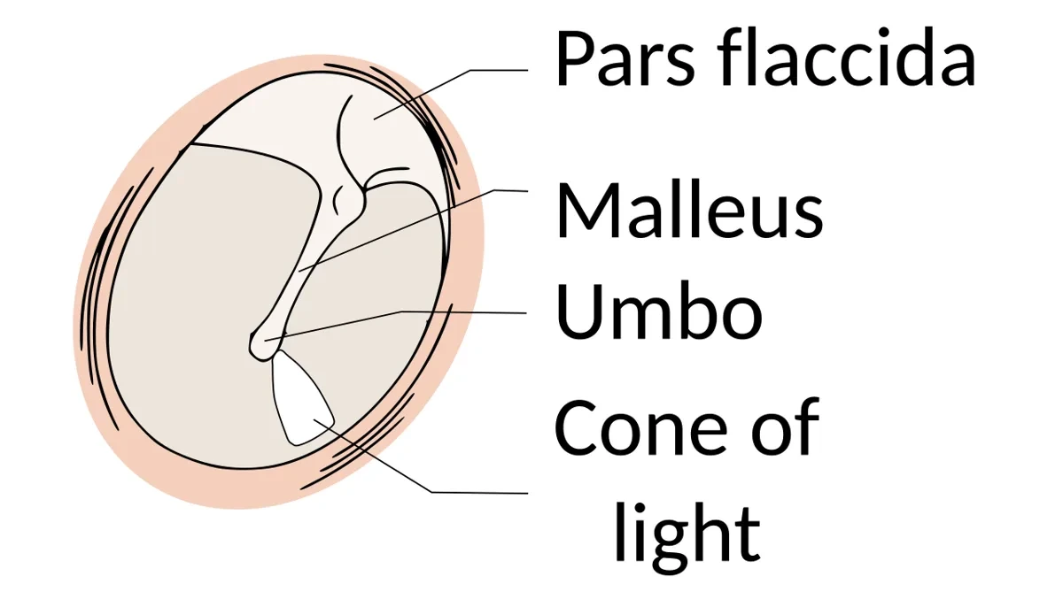
外耳道は外耳孔から鼓膜までの長さ約2.5 cm の管である。外側1/3 が軟骨部(耳介軟骨の続き)、内側2/3 が骨部(側頭骨の鼓室部)で、皮膚には毛と耳垢腺 glandulae ceruminosae(変形したアポクリン汗腺)があり、どちらも軟骨部に多い。毛と耳垢が異物の入口をふさぐ。管はまっすぐではなく、外から前上方へ進んでやや後ろへ曲がり、最後に前下方へ下る。成人では耳介を後上方へ引くとまっすぐになって鼓膜が見える。骨部がまだ短い乳幼児では、後下方へ引く。
鼓膜 membrana tympanicaは直径約1 cm の楕円形の薄い膜で、外耳道の奥に斜めに張り、外面は外下方を向く。外面は浅い漏斗のようにへこみ、中心の鼓膜臍 umboにツチ骨柄の先が付いている。下の大部分は張りのある緊張部 pars tensaで、縁の線維軟骨輪が側頭骨の溝にはまる。上の小さな部分は線維層を欠いて薄くゆるい弛緩部 pars flaccidaである。3層の組織は中耳と耳管(組織学)にある。耳鏡では、ツチ骨柄が透けて見えるツチ骨条、その上端のツチ骨隆起、そして前下方に光錐(光の反射)が見える。
光錐が前下方に出るのは、鼓膜が傾いた漏斗だからである。斜めに張ったへこんだ面のうち、耳鏡の光をまっすぐ返す向きの部分は前下方にしかない。したがって光錐が見えないことは、鼓膜の形や位置が変わった(膨らんだ・引き込まれた)サインになる。右耳では5時、左耳では7時の方向に出る。
鼓膜はツチ骨柄の延長線と、それに直角な線で4つの象限に分ける。後上象限の裏にはキヌタ骨長脚・アブミ骨・鼓索神経がある。このため、中耳の液を出す鼓膜切開は下方の象限で行い、後上象限は避ける。
外耳道の皮膚は、前上壁が耳介側頭神経(V3)、後下壁が迷走神経の耳介枝、鼓膜の内面は舌咽神経(鼓室神経)が担う。外耳道の前壁は顎関節の下顎頭のすぐ後ろにあり、口を開け閉めすると外耳道に入れた指で動きが分かる。
耳を見ても異常の無い耳痛は、ほかの場所からの関連痛を考える。耳の皮膚の神経(V3・IX・X)は、歯・顎関節(V3)、扁桃・咽頭(IX)、喉頭・下咽頭(X)とも共通である。成人の片側の耳痛で鼓膜が正常なら、咽頭・喉頭の癌を見落とさないようにする。
弛緩部は線維層が無く弱いので、耳管の働きが悪く中耳が陰圧になると、ここが内側へ引き込まれてポケットをつくる。そこに角化した皮膚がたまって骨を壊していくのが真珠腫性中耳炎で、多くは弛緩部から始まる。小さな鼓膜の穿孔は自然に閉じることが多い。
English version
Core
The external acoustic meatus is a canal about 2.5 cm long from the opening to the eardrum. Its outer third is cartilage (continuous with the auricle) and its inner two-thirds is bone (tympanic part of the temporal bone). The skin has hairs and ceruminous glands (modified apocrine sweat glands), mostly in the cartilaginous part; hair and wax block foreign bodies. The canal is not straight: it runs forward and up, turns a little back, then goes forward and down. In adults, pulling the auricle up and back straightens it so the eardrum can be seen; in infants, whose bony part is short, pull down and back.
The tympanic membrane is a thin oval membrane about 1 cm across. It lies obliquely at the end of the canal, its outer surface facing down and out. The outer surface is a shallow funnel; at its centre, the umbo, the tip of the handle of the malleus is attached. Most of it is the tight pars tensa, whose fibrocartilaginous ring sits in a groove of the temporal bone. The small upper part, without a fibrous layer, is the thin, loose pars flaccida. With an otoscope you see the malleus handle (stria), the lateral process at its top, and a cone of light in the anterior-inferior part.
Going further
The cone of light is anterior-inferior because the eardrum is a tilted funnel. Only that part of the oblique, concave surface sends the otoscope light straight back. So a missing light reflex means the shape or position of the drum has changed (bulging or retracted). It is at 5 o’clock in the right ear and 7 o’clock in the left.
The drum is divided into four quadrants by a line along the malleus handle and a line at right angles to it. Behind the posterior-superior quadrant lie the long process of the incus, the stapes and the chorda tympani. Myringotomy to drain fluid is therefore done in a lower quadrant, avoiding the posterior-superior one.
The canal skin is supplied by the auriculotemporal nerve (V3) at the front and top, the auricular branch of the vagus at the back and bottom, and the inner surface of the drum by the glossopharyngeal nerve (tympanic nerve). The front wall of the canal lies just behind the head of the mandible: with a finger in the canal you can feel the jaw move.
In the clinic
Ear pain with a normal-looking ear suggests referred pain. The nerves of the ear (V3, IX, X) also supply the teeth and jaw joint (V3), tonsils and pharynx (IX), and larynx and hypopharynx (X). In an adult with one-sided ear pain and a normal drum, do not miss a cancer of the pharynx or larynx.
The pars flaccida has no fibrous layer and is weak, so when poor tubal function makes the middle ear negative, it is pulled in and forms a pocket. Keratinised skin collects there and erodes bone — cholesteatoma — and most cases start in the pars flaccida. Small perforations of the drum often heal by themselves.
鼓室 — 6つの壁と、その向こう側Cavitas tympani
中耳 auris media は鼓室 cavitas tympaniと耳管 tuba auditivaからなる。鼓室は側頭骨錐体の中の、縦に長く横に薄い空気の部屋である(高さ約15 mm、中央の幅はわずか約2 mm)。粘膜に覆われ、3つの耳小骨・2つの筋・空気を入れる。鼓膜の高さを中鼓室、それより上を上鼓室 recessus epitympanicus(ツチ骨頭とキヌタ骨体が入る)、下を下鼓室と呼ぶ。
| 壁 | ラテン語 | 主な構造 | 壁の向こう |
|---|---|---|---|
| 上壁 | paries tegmentalis | 鼓室蓋 tegmen tympani(薄い骨板) | 中頭蓋窩・側頭葉 |
| 下壁 | paries jugularis | 頸静脈窩の上の薄い骨 | 内頸静脈の上球 |
| 前壁 | paries caroticus | 上に鼓膜張筋半管、下に耳管半管(耳管鼓室口) | 内頸動脈(頸動脈管) |
| 後壁 | paries mastoideus | 乳突洞口 aditus ad antrum、錐体隆起(中からアブミ骨筋の腱が出る) | 乳突洞・乳突蜂巣、顔面神経管の下行部 |
| 外側壁 | paries membranaceus | 鼓膜(その上は上鼓室の骨の外壁) | 外耳道 |
| 内側壁 | paries labyrinthicus | 岬角 promontorium、前庭窓(アブミ骨底)、蝸牛窓(第二鼓膜)、顔面神経管隆起、外側半規管隆起 | 内耳 |
内側壁の中央のふくらみ岬角は蝸牛の基底回転が押し出したもので、表面に舌咽神経の鼓室神経がつくる鼓室神経叢が乗る(小錐体神経となって耳神経節 → 耳下腺へ)。その後上に前庭窓 fenestra vestibuli(卵円窓、アブミ骨底がはまる)、後下に蝸牛窓 fenestra cochleae(正円窓、第二鼓膜で閉じ、鼓室階に通じる)がある。外側壁の上部では、顔面神経の枝の鼓索神経がツチ骨とキヌタ骨の間を横切っていく。
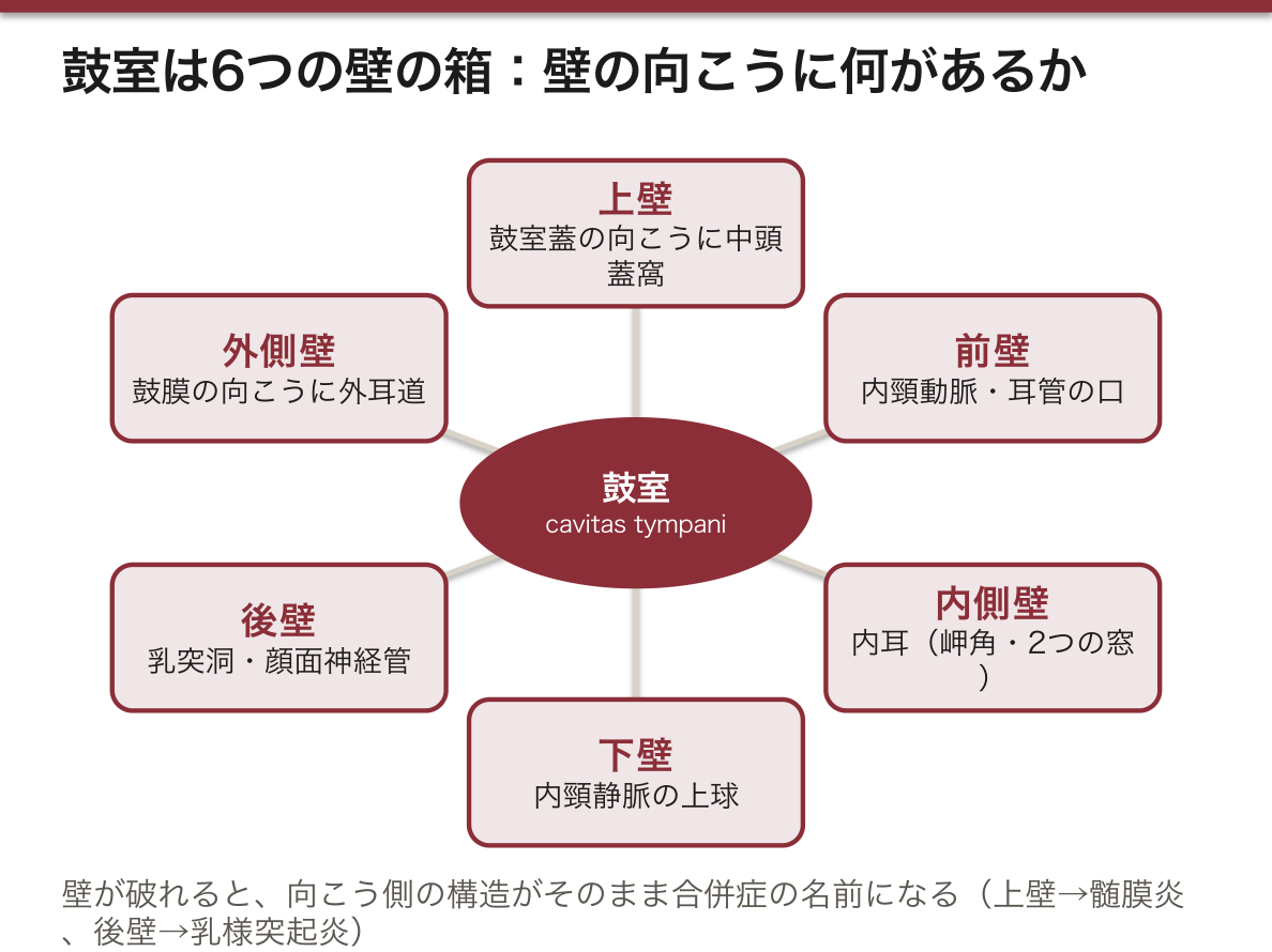
顔面神経は鼓室の壁の中を通っている。顔面神経管 canalis n. facialis は、内耳道 → 膝神経節(ここで急に後ろへ曲がる)→ 鼓室の内側壁を前庭窓の上で後ろへ(水平部)→ 後壁の中を下へ(垂直部、途中でアブミ骨筋神経と鼓索神経を出す)→ 茎乳突孔、と走る。管の骨は薄く、生まれつき一部が欠けている人もいる。そのため中耳炎・真珠腫・中耳の手術で顔面神経麻痺が起こりうる。鼓索神経が鼓室を横切ることは、中耳の手術後に舌の前2/3 の味覚が変わることがある理由になる。
6つの壁は「破れたら何が起こるか」で覚えると忘れない。壁ごとに、向こう側の構造がそのまま合併症の名前になっている。
| 壁 | 壁が破れる・病気が越えると |
|---|---|
| 上壁 | 髄膜炎・側頭葉の膿瘍。頭蓋底骨折が鼓室蓋と鼓膜を裂くと、外耳道から血液と髄液が出る(髄液耳漏) |
| 下壁 | 内頸静脈の上球が鼓室へ張り出していると、鼓膜の向こうに青い影が見える。グロムス腫瘍は拍動性の耳鳴を起こす |
| 後壁 | 乳様突起炎、さらにS状静脈洞の血栓症 |
| 内側壁 | 内耳炎・迷路瘻孔(めまい・感音難聴)、顔面神経麻痺 |
乳様突起とS状静脈洞の位置関係は乳様突起の項目にもある。
English version
Core
The middle ear consists of the tympanic cavity and the auditory tube. The tympanic cavity is a tall, very narrow air space in the petrous temporal bone (about 15 mm high, only about 2 mm wide in the middle). It is lined with mucosa and contains three ossicles, two muscles and air. The level of the drum is the mesotympanum; above it is the epitympanic recess (with the head of the malleus and body of the incus); below it is the hypotympanum.
| Wall | Latin | Main features | On the other side |
|---|---|---|---|
| Roof | paries tegmentalis | tegmen tympani (thin bone) | middle cranial fossa, temporal lobe |
| Floor | paries jugularis | thin bone over the jugular fossa | bulb of the internal jugular vein |
| Anterior | paries caroticus | canal for tensor tympani above, canal for the auditory tube below | internal carotid artery |
| Posterior | paries mastoideus | aditus to the mastoid antrum; pyramidal eminence (stapedius tendon) | mastoid antrum and air cells; descending facial canal |
| Lateral | paries membranaceus | tympanic membrane (bone above it) | external acoustic meatus |
| Medial | paries labyrinthicus | promontory, oval window (stapes), round window (secondary tympanic membrane), facial canal prominence, lateral canal prominence | internal ear |
The promontory on the medial wall is made by the basal turn of the cochlea; the tympanic plexus (from the tympanic nerve of IX) lies on it and continues as the lesser petrosal nerve to the otic ganglion and parotid gland. Above and behind it is the oval window (stapes footplate), below and behind it the round window (secondary tympanic membrane, leading to the scala tympani). On the upper lateral wall the chorda tympani crosses between malleus and incus.
Going further
The facial nerve runs inside the walls of the tympanic cavity. The facial canal goes: internal acoustic meatus → geniculate ganglion (sharp turn backward) → along the medial wall above the oval window (horizontal part) → down inside the posterior wall (vertical part, giving the nerve to stapedius and the chorda tympani) → stylomastoid foramen. The bony canal is thin and in some people partly missing, so otitis media, cholesteatoma and middle ear surgery can cause facial palsy. Because the chorda tympani crosses the cavity, taste on the front two-thirds of the tongue may change after middle ear surgery.
Learn the six walls by asking what happens if each one is broken: the structure on the other side gives the name of the complication.
In the clinic
| Wall | If disease or injury crosses it |
|---|---|
| Roof | meningitis, temporal lobe abscess; a skull base fracture through the tegmen and drum lets blood and CSF leak from the ear (CSF otorrhoea) |
| Floor | a high jugular bulb shows as a blue shadow behind the drum; a glomus tumour causes pulsatile tinnitus |
| Posterior | mastoiditis, then sigmoid sinus thrombosis |
| Medial | labyrinthitis, labyrinthine fistula (vertigo, sensorineural hearing loss), facial palsy |
耳小骨と鼓室の筋 — 音を渡す鎖と、そのブレーキOssicula auditus
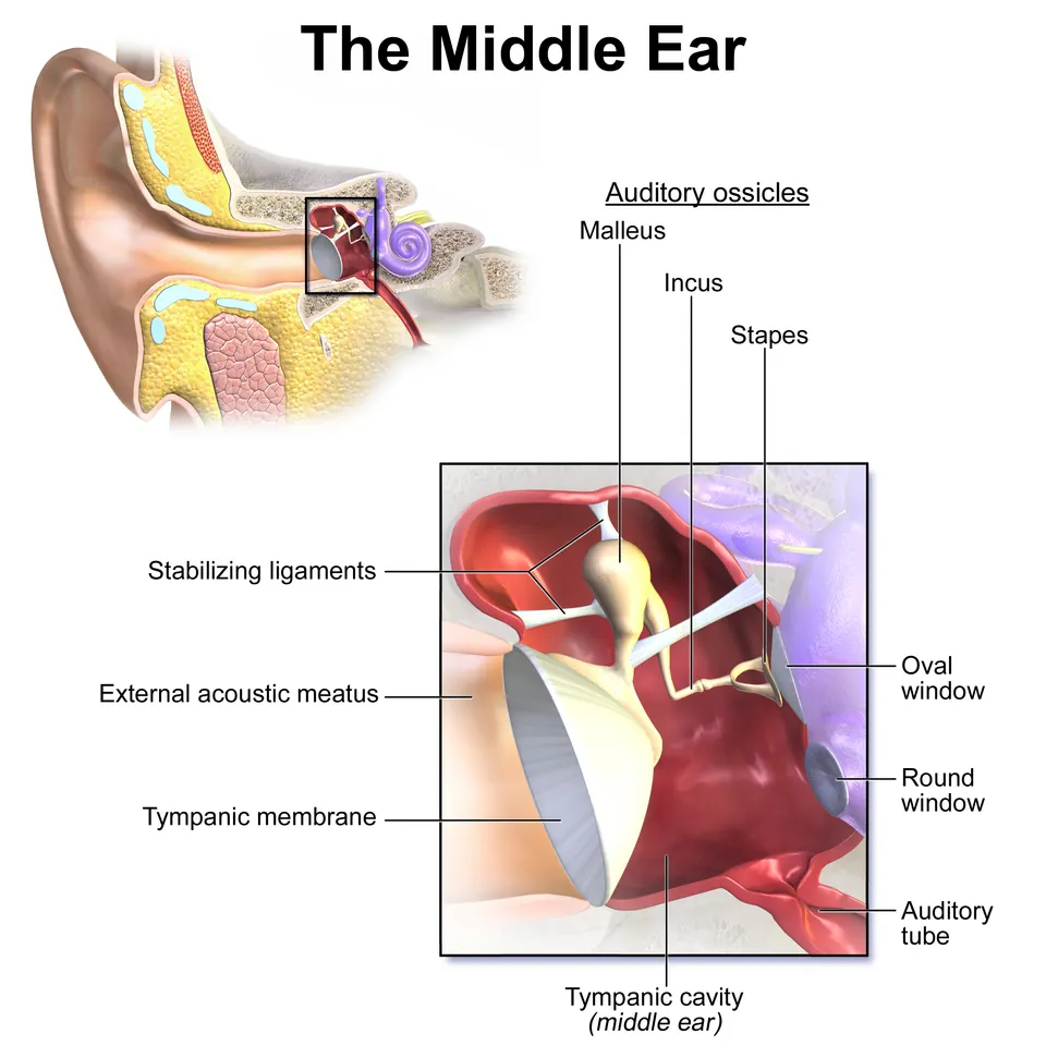
3つの耳小骨が鼓膜から前庭窓まで鎖をつくる。
| 骨 | 部位 | つながる相手 |
|---|---|---|
| ツチ骨 malleus | 頭 caput・頸 collum・柄 manubrium・外側突起・前突起 | 柄が鼓膜に埋まり鼓膜臍をつくる。頭は上鼓室でキヌタ骨体と関節 |
| キヌタ骨 incus | 体 corpus・短脚 crus breve・長脚 crus longum(先端に豆状突起) | 短脚は後方で靱帯により固定。長脚の豆状突起がアブミ骨頭と関節 |
| アブミ骨 stapes | 頭 caput・前脚・後脚・底 basis | 底が前庭窓にはまり、輪状靱帯で窓の縁につながる(鼓室アブミ骨結合) |
関節は2つの滑膜関節で、キヌタ・ツチ関節 art. incudomallearis は鞍関節、キヌタ・アブミ関節 art. incudostapedialis は小さな関節で、ヨーロッパの教科書では平面関節とし、球関節とする本もある。
| 筋(横紋筋) | 起始 | 停止 | 支配 | 働き |
|---|---|---|---|---|
| 鼓膜張筋 m. tensor tympani | 鼓膜張筋半管(耳管の上) | ツチ骨柄の根元 | 下顎神経(V3) | ツチ骨柄を内側へ引き、鼓膜を張る |
| アブミ骨筋 m. stapedius | 後壁の錐体隆起の中 | アブミ骨頸 | 顔面神経(VII)のアブミ骨筋神経 | アブミ骨を後ろへ傾け、底の動きを抑える |
どちらも大きな音で反射的に縮んで耳小骨の振動を弱める。アブミ骨筋は体でいちばん小さな骨格筋である。
2つの筋の神経が違うのは、出身の咽頭弓が違うからである。ツチ骨・キヌタ骨と鼓膜張筋は第1咽頭弓(下顎の弓)から生じ、咀嚼筋と同じく三叉神経第3枝に支配される。アブミ骨(大部分)とアブミ骨筋は第2咽頭弓から生じ、表情筋と同じく顔面神経に支配される。口蓋帆張筋が V3、口蓋帆挙筋が迷走神経の咽頭神経叢、と分かれているのと同じ理屈で、「筋の神経は、その筋が生まれた弓の神経」と覚えれば暗記は要らない。
アブミ骨筋反射の回路は、蝸牛 → 蝸牛神経 → 蝸牛神経核 → 上オリーブ核 → 両側の顔面神経核 → アブミ骨筋である。片耳に大きな音を入れると両耳のアブミ骨筋が縮むので、音を入れる耳と測る耳を分けて、蝸牛・聴神経・顔面神経のどこが傷んでいるかを調べられる(ティンパノメトリーの一部として行う)。中耳が音を増幅する理屈(面積比とてこ比)は中耳と耳管(組織学)にある。
耳硬化症ではアブミ骨底のまわりに異常な骨ができて前庭窓に固着し、鎖の最後が動かなくなって伝音難聴になる。アブミ骨の上部構造を外して底に小さな穴を開け、人工のピストンを立てる手術(アブミ骨手術)で聞こえが戻る。慢性中耳炎で最も壊れやすいのはキヌタ骨の長脚の先である。血液の供給が細い終末部のため、炎症で溶けて鎖が切れやすい。顔面神経麻痺で音が響いてうるさく感じる(聴覚過敏)なら、アブミ骨筋神経の分岐より中枢側の障害を考える。
English version
Core
Three ossicles form a chain from the eardrum to the oval window.
| Bone | Parts | Connections |
|---|---|---|
| Malleus | head, neck, handle (manubrium), lateral and anterior processes | the handle is in the drum and forms the umbo; the head articulates with the incus in the epitympanic recess |
| Incus | body, short limb, long limb (lenticular process at its tip) | short limb is held behind by a ligament; the lenticular process articulates with the head of the stapes |
| Stapes | head, anterior and posterior limbs, base (footplate) | the footplate fits the oval window, joined to its rim by the annular ligament (tympanostapedial syndesmosis) |
The incudomallear joint is a saddle joint. The incudostapedial joint is small; European textbooks call it a plane joint, and some books call it a ball-and-socket joint.
| Muscle (striated) | Origin | Insertion | Nerve | Action |
|---|---|---|---|---|
| Tensor tympani | canal above the auditory tube | root of the malleus handle | mandibular nerve (V3) | pulls the handle medially, tenses the drum |
| Stapedius | inside the pyramidal eminence, posterior wall | neck of stapes | nerve to stapedius (VII) | tilts the stapes back and limits footplate movement |
Both contract by reflex to loud sound and damp the vibration of the ossicles. The stapedius is the smallest skeletal muscle in the body.
Going further
The two muscles have different nerves because they come from different pharyngeal arches. The malleus, incus and tensor tympani come from the first (mandibular) arch and, like the muscles of mastication, are supplied by V3. The stapes (mostly) and stapedius come from the second arch and, like the muscles of facial expression, are supplied by VII. “A muscle keeps the nerve of the arch it came from” — no memorising needed.
The stapedius reflex runs: cochlea → cochlear nerve → cochlear nuclei → superior olivary nucleus → facial nuclei on both sides → stapedius. Sound in one ear contracts the stapedius in both ears, so by separating the stimulated ear and the measured ear you can find whether the cochlea, the auditory nerve or the facial nerve is damaged (done as part of tympanometry).
In the clinic
In otosclerosis abnormal bone around the stapes footplate fixes it in the oval window, and the end of the chain stops moving: conductive hearing loss. Removing the upper stapes, making a small hole in the footplate and placing a prosthetic piston (stapes surgery) restores hearing. In chronic otitis media the part most easily destroyed is the tip of the long limb of the incus, an end region with a thin blood supply, so the chain breaks there. If a patient with facial palsy finds sounds painfully loud (hyperacusis), the lesion is proximal to the branch to stapedius.
耳管と乳突蜂巣 — 中耳の換気口と、奥の空洞Tuba auditiva
耳管 tuba auditiva(Eustachius 管)は鼓室の前壁の耳管鼓室口から前内下方へ進み、咽頭鼻部の外側壁の耳管咽頭口に開く、長さ約3.5〜4 cm の管である。後外側の約1/3 が骨部 pars ossea(筋耳管管の下半分=耳管半管の中)、前内側の約2/3 が軟骨部 pars cartilaginea(弾性軟骨と膜でできる)で、両者の境のいちばん細い所を耳管峡 isthmusという。粘膜は線毛をもつ上皮で、線毛は咽頭の向きに掃いている。咽頭口の後上縁は軟骨が盛り上げた耳管隆起で、まわりに耳管扁桃がある(耳管咽頭口)。
耳管は普段は閉じていて、嚥下やあくびのときに開く。開けるのは主に口蓋帆張筋(V3)で、軟骨部の外側の壁を引いて口を開く。口蓋帆挙筋と耳管咽頭筋も加わる。
鼓室の後ろ上方は、乳突洞口 aditus ad antrum → 乳突洞 antrum mastoideum → 乳突蜂巣 cellulae mastoideae(乳様突起の中の多数の小さな空気の部屋)へと続く。粘膜は鼓室と一続きで、耳管 → 鼓室 → 乳突蜂巣は1本の空気の通り道になっている。
なぜ耳管が開かないと液がたまるのか。中耳の空気は粘膜の血管に少しずつ吸収されていく。耳管がときどき開いて空気を補わないと、中耳は陰圧になり、鼓膜が内側へ引き込まれ、やがて粘膜から液がしみ出してたまる(滲出性中耳炎)。
口蓋裂の子どもに滲出性中耳炎が非常に多いのはこの仕組みによる。耳管を開ける口蓋帆張筋は、口蓋の正中の腱膜に付いて初めて引く力を出せるが、口蓋裂では左右の筋が正中でつながらず、耳管を開けられないからである。小児の耳管が短く水平に近いことと急性中耳炎の関係は中耳と耳管(組織学)にある。
乳様突起は生まれたときにはほとんど無く、生後、胸鎖乳突筋に引かれて育ち、中に蜂巣が広がっていく。そのため新生児では茎乳突孔が浅く、顔面神経が皮膚のすぐ下に出ていて、鉗子分娩や耳の後ろの切開で傷つきやすい。
乳様突起炎は中耳炎が乳突蜂巣へ広がったもので、耳の後ろが赤く腫れて押すと痛み、耳介が前へ押し出される。蜂巣のすぐ内側にはS状静脈洞、上には中頭蓋窩、後ろには後頭蓋窩があるので、静脈洞血栓症・髄膜炎・脳膿瘍へ進むことがある。成人の片側だけの滲出性中耳炎では、耳管咽頭口の近くにできた上咽頭癌が耳管を塞いでいないかを確かめる(咽頭陥凹)。
English version
Core
The auditory (Eustachian) tube runs from the tympanic opening in the anterior wall of the tympanic cavity forward, medially and down to the pharyngeal opening in the lateral wall of the nasopharynx. It is about 3.5–4 cm long. The back third is the bony part (in the lower half of the musculotubal canal) and the front two-thirds is the cartilaginous part (elastic cartilage and membrane). The narrowest point where they meet is the isthmus. The lining has ciliated epithelium that beats toward the pharynx. The cartilage raises the torus tubarius behind the pharyngeal opening, with the tubal tonsil around it.
The tube is normally closed and opens during swallowing and yawning, mainly by the tensor veli palatini (V3), which pulls the lateral wall of the cartilaginous part. The levator veli palatini and salpingopharyngeus also help.
Behind and above the tympanic cavity the air space continues through the aditus to the mastoid antrum and the mastoid air cells in the mastoid process. The mucosa is continuous, so tube → tympanic cavity → mastoid cells is one air passage.
Going further
Why does fluid collect when the tube does not open? Air in the middle ear is slowly absorbed by mucosal vessels. Unless the tube opens from time to time to refill it, the pressure becomes negative, the drum is pulled in, and fluid leaks from the mucosa (otitis media with effusion).
This is why children with cleft palate very often have otitis media with effusion. The tensor veli palatini can only pull when it is anchored to the palatine aponeurosis in the midline; in a cleft palate the two sides do not join, so the tube cannot be opened.
The mastoid process is almost absent at birth; it grows after birth under the pull of sternocleidomastoid, and the air cells spread inside it. In newborns the stylomastoid foramen is shallow and the facial nerve comes out just under the skin, so it is easily injured by forceps delivery or an incision behind the ear.
In the clinic
Mastoiditis is the spread of otitis media into the mastoid cells: redness, swelling and tenderness behind the ear, and the auricle pushed forward. The sigmoid sinus lies just medial to the cells, the middle cranial fossa above and the posterior cranial fossa behind, so it can lead to sinus thrombosis, meningitis or brain abscess. In an adult with one-sided otitis media with effusion, check for a nasopharyngeal cancer near the tubal opening blocking the tube.
骨迷路 — 前庭・半規管・蝸牛Labyrinthus osseus
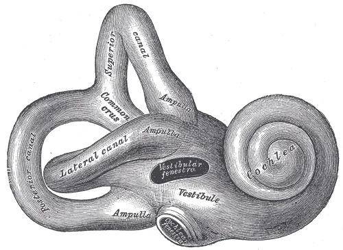
内耳 auris interna は錐体の中、外側の中耳と内側の内耳道の間にある。骨でできた空洞の系骨迷路 labyrinthus osseusの中に、膜でできた袋の系膜迷路が浮かび、その間を外リンパ perilymphaが満たす。骨迷路は前から蝸牛・前庭・半規管の3部である。
前庭 vestibulumは中央の小部屋。外側壁(鼓室側)に前庭窓が開く。内側壁には前の球形陥凹 recessus sphericus(球形嚢が入る)と後ろの楕円陥凹 recessus ellipticus(卵形嚢が入り、前庭小管の内口がある)があり、間を前庭稜が隔てる。小さな孔の集まり(篩斑)を前庭神経の線維が通る。後ろに半規管の5つの口、前に蝸牛の前庭階が開く。
半規管 canales semicircularesは前・後・外側の3本で、互いにほぼ直角に向く。それぞれ2/3 周の弧で、一方の端が膨らんで骨膨大部 ampulla ossea、もう一方が単脚になる。前と後の単脚が合わさって総脚 crus communeになるので、前庭への口は6つでなく5つである。
蝸牛 cochleaは2回半巻くカタツムリの殻である。中心の骨の柱蝸牛軸 modiolusのまわりをらせん管が巻き、軸から張り出す骨らせん板 lamina spiralis osseaが、膜迷路と一緒に管を上の前庭階 scala vestibuliと下の鼓室階 scala tympaniに分ける。両階は頂上の蝸牛孔 helicotremaでつながる。蝸牛の底は後内側(内耳道の側)、頂は前外側を向く。鼓室階の始まりの近くから蝸牛小管が出て、外リンパの通り道(外リンパ管)がくも膜下腔へつながる。
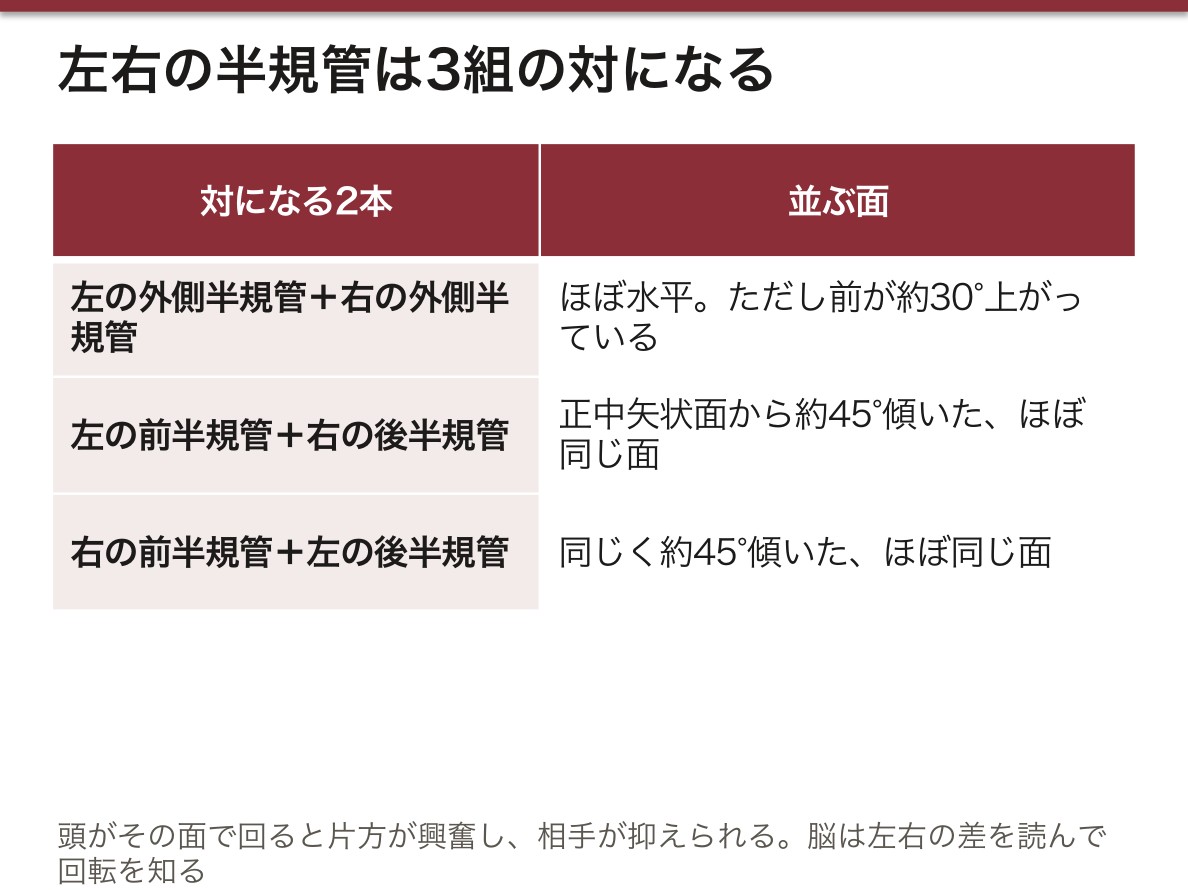
半規管の向きの本当の姿。教科書ではよく前半規管を矢状面、後半規管を前頭面、外側半規管を水平面と単純化する。実際には前・後半規管はどちらも正中矢状面から約45°傾いていて、片側の前半規管と反対側の後半規管がほぼ同じ面に並ぶ(左前と右後、右前と左後)。外側半規管も水平ではなく、前が約30°上がっている。こうして左右の6本が3組の対になり、頭がある面で回ると片方が興奮し、相手が抑えられる。脳は左右の差を読んで回転を知る。
外側半規管が30°傾いていることは、温度眼振検査(カロリック検査)の姿勢を決めている。仰向けで頭を30°起こすと外側半規管が垂直に立つ。そこで外耳道に冷水や温水を入れると、近い側の内リンパだけが冷やされ(温められ)て対流が起こり、回転したのと同じ刺激が片側の半規管だけに生まれる。
骨迷路の窓は本来2つだけである。前庭窓で押し込んだ分を蝸牛窓が外へ逃がす。液体は縮まないので、逃げ道が無ければ内耳の液は動けない。
前半規管の上の骨が薄くなって欠けると、骨迷路に第3の窓ができる(上半規管裂隙症候群)。大きな音やいきみで外リンパが動いて半規管が刺激され、めまいや眼振が起こり、自分の声や脈の音が響いて聞こえる。外リンパが蝸牛小管でくも膜下腔とつながっていることは、髄膜炎が内耳へ広がって難聴を残す道の一つと考えられている。
English version
Core
The internal ear lies in the petrous bone, between the middle ear laterally and the internal acoustic meatus medially. A system of bony cavities, the bony labyrinth, contains a system of membranous sacs, the membranous labyrinth, with perilymph between them. The bony labyrinth has three parts: cochlea, vestibule and semicircular canals.
The vestibule is the central chamber. The oval window opens in its lateral wall. Its medial wall has the anterior spherical recess (for the saccule) and the posterior elliptical recess (for the utricle, with the opening of the vestibular aqueduct), separated by the vestibular crest. Groups of small holes let vestibular nerve fibres through. Behind are five openings of the semicircular canals; in front is the scala vestibuli of the cochlea.
The three semicircular canals (anterior, posterior, lateral) are roughly at right angles. Each is two-thirds of a circle with a swollen end, the bony ampulla, and a simple end. The simple ends of the anterior and posterior canals join as the common limb, so there are five openings, not six.
The cochlea is a snail shell of two and a half turns. A spiral canal winds around the central bony column, the modiolus. The osseous spiral lamina projects from it and, together with the membranous labyrinth, divides the canal into the upper scala vestibuli and lower scala tympani, which join at the apex through the helicotrema. The base faces back and medially (toward the internal meatus), the apex forward and laterally. Near the start of the scala tympani the cochlear aqueduct connects the perilymph to the subarachnoid space.
Going further
The real orientation of the canals. Textbooks often say the anterior canal is sagittal, the posterior frontal and the lateral horizontal. In fact the anterior and posterior canals are both about 45° from the midsagittal plane, and the anterior canal of one side lies almost in the same plane as the posterior canal of the other. The lateral canal is tilted up about 30° at the front. So the six canals form three pairs: rotation in one plane excites one canal and inhibits its partner, and the brain reads the difference.
The 30° tilt of the lateral canal sets the position for the caloric test. Lying supine with the head raised 30° makes the lateral canal vertical. Cold or warm water in the ear canal then cools or warms the nearby endolymph and causes convection, giving one canal the same signal as a rotation.
The bony labyrinth normally has only two windows. What the oval window pushes in, the round window lets out. Fluid cannot be compressed, so without this outlet the inner ear fluid could not move.
In the clinic
If the bone over the anterior canal is thin or missing, a third window appears (superior canal dehiscence syndrome). Loud sounds or straining move the perilymph and stimulate the canal, causing vertigo and nystagmus, and patients hear their own voice and pulse too loudly. The link between perilymph and the subarachnoid space through the cochlear aqueduct is thought to be one way meningitis spreads to the inner ear and leaves deafness.
膜迷路 — 内リンパの閉じた袋と、3種類の受容器Labyrinthus membranaceus
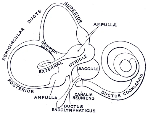
膜迷路は骨迷路の中に浮かぶ閉じた袋の系で、中は内リンパ endolymphaで満たされ、外を外リンパが囲む。骨迷路の各部に、対応する膜迷路と受容器がある。
| 骨迷路 | 中の膜迷路 | 受容器 | 感じるもの |
|---|---|---|---|
| 前庭 | 卵形嚢 utriculus(大きい・楕円陥凹)と球形嚢 sacculus(小さい・球形陥凹) | 卵形嚢斑(ほぼ水平)と球形嚢斑(ほぼ垂直) | 重力・直線の加速度(頭の傾き) |
| 半規管 | 半規管 ductus semicirculares(3本。骨の管よりずっと細い) | 膨大部稜 crista ampullaris | 回転(角加速度) |
| 蝸牛 | 蝸牛管 ductus cochlearis | らせん器(Corti 器) | 音 |
袋どうしはつながっている。卵形嚢と球形嚢は連嚢管で結ばれ、そこから内リンパ管 ductus endolymphaticusが前庭小管(前庭水管)を通って、錐体の後面の硬膜の間にある行き止まりの内リンパ嚢 saccus endolymphaticusに終わる。球形嚢と蝸牛管は結合管 ductus reuniensでつながり、半規管は5つの口で卵形嚢に開く。
蝸牛管は断面が三角形で、上壁が前庭膜(Reissner 膜)、外壁が内リンパを作る血管条とらせん靱帯、下壁が骨らせん板の先から外壁へ張る基底板(らせん膜)である。基底板の上にらせん器が乗る。蝸牛管が入ることで、らせん管は上から前庭階・蝸牛管・鼓室階の3段になる。有毛細胞と内リンパの組成はコルチ器と前庭にある。
音の通り道:鼓膜 → 耳小骨 → アブミ骨底が前庭窓を押す → 前庭階の外リンパに波 → 蝸牛孔 → 鼓室階 → 蝸牛窓の第二鼓膜が外へふくらむ。途中で前庭膜と基底板にはさまれた蝸牛管が揺すられ、基底板が振動してらせん器の有毛細胞が刺激される。空気の振幅の大きい弱い波が、外リンパの振幅の小さい強い波に変えられて内耳へ入る。
内リンパの出口は内リンパ嚢しかない。膜迷路は外とつながらない閉じた袋で、血管条が作った内リンパは内リンパ嚢で吸収される。作る量と吸収の釣り合いが崩れると袋がふくらむ(内リンパ水腫)。蝸牛管と前庭が結合管で同じ袋を共有しているので、難聴・耳鳴りとめまいが同時に起こるメニエール病になる。
良性発作性頭位めまい症(BPPV)の大半が後半規管で起こるのは、解剖の位置による。立っていても寝ていても、後半規管は膜迷路の中でいちばん低い場所にあり、その口は卵形嚢に開いている。卵形嚢斑から剥がれた耳石は、重力でまず後半規管へ落ちこむ。耳石を卵形嚢へ戻す頭位治療の考え方は前庭(組織学)にある。メニエール病に対して内リンパ嚢を開放する手術が行われるのは、ここが唯一の出口だからである。
English version
Core
The membranous labyrinth is a closed system of sacs inside the bony labyrinth. It is filled with endolymph and surrounded by perilymph. Each part of the bony labyrinth contains its own membranous part and receptor.
| Bony labyrinth | Membranous part | Receptor | Senses |
|---|---|---|---|
| Vestibule | utricle (larger, elliptical recess) and saccule (smaller, spherical recess) | macula of utricle (about horizontal), macula of saccule (about vertical) | gravity and linear acceleration (head tilt) |
| Semicircular canals | semicircular ducts (three; much thinner than the bony canals) | crista ampullaris | rotation (angular acceleration) |
| Cochlea | cochlear duct | spiral organ (organ of Corti) | sound |
The sacs are connected. The utricle and saccule are joined by the utriculosaccular duct, from which the endolymphatic duct runs through the vestibular aqueduct to the blind endolymphatic sac between the layers of dura on the back of the petrous bone. The saccule joins the cochlear duct by the ductus reuniens, and the semicircular ducts open into the utricle by five openings.
The cochlear duct is triangular in section: its upper wall is the vestibular (Reissner’s) membrane, its outer wall the stria vascularis (which makes endolymph) and spiral ligament, and its lower wall the basilar membrane stretching from the osseous spiral lamina to the outer wall. The spiral organ sits on the basilar membrane. With the cochlear duct in place, the spiral canal has three levels: scala vestibuli, cochlear duct, scala tympani.
Going further
The path of sound: eardrum → ossicles → stapes footplate pushes the oval window → wave in the perilymph of the scala vestibuli → helicotrema → scala tympani → round window bulges out. On the way the cochlear duct between the vestibular and basilar membranes is moved; the basilar membrane vibrates and the hair cells of the spiral organ are stimulated. A large, weak wave in air becomes a small, strong wave in the perilymph.
The only exit for endolymph is the endolymphatic sac. The membranous labyrinth is a closed bag; endolymph made by the stria vascularis is absorbed in the sac. If production and absorption lose balance, the bag swells (endolymphatic hydrops). Because the cochlear duct and vestibule share the same bag through the ductus reuniens, hearing loss and tinnitus come together with vertigo — Ménière disease.
In the clinic
Most benign paroxysmal positional vertigo (BPPV) involves the posterior canal because of its position. Whether standing or lying, the posterior canal is the lowest part of the membranous labyrinth, and it opens into the utricle. Otoconia that come loose from the utricular macula fall into it first by gravity. Surgery to open the endolymphatic sac is used for Ménière disease because the sac is the only exit.
聴覚路と平衡覚路 — 両側へ上る音、4方向へ散る平衡Via acustica et via vestibularis
聴覚路:1次ニューロンは蝸牛軸の中のらせん神経節の双極細胞で、末梢の突起がらせん器の有毛細胞から始まる。中枢の突起は蝸牛神経となり、前庭神経と合わさって内耳神経(VIII)として内耳道を通り(顔面神経・中間神経・迷路動脈と一緒)、橋と延髄の境(小脳橋角)から脳幹に入る。
| 中継 | 場所 | 次へ |
|---|---|---|
| 蝸牛神経核(背側・腹側) | 菱形窩の外側角 | 多くが台形体(腹側)や背側の聴条を通って反対側へ渡る。同じ側にも上る |
| 上オリーブ核・台形体核 | 橋の被蓋 | 外側毛帯を上る |
| 下丘 | 中脳背側 | 下丘腕 |
| 内側膝状体 | 視床 | 聴放線(内包後脚の後部) |
| 横側頭回(Heschl 回) | 上側頭回の上面(41・42野) | — |
背側の聴条は菱形窩の表面近くを横切るが、第四脳室髄条とは別物である。ヨーロッパの教科書ではこの経路を4つのニューロン(らせん神経節・蝸牛神経核・台形体核・内側膝状体)にまとめて数える。
平衡覚路:1次ニューロンは内耳道の底の前庭神経節で、末梢の突起は卵形嚢斑・球形嚢斑・膨大部稜から始まる。前庭神経として蝸牛神経と並んで脳幹に入り、菱形窩の前庭野の下の前庭神経核(上・下・内側・外側)で乗り換える。そこから4方向へ広がる。
| 行き先 | 経路 | 役割 |
|---|---|---|
| 脊髄 | 前庭脊髄路(外側・内側) | 抗重力筋の緊張と、頭頸部・体幹の姿勢 |
| 小脳 | 前庭小脳路(片葉小節葉と室頂核へ) | 平衡と歩行の自動的な調整 |
| 眼筋の核 | 内側縦束 → 外転神経核・動眼神経核・滑車神経核 | 前庭動眼反射 |
| 大脳皮質 | 視床を経由 | 回転・傾きの自覚 |
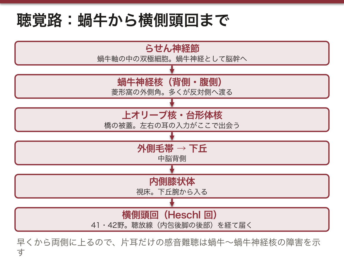
聴覚路は早い段階から両側に上る。蝸牛神経核の先、上オリーブ核ですでに左右の耳からの入力が出会い、届く時間の差と音の強さの差を比べて、音のする方向を割り出す。その結果、片側の脳の病変(脳幹の上部・内包・側頭葉)で片耳だけ聞こえなくなることは、ほとんど無い。片耳だけの感音難聴は、蝸牛・蝸牛神経・蝸牛神経核という「左右が出会う前」の障害を示す。
前庭動眼反射は、頭が動いても見ている物の像を網膜の上に止めるための反射である。頭が右へ回ると右の外側半規管が興奮し、前庭神経核から左の外転神経核(→ 左の外側直筋)と、内側縦束を上って右の動眼神経核(→ 右の内側直筋)へ信号が行き、両眼が左へ同じ速さで動く。経路が3つのニューロンしかないので、頭の動きにほとんど遅れずに追従できる。
前庭の情報が届く大脳皮質は、古典的な教科書では中側頭回とされるが、現在は頭頂葉と島の境のあたりが中心で、一次聴覚野や視覚野のような1つの場所は無いと考えられている。
片側の感音難聴と耳鳴りでは、内耳道から小脳橋角を MRI で調べる。前庭神経から生じる良性腫瘍(前庭神経鞘腫、いわゆる聴神経腫瘍)が、内耳道の中で蝸牛神経を圧迫するためである(内耳道)。大きくなると顔面神経・三叉神経・小脳が押される。
前庭動眼反射は意識の無い患者でも調べられる。頭を左右に回して眼が反対へ動けば(人形の目現象)、前庭神経核から内側縦束・動眼神経核・外転神経核までの脳幹は働いている。めまいの患者で頭を素早く回す検査(head impulse test)で眼が遅れて追いつくなら末梢(前庭神経炎など)の障害、遅れが無いのにめまいと眼振が強いなら、小脳や脳幹の脳卒中を疑う。
English version
Core
Auditory pathway: the first neurons are bipolar cells of the spiral ganglion in the modiolus; their peripheral processes start at the hair cells of the spiral organ. Their central processes form the cochlear nerve, which joins the vestibular nerve as the vestibulocochlear nerve (VIII), passes through the internal acoustic meatus (with the facial nerve, nervus intermedius and labyrinthine artery) and enters the brainstem at the junction of pons and medulla (cerebellopontine angle).
| Relay | Location | Next |
|---|---|---|
| Cochlear nuclei (dorsal, ventral) | lateral angle of the rhomboid fossa | many fibres cross through the trapezoid body (ventral) or dorsal acoustic striae; some stay on the same side |
| Superior olivary and trapezoid nuclei | pontine tegmentum | ascend in the lateral lemniscus |
| Inferior colliculus | dorsal midbrain | brachium of the inferior colliculus |
| Medial geniculate body | thalamus | acoustic radiation (back of the posterior limb of the internal capsule) |
| Transverse temporal gyri (Heschl) | upper surface of the superior temporal gyrus (areas 41, 42) | — |
The dorsal acoustic striae cross near the floor of the rhomboid fossa but are not the striae medullares of the fourth ventricle. European textbooks count this pathway as four neurons (spiral ganglion, cochlear nuclei, trapezoid nuclei, medial geniculate body).
Vestibular pathway: the first neurons are in the vestibular ganglion at the bottom of the internal meatus; their peripheral processes start at the maculae of the utricle and saccule and the ampullary crests. The vestibular nerve enters the brainstem with the cochlear nerve and synapses in the vestibular nuclei (superior, inferior, medial, lateral) under the vestibular area of the rhomboid fossa. From there signals go in four directions.
| Target | Route | Role |
|---|---|---|
| Spinal cord | vestibulospinal tracts (lateral, medial) | tone of antigravity muscles; posture of head, neck and trunk |
| Cerebellum | vestibulocerebellar fibres (to the flocculonodular lobe and fastigial nucleus) | automatic control of balance and gait |
| Eye muscle nuclei | medial longitudinal fasciculus → abducens, oculomotor and trochlear nuclei | vestibulo-ocular reflex |
| Cerebral cortex | via the thalamus | awareness of rotation and tilt |
Going further
The auditory pathway becomes bilateral early. Already at the superior olivary nucleus, input from both ears meets, and differences in arrival time and loudness are compared to locate the sound. As a result, a lesion on one side of the brain (upper brainstem, internal capsule, temporal lobe) almost never makes one ear deaf. One-sided sensorineural hearing loss points to the cochlea, the cochlear nerve or the cochlear nuclei — before the two sides meet.
The vestibulo-ocular reflex keeps the image of what we look at still on the retina while the head moves. When the head turns right, the right lateral canal is excited; signals go from the vestibular nuclei to the left abducens nucleus (left lateral rectus) and, through the medial longitudinal fasciculus, to the right oculomotor nucleus (right medial rectus), and both eyes move left at the same speed. With only three neurons, the reflex follows the head almost without delay.
Classical textbooks place vestibular cortex in the middle temporal gyrus; today the main region is thought to be around the border of the parietal lobe and insula, and there is no single primary area like the auditory or visual cortex.
In the clinic
For one-sided sensorineural hearing loss and tinnitus, image the internal acoustic meatus and cerebellopontine angle with MRI. A benign tumour of the vestibular nerve (vestibular schwannoma, “acoustic neuroma”) compresses the cochlear nerve in the meatus. As it grows it presses on the facial and trigeminal nerves and the cerebellum.
The vestibulo-ocular reflex can be tested in unconscious patients. If the eyes move opposite to a head turn (doll’s eye phenomenon), the brainstem from the vestibular nuclei through the medial longitudinal fasciculus to the oculomotor and abducens nuclei is working. In a dizzy patient, if the eyes fall behind and catch up during a quick head turn (head impulse test), the lesion is peripheral (such as vestibular neuritis); if there is no catch-up but strong vertigo and nystagmus, suspect a stroke in the cerebellum or brainstem.
まとめ
| 外耳と耳介 — 形の名前と、皮膚の神経 | 耳は外耳 auris externa・中耳 auris media・内耳 auris internaの3部に分かれる。 |
|---|---|
| 外耳道と鼓膜 — 曲がった管と、傾いた膜 | 外耳道は外耳孔から鼓膜までの長さ約2.5 cm の管である。 |
| 鼓室 — 6つの壁と、その向こう側 | 中耳 auris media は鼓室 cavitas tympaniと耳管 tuba auditivaからなる。 |
| 耳小骨と鼓室の筋 — 音を渡す鎖と、そのブレーキ | 3つの耳小骨が鼓膜から前庭窓まで鎖をつくる。 |
| 耳管と乳突蜂巣 — 中耳の換気口と、奥の空洞 | 耳管 tuba auditiva(Eustachius 管)は鼓室の前壁の耳管鼓室口から前内下方へ進み、咽頭鼻部の外側壁の耳管咽頭口に開く、長さ約3.5〜4 cm の管である。 |
| 骨迷路 — 前庭・半規管・蝸牛 | 内耳 auris interna は錐体の中、外側の中耳と内側の内耳道の間にある。 |
| 膜迷路 — 内リンパの閉じた袋と、3種類の受容器 | 膜迷路は骨迷路の中に浮かぶ閉じた袋の系で、中は内リンパ endolymphaで満たされ、外を外リンパが囲む。 |
| 聴覚路と平衡覚路 — 両側へ上る音、4方向へ散る平衡 | 聴覚路:1次ニューロンは蝸牛軸の中のらせん神経節の双極細胞で、末梢の突起がらせん器の有毛細胞から始まる。 |
このノートは医学生が授業の内容を自分の言葉でまとめたもので、診療の判断に使うものではありません。誤りに気づいたらお知らせください。