蛋白質については、「配列が形を決め、形が働きを決める」という一本の筋だけ押さえればよい。だから形が壊れることが、そのまま病気になる。
構造は4段階に分ける。
- 一次——アミノ酸の並び。ペプチド結合
- 二次——αヘリックス・βシート。主鎖どうしの水素結合
- 三次——1本の鎖の立体構造。疎水性相互作用が主役、加えてジスルフィド結合
- 四次——複数の鎖の組み合わせ。ヘモグロビンの α2β2
この章では、形と働きの関係が最もはっきり見える3つを扱う——ヘモグロビン(四次構造が働きを作る)、コラーゲン(一次配列の1文字が全体を壊す)、折り畳みの失敗(正しい配列でも形を間違える)。
運動器を学ぶうえではコラーゲンの項が特に重要。骨・腱・軟骨・靱帯はすべてコラーゲンの種類と架橋の話に還元できる。
- ヘモグロビン — 四次構造が協同性を生む
- コラーゲン — 3本鎖と、グリシンでなければならない理由
- 折り畳みの失敗 — シャペロンとアミロイド
- 血漿蛋白 — 濃度が語ること
- 蛋白質の構造の4階層 — 配列が形を決め、形が働きを決める
ヘモグロビン — 四次構造が協同性を生む
α2β2 の四量体。各サブユニットがヘムを1つ持ち、鉄(2価)に酸素が結合する。
- T型(緊張型)——酸素と結びにくい。組織で優勢
- R型(弛緩型)——結びやすい。肺で優勢
1つ結合すると残りも結合しやすくなる(協同性)。だから酸素解離曲線はS字になる。
右方移動=離しやすい:二酸化炭素↑・水素イオン↑(pH低下)・温度↑・2,3-ビスホスホグリセリン酸↑。
胎児ヘモグロビン(α2γ2)は 2,3-BPG と結びにくいので左方移動し、母体から酸素を奪える。
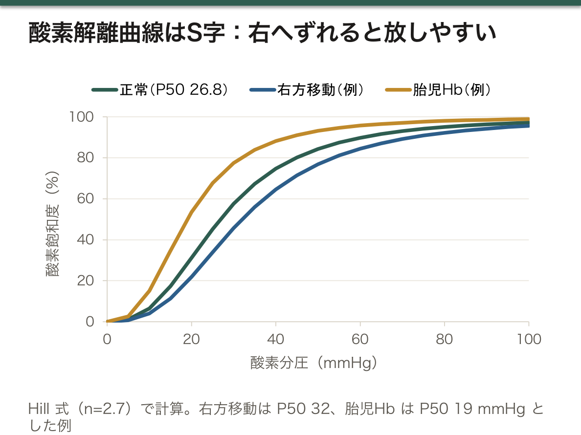
S字曲線であること自体が機能である。もし直線なら、肺で満たしても組織でわずかしか放せない。急な部分が組織の酸素分圧の範囲に来るように設計されているので、少しの分圧低下で大量に放出できる。上が平らなのも重要で、肺の酸素分圧が多少下がっても飽和度はほとんど落ちない——予備能である。
この形が、パルスオキシメータの読み方を決める。飽和度90%が酸素分圧60Torr に当たり、そこから先は崖になる。「90%を切ったら急に悪くなる」のは臨床の経験則ではなく、曲線の形そのものである。
右方移動の意味も統一的に読める——組織が激しく働いている印(酸性・高二酸化炭素・高温)が揃った場所ほど、酸素を多く手放す(Bohr 効果)。需要のある場所に自動的に配る仕組みが、蛋白の構造変化だけで実現している。
一酸化炭素中毒が危険なのは2重の理由による。結合力が酸素の200倍以上であることに加え、残りのサブユニットを R型に固定して曲線を左方へずらすため、運べる量が減るうえに放せなくなる。酸素分圧は正常なのでパルスオキシメータは正常に見える——測っているのが飽和度であって内容量ではないことを知らないと見落とす。
鎌状赤血球症は β鎖6番目のグルタミン酸がバリンに置き換わる1アミノ酸の変異である。疎水性のバリンが表面に出るため、脱酸素化した状態で分子どうしが重合して線維を作り、赤血球が鎌状に変形する。
脱酸素化したときにだけ起こるので、低酸素・脱水・アシドーシス・感染が発作の引き金になる。形が変わる条件が、そのまま予防すべき条件である。
メトヘモグロビン血症では鉄が3価になって酸素を運べない。チアノーゼがあるのに酸素分圧は正常で、酸素投与に反応しない。治療はメチレンブルー(還元を助ける)。「酸素が足りない」のではなく「運び手が壊れている」という区別が要る。
English version
Core
A tetramer, α2β2. Each subunit carries one haem, and oxygen binds to the iron (ferrous).
- T state (tense) — binds oxygen poorly. Predominant in the tissues.
- R state (relaxed) — binds readily. Predominant in the lung.
One binding makes the rest bind more easily (cooperativity), so the dissociation curve is sigmoid.
Right shift = releases more readily: raised CO₂, raised H⁺ (falling pH), raised temperature, raised 2,3-bisphosphoglycerate.
Fetal haemoglobin (α2γ2) binds 2,3-BPG poorly and so is shifted left, and can take oxygen from the mother.
Going further
That the curve is sigmoid is itself the function. Were it a straight line, the lung could fill it but the tissues could unload only a little. The steep part is placed within the range of tissue oxygen tension, so a small fall in tension releases a large amount. That the top is flat also matters — a moderate fall in alveolar oxygen tension barely reduces the saturation: a reserve.
That shape decides how a pulse oximeter is read. A saturation of 90 per cent corresponds to a tension of 60 Torr, and beyond that is a cliff. "Below 90 per cent it deteriorates suddenly" is not clinical folklore but the shape of the curve itself.
The right shift can be read as one idea — the more the marks of hard-working tissue are present (acid, high CO₂, warm), the more oxygen is released (the Bohr effect). A mechanism that distributes automatically to where the demand is, achieved by a change in the shape of a protein alone.
Carbon monoxide poisoning is dangerous for two reasons. Besides binding over 200 times more tightly than oxygen, it fixes the remaining subunits in the R state and shifts the curve to the left, so less can be carried and what is carried cannot be released. The oxygen tension is normal, so the pulse oximeter reads normal — and this is missed unless one knows that it measures saturation and not content.
In the clinic
Sickle cell disease is a single amino acid change, glutamate to valine at position 6 of the β chain. The hydrophobic valine is exposed at the surface, so in the deoxygenated state the molecules polymerise into fibres and the red cell deforms into a sickle.
It happens only when deoxygenated, so hypoxia, dehydration, acidosis and infection trigger a crisis. The conditions under which the shape changes are exactly the conditions to be prevented.
In methaemoglobinaemia the iron is ferric and cannot carry oxygen. There is cyanosis with a normal oxygen tension and no response to oxygen. Treatment is methylene blue (assisting reduction). The distinction required is not "there is too little oxygen" but "the carrier is broken".
コラーゲン — 3本鎖と、グリシンでなければならない理由
体の蛋白質の約30%を占める。3本のポリペプチドが右巻きに撚り合わさった三重らせん。
配列は(グリシン-X-Y)の繰り返しで、X にプロリン、Y にヒドロキシプロリンが多い。
合成の流れ。
- 粗面小胞体で プレプロコラーゲン → 水酸化(ビタミンC が必要)→ 糖鎖付加 → 三重らせん形成(プロコラーゲン)
- 分泌後に両端が切断されてトロポコラーゲンに
- リシルオキシダーゼが架橋を作って線維になる(銅が必要)
3残基ごとにグリシンでなければならないのは、三重らせんの中心軸に側鎖が入る余地がないためである。グリシンの側鎖は水素原子1個しかない。他のアミノ酸に置き換わると、その1か所でらせんがねじれて全体が歪む。
だから骨形成不全症は、1文字の置換なのに重症になる。しかも3本のうち1本でも異常なら三重らせんが作れないので、正常な遺伝子を1つ持っていても発症する(優性阻害)。「劣性のはずの酵素欠損」と違って、構造蛋白の変異は優性になりやすい——構造は多数決で決まらない。
補酵素と微量元素の役割もここで一貫する。
- ビタミンC欠乏(壊血病)——水酸化ができずらせんが不安定になる。歯肉出血・創が治らない・毛細血管の脆弱性。「古い傷が開く」のは、絶えず作り直しているコラーゲンが作れなくなるため
- 銅欠乏(Menkes 病)——架橋ができず、血管も骨も脆くなる
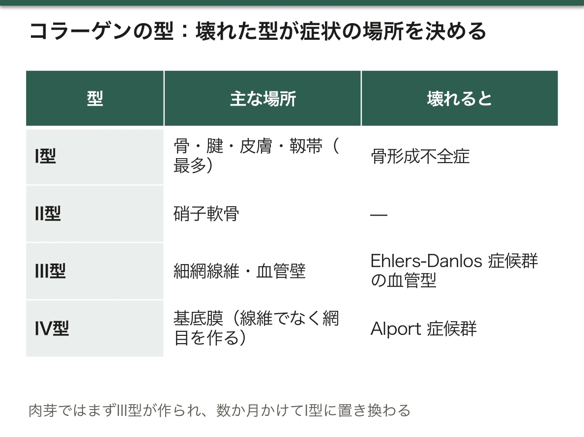
種類と場所の対応も押さえる。I型=骨・腱・皮膚・靱帯(最多)、II型=硝子軟骨、III型=細網線維・血管壁、IV型=基底膜(線維を作らず網目を作る)。Alport 症候群がIV型(基底膜)、Ehlers-Danlos 症候群の血管型がIII型——壊れた型が、症状の出る場所を決めている。
Ehlers-Danlos 症候群は関節過可動性・皮膚の伸展性・易出血。血管型(III型コラーゲン)だけは動脈・腸管の破裂を起こすため予後が違う。整形外科では反復性脱臼で出会う。
骨形成不全症は易骨折・青色強膜・難聴が三徴。強膜が青いのはコラーゲンが薄く、下の脈絡膜が透けて見えるためで、症状ではなく構造がそのまま見えている。
創傷治癒でも型が入れ替わる。肉芽組織ではまずIII型が作られ、時間をかけてI型に置き換わる。だから術後の創が強度を取り戻すのに数か月かかる——抜糸してもまだ弱い理由がここにある。
English version
Core
About 30 per cent of the protein of the body. Three polypeptides wound into a right-handed triple helix. The sequence is a repeat of (glycine-X-Y), with proline common at X and hydroxyproline at Y.
The course of synthesis.
- In the rough endoplasmic reticulum, preprocollagen → hydroxylation (requiring vitamin C) → glycosylation → formation of the triple helix (procollagen).
- After secretion the ends are cleaved to give tropocollagen.
- Lysyl oxidase forms the cross-links that make fibres (requiring copper).
Going further
Every third residue must be glycine because there is no room for a side chain on the central axis of the triple helix. The side chain of glycine is a single hydrogen atom. Replace it with another amino acid and the helix twists at that point and the whole is distorted.
So osteogenesis imperfecta is severe despite being a single-letter substitution. And since an abnormality in even one of the three chains prevents the triple helix forming, disease occurs even with one normal gene (dominant negative). Unlike a "recessive" enzyme deficiency, a mutation of a structural protein tends to be dominant — structure is not settled by majority vote.
The roles of coenzymes and trace elements are consistent here.
- Vitamin C deficiency (scurvy) — hydroxylation fails and the helix is unstable. Bleeding gums, wounds that will not heal, fragile capillaries. That old wounds reopen is because collagen, being continuously renewed, can no longer be made.
- Copper deficiency (Menkes disease) — no cross-links form, and vessels and bone become fragile.
Fix the types and their places. Type I = bone, tendon, skin, ligament (the commonest); type II = hyaline cartilage; type III = reticular fibres, vessel walls; type IV = basement membrane (forming a mesh rather than fibres). Alport syndrome is type IV (basement membrane), the vascular type of Ehlers–Danlos syndrome type III — the type that is broken decides where the symptoms appear.
In the clinic
Ehlers–Danlos syndrome — hypermobile joints, extensible skin, easy bruising. The vascular type (type III collagen) alone causes rupture of arteries and bowel and has a different prognosis. In orthopaedics it is met as recurrent dislocation.
Osteogenesis imperfecta — the triad of fragile bones, blue sclerae and deafness. The sclerae are blue because the collagen is thin and the choroid shows through — the structure itself made visible, not a symptom.
The types change over in wound healing too. Granulation tissue makes type III first, replaced over time by type I. So a surgical wound takes months to regain its strength — here is why it is still weak when the sutures come out.
折り畳みの失敗 — シャペロンとアミロイド
正しい配列でも、正しい形になるとは限らない。
- シャペロン——折り畳みを助ける。誤った凝集を防ぐ。熱ショック蛋白
- ユビキチン・プロテアソーム系——目印(ユビキチン)を付けて壊す。短命の蛋白と異常蛋白
- オートファジー・リソソーム系——大きな塊や細胞小器官ごと
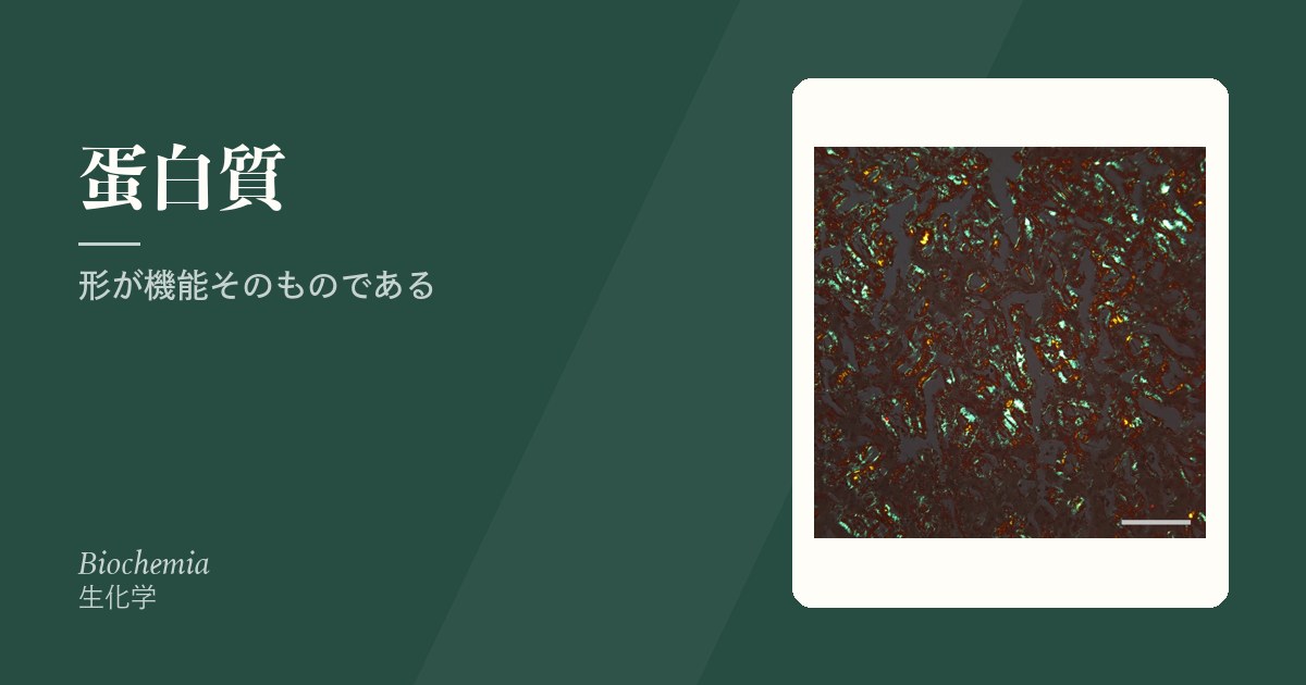
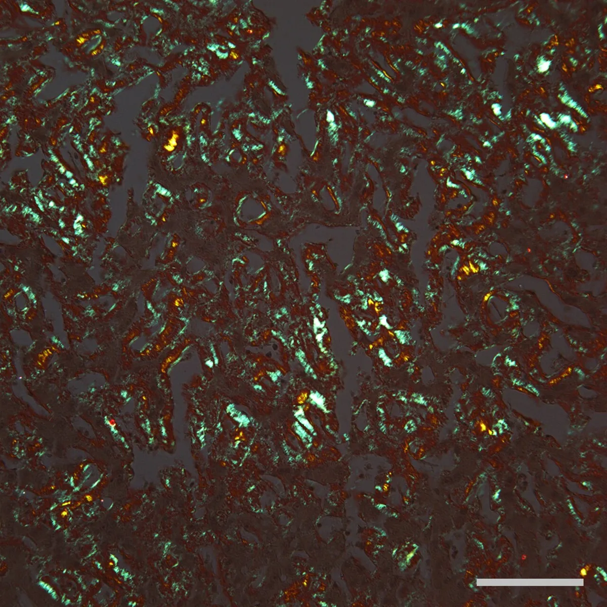
折り畳みに失敗した蛋白がβシートを主体とする線維を作って沈着したものがアミロイド。Congo red で染めて偏光顕微鏡で緑色の複屈折を示す。
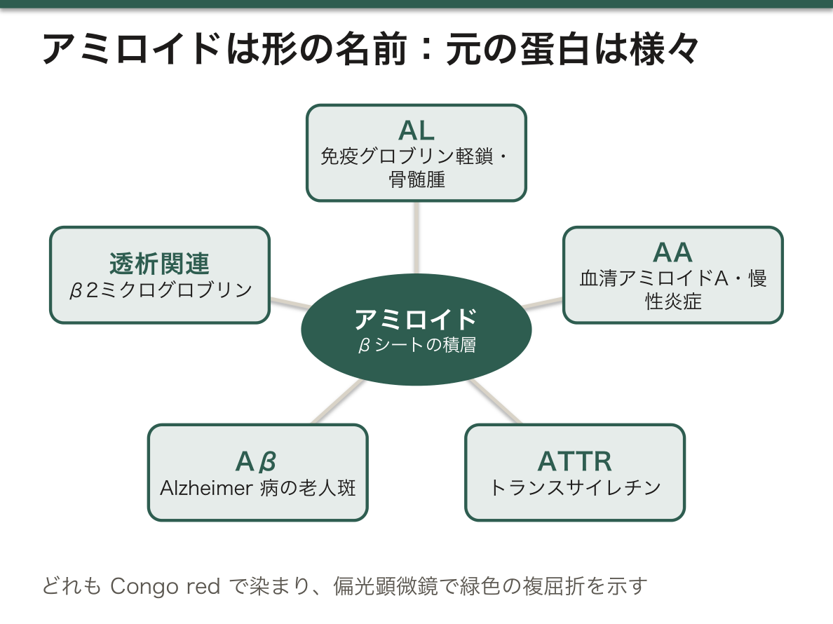
アミロイドは1つの物質の名前ではなく、形の名前である。もとの蛋白は疾患ごとに違うのに、沈着したときの構造(βシートの積層)が同じだから、同じ染色で染まり同じ見え方をする。
- AL——免疫グロブリン軽鎖。多発性骨髄腫など形質細胞の疾患
- AA——血清アミロイドA。関節リウマチ・結核など慢性炎症が続いた結果(続発性)
- ATTR——トランスサイレチン。家族性アミロイドポリニューロパチーと老年性の心アミロイドーシス
- Aβ——Alzheimer 病の老人斑
- β2ミクログロブリン——長期透析。手根管症候群として整形外科で出会う
「原因蛋白が違えば臓器が違う」——ALは心・腎・舌、AAは腎、ATTRは心と末梢神経、透析関連は骨・関節。だから治療も「沈着物を溶かす」ではなく「もとの蛋白の産生を止める」方向になる(ALには化学療法、AAには原疾患の炎症制御、ATTRには安定化薬や遺伝子サイレンシング)。
折り畳み異常が別の形で現れることもある。α1アンチトリプシン欠損症では、変異蛋白が肝細胞の中で重合して出られなくなる。結果、肝には溜まって肝硬変を起こし、肺には届かず肺気腫を起こす——1つの折り畳み異常が、溜まる側と足りない側の両方で病気を作る。
English version
Core
The right sequence does not guarantee the right shape.
- Chaperones — assist folding and prevent wrong aggregation. The heat shock proteins.
- The ubiquitin–proteasome system — tags with ubiquitin and destroys. Short-lived and abnormal proteins.
- Autophagy and the lysosome — for large aggregates and whole organelles.
A protein that has failed to fold and has formed fibres of predominantly β sheet, deposited in tissue, is amyloid. It stains with Congo red and shows green birefringence under polarised light.
Going further
Amyloid is not the name of one substance but the name of a shape. The parent protein differs from disease to disease, but the structure when deposited (stacked β sheets) is the same, so it takes the same stain and looks the same.
- AL — immunoglobulin light chain. Plasma cell diseases such as myeloma.
- AA — serum amyloid A. The result of continuing chronic inflammation (rheumatoid arthritis, tuberculosis) — secondary.
- ATTR — transthyretin. Familial amyloid polyneuropathy and senile cardiac amyloidosis.
- Aβ — the senile plaques of Alzheimer’s disease.
- β2 microglobulin — long-term dialysis. Met in orthopaedics as carpal tunnel syndrome.
A different parent protein means a different organ — AL heart, kidney and tongue; AA kidney; ATTR heart and peripheral nerve; dialysis-related bone and joint. So treatment is directed not at dissolving the deposit but at stopping the production of the parent protein (chemotherapy for AL, control of the underlying inflammation for AA, stabilisers or gene silencing for ATTR).
Misfolding can present in another form altogether. In α1-antitrypsin deficiency the mutant protein polymerises within the hepatocyte and cannot get out. The result is accumulation in the liver causing cirrhosis and failure to reach the lung causing emphysema — one folding defect making disease both where it accumulates and where it is lacking.
血漿蛋白 — 濃度が語ること
血漿蛋白の大半は肝臓が作る(免疫グロブリンを除く)。
- アルブミン——全体の約60%。膠質浸透圧の8割を担い、薬物・ビリルビン・カルシウム・脂肪酸を運ぶ。半減期約3週間
- グロブリン——α・β・γ に分ける。γ が免疫グロブリン(形質細胞が作る)
- トランスサイレチン(プレアルブミン)——半減期約2日
電気泳動でパターンを読む。
半減期の違いが、検査の使い分けを決める。アルブミンは3週間かかるので今日の栄養状態は映さない。短期の評価にはトランスサイレチンやレチノール結合蛋白を使う。「アルブミンが低い=低栄養」とは限らない——炎症でも作られなくなる(負の急性期蛋白)ので、CRPと並べて読む。
運搬役であることから、実用的な補正が2つ出る。
- カルシウム——測っているのは総カルシウムだが、生理活性があるのはイオン化カルシウム。アルブミンが低いと総値だけ下がる。補正カルシウム=実測Ca +(4 − Alb)
- 薬の効き——結合していない遊離型だけが効く。低アルブミン血症では同じ投与量でも効きすぎる(フェニトイン、ワルファリン)
電気泳動のパターンにも読み方がある。γ領域に鋭い山(Mピーク)が立てば単クローン性——多発性骨髄腫。γ全体がなだらかに増えるのは多クローン性で、慢性炎症・肝硬変・膠原病。肝硬変では β と γ が繋がって見える(β-γ ブリッジ)。山の形が、増やしている細胞の数を教えている。
English version
Core
Most plasma proteins are made by the liver (immunoglobulins excepted).
- Albumin — about 60 per cent of the total. It provides 80 per cent of the colloid osmotic pressure and carries drugs, bilirubin, calcium and fatty acids. Half-life about three weeks.
- Globulins — divided into α, β and γ; the γ are the immunoglobulins (made by plasma cells).
- Transthyretin (prealbumin) — half-life about two days.
The pattern is read by electrophoresis.
Going further
The difference in half-life decides which test is used for what. Albumin takes three weeks and does not reflect today’s nutrition; for short-term assessment, transthyretin or retinol-binding protein. "Low albumin = malnutrition" is not necessarily so — it is also a negative acute-phase protein and falls with inflammation, so read it alongside the CRP.
Two practical corrections follow from its being a carrier.
- Calcium — what is measured is total calcium, but what is active is the ionised fraction. With a low albumin only the total falls. Corrected calcium = measured Ca + (4 − Alb).
- Drug effect — only the unbound fraction acts. In hypoalbuminaemia the same dose acts too strongly (phenytoin, warfarin).
The electrophoretic pattern is read too. A sharp peak in the γ region (an M peak) means monoclonal — myeloma. A broad increase across γ is polyclonal — chronic inflammation, cirrhosis, connective tissue disease. In cirrhosis β and γ run together (β-γ bridging). The shape of the peak tells you how many clones of cell are producing it.
蛋白質の構造の4階層 — 配列が形を決め、形が働きを決める
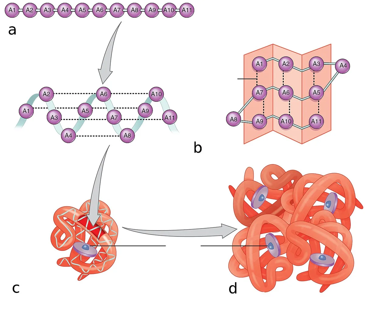
蛋白の構造は4段階で記述する。上の階層は下の階層から自動的に決まる(Anfinsen の原理:一次構造が立体構造を決定する)。
| 階層 | 内容 | 支える結合 |
|---|---|---|
| 一次構造 | アミノ酸の配列 | ペプチド結合(部分二重結合性で平面、回転しない) |
| 二次構造 | αヘリックス・βシート・ターン | 主鎖どうしの水素結合(側鎖は関与しない) |
| 三次構造 | 1本のポリペプチドの立体的な折り畳み | 疎水性相互作用(最大)・水素結合・イオン結合・ジスルフィド結合 |
| 四次構造 | 複数のサブユニットの会合 | 三次構造と同じ非共有結合 |
αヘリックスは1回転3.6残基で、n 番目と n+4 番目の主鎖が水素結合する。βシートは並行・逆平行があり、隣り合う鎖の間で水素結合する。
「疎水性相互作用が三次構造の主役」という点が要点。水の中で疎水性側鎖が内側に集まる——これは疎水基どうしが引き合うのではなく、水分子が秩序を失うのを避けようとする(エントロピー)結果として起こる。だから折り畳みは温度に敏感で、加熱で変性する。
変性〈denaturation〉は立体構造だけが壊れ、一次構造は保たれる状態。原因は熱・極端なpH・尿素やグアニジン・有機溶媒・重金属・界面活性剤。手指消毒のアルコール、器具の加熱滅菌、胃酸による蛋白の変性はすべて同じ現象。
| 階層の異常が起こす病気 | 内容 |
|---|---|
| 一次構造の1残基置換 | 鎌状赤血球症——β鎖6番目 Glu→Val。表面に疎水性のパッチができ、脱酸素時に重合して線維になる。たった1個のアミノ酸が、細胞の形と患者の一生を変える |
| 二次構造の転換 | プリオン病——同じ配列のままαヘリックスがβシートへ変わる。βシート型が正常型を鋳型として変換していく(感染性を持つ蛋白) |
| 折り畳みの失敗 | 嚢胞性線維症(CFTR ΔF508 は折り畳めず分解される)、α1-アンチトリプシン欠損症(重合して肝細胞に溜まり、肺では不足する) |
| 凝集 | アミロイドーシス——βシートに富む線維。Congo red で赤染し、偏光顕微鏡で緑色の複屈折 |
アミロイドはどれも「βシートの積み重なり」という共通の形をとる——元の蛋白が何であっても(AL=免疫グロブリン軽鎖、AA=血清アミロイドA、ATTR=トランスサイレチン、Aβ=Alzheimer)。配列が違うのに同じ染色性を示すのは、二次構造が共通だから。
English version
Core
Protein structure is described at four levels, each determined automatically by the one below (Anfinsen’s principle: the primary structure determines the conformation).
| Level | Content | Bonds |
|---|---|---|
| Primary | The sequence of amino acids | Peptide bonds (partly double-bonded, planar, not rotating) |
| Secondary | α helix, β sheet, turns | Hydrogen bonds between main chains (side chains take no part) |
| Tertiary | The folding of one polypeptide in space | Hydrophobic interactions (greatest), hydrogen bonds, ionic bonds, disulphide bonds |
| Quaternary | The association of several subunits | The same non-covalent bonds as tertiary |
The α helix has 3.6 residues per turn, the main chain of residue n hydrogen-bonding to n+4. The β sheet may be parallel or antiparallel, hydrogen-bonded between adjacent strands.
Going further
The point is that hydrophobic interaction is the protagonist of tertiary structure. In water the hydrophobic side chains gather inwards — not because they attract one another but because the water molecules avoid losing their order (an entropic effect). So folding is sensitive to temperature and denatures on heating.
Denaturation destroys the conformation only; the primary structure is preserved. Causes are heat, extremes of pH, urea and guanidine, organic solvents, heavy metals and detergents. Alcohol hand rub, heat sterilisation of instruments and the denaturation of protein by gastric acid are all the same phenomenon.
| Level of the defect | Disease |
|---|---|
| One residue of the primary structure | Sickle cell disease — β chain position 6, Glu→Val. A hydrophobic patch appears at the surface and the molecules polymerise into fibres when deoxygenated. One amino acid changing the shape of a cell and the whole of a patient’s life |
| Conversion of secondary structure | Prion disease — with the sequence unchanged, α helix becomes β sheet, and the β form converts the normal form using itself as a template (an infectious protein) |
| Failure of folding | Cystic fibrosis (CFTR ΔF508 cannot fold and is degraded), α1-antitrypsin deficiency (polymerises and accumulates in the hepatocyte while the lung goes short) |
| Aggregation | Amyloidosis — fibres rich in β sheet. Red with Congo red, green birefringence under polarised light |
Every amyloid takes the same form — stacked β sheets — whatever the parent protein (AL immunoglobulin light chain, AA serum amyloid A, ATTR transthyretin, Aβ Alzheimer). Different sequences give the same staining because the secondary structure is shared.
まとめ
| ヘモグロビン — 四次構造が協同性を生む | α2β2 の四量体。各サブユニットがヘムを1つ持ち、鉄(2価)に酸素が結合する。 |
|---|---|
| コラーゲン — 3本鎖と、グリシンでなければならない理由 | 体の蛋白質の約30%を占める。3本のポリペプチドが右巻きに撚り合わさった三重らせん。 |
| 折り畳みの失敗 — シャペロンとアミロイド | 正しい配列でも、正しい形になるとは限らない。 |
| 血漿蛋白 — 濃度が語ること | 血漿蛋白の大半は肝臓が作る(免疫グロブリンを除く)。 |
| 蛋白質の構造の4階層 — 配列が形を決め、形が働きを決める | 蛋白の構造は4段階で記述する。上の階層は下の階層から自動的に決まる(Anfinsen の原理:一次構造が立体構造を決定する)。 |
このノートは医学生が授業の内容を自分の言葉でまとめたもので、診療の判断に使うものではありません。誤りに気づいたらお知らせください。