分子細胞生物学で学んだ仕組みは、そのまま検査と治療の道具になっている。PCRはDNA複製を試験管で再現したものであり、シーケンスは「3′-OHが無ければ鎖は伸びない」という性質を利用し、分子標的薬は受容体とシグナル伝達の知識から設計されている。この章では、顕微鏡から始めて、PCR・電気泳動・シーケンス・オミクスといった実験の手技、そしてそれを使う精密医療・分子標的治療・バイオテクノロジーまでをつなげて扱う。
手技を覚えるときは、手順の暗記より「その手順は細胞の中の何の代わりをしているのか」と「結果が出なかったら何を疑うか」を考えるとよい。対照(コントロール)の意味がわかれば、自分の結果も他人の結果も正しく読めるようになる。
- 光学顕微鏡と染色 — 低倍から始め、油浸で解像度を稼ぐ
- PCR — DNA複製を、温度で操って試験管で繰り返す
- ゲル電気泳動 — 網目の中を、短いものほど速く走る
- シーケンシング — 1本を正確に、何百万本を同時に、長いまま読む
- オミクスとRT-PCR — 遺伝子から代謝物まで、層ごとに測る
- 精密医療と個別化医療 — 変異の種類から、効く薬が決まる
- 癌の分子標的治療 — 大きさで、狙える場所が決まる
- バイオテクノロジーと組換えインスリン — 細胞に、ヒトの薬を作らせる
光学顕微鏡と染色 — 低倍から始め、油浸で解像度を稼ぐ
光学顕微鏡では、必ず低倍率の対物レンズ(10×)で標本を探して焦点を合わせてから、40×、さらに100×の油浸レンズへ進む。低倍率は視野が広く、目的の細胞を見つけやすい。また高倍率のレンズほど標本との距離(作動距離)が短いので、いきなり高倍率でピントを探すと、レンズでスライドガラスを割ったり標本を傷つけたりしやすい。
細胞の多くは無色透明なので、染色して見る。メチレンブルーは正に荷電した塩基性色素で、負に荷電した分子、つまりDNAやRNAに強く結合する。だから核が濃い青に染まる。口の粘膜をこすりとった標本では、細胞のまわりに小さな濃い点が多数見えることがあり、これは口の中に住む細菌である。
染色体を見るには、培養した細胞にコルヒチンなどを加えて微小管の重合を止め、分裂中期で止めた細胞を広げる。中期の染色体は、動原体の位置で中部動原体型・次中部動原体型・端部動原体型に分けられる。
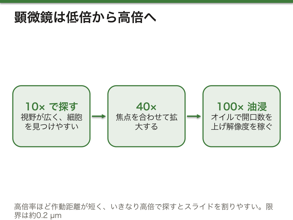
なぜ100×では油を垂らすのか。標本から出た光は、ガラスから空気に出るところで大きく屈折し、レンズに入らずに逃げてしまう。ガラスとほぼ同じ屈折率(約1.5)のイマージョンオイルでガラスとレンズの間を満たすと、斜めの光まで逃がさずに集められる。集める光の角度の広さを開口数といい、解像度(見分けられる2点の最小の距離)は開口数に反比例する。油浸で開口数を1.4程度まで上げても、光学顕微鏡の限界はおよそ0.2 µmである。
細胞のDNAの量を比べる問題は、核の数と倍数性で考えると解ける。成熟赤血球は核が無いのでゼロ、精子は一倍体(n)、ふつうの体細胞は二倍体(2n)、骨格筋線維は多数の核をもつので最も多い。
出生前の染色体検査では、羊水に含まれる胎児由来の細胞を培養し、分裂中期の染色体を染めて核型を調べる。21番染色体が3本あれば47,XY,+21(または47,XX,+21)と書き、21トリソミーと判断する。培養に時間がかかるため、急ぐ場合はFISHやPCRを使った迅速な方法が併用される。
English version
Core
With a light microscope, always find the specimen and focus with the low-power objective (10×) first, then move to 40× and finally the 100× oil immersion lens. Low power gives a wide field of view, so the target cell is easy to find. Also, higher-power lenses sit closer to the specimen (shorter working distance). If you search for focus at high power straight away, you can easily crack the slide or damage the specimen with the lens.
Most cells are colourless and transparent, so they are stained. Methylene blue is a positively charged basic dye that binds strongly to negatively charged molecules: DNA and RNA. So the nucleus stains dark blue. In a scraping of the cheek lining, many small dark dots may appear around the cells; these are bacteria that live in the mouth.
To see chromosomes, cultured cells are treated with a drug such as colchicine that stops microtubules from forming, so cells are held in metaphase, and then spread out. Metaphase chromosomes are classified by centromere position as metacentric, submetacentric or acrocentric.
Going further
Why add oil at 100×? Light from the specimen bends sharply as it leaves the glass into air, and much of it escapes the lens. Immersion oil, with almost the same refractive index as glass (about 1.5), fills the space between glass and lens, so even light at a steep angle is collected. The width of the angle of light collected is the numerical aperture, and resolution (the smallest distance between two points that can be told apart) is inversely proportional to it. Even with oil raising the numerical aperture to about 1.4, the limit of the light microscope is about 0.2 µm.
Questions that compare the amount of DNA in cells can be solved from the number of nuclei and ploidy. A mature red blood cell has no nucleus, so zero; a sperm is haploid (n); an ordinary somatic cell is diploid (2n); and a skeletal muscle fibre has many nuclei, so it has the most.
In the clinic
In prenatal chromosome testing, fetal cells in amniotic fluid are cultured, and metaphase chromosomes are stained to examine the karyotype. Three copies of chromosome 21 are written 47,XY,+21 (or 47,XX,+21) and mean trisomy 21. Culture takes time, so when speed matters, rapid methods using FISH or PCR are used as well.
PCR — DNA複製を、温度で操って試験管で繰り返す
PCR(ポリメラーゼ連鎖反応)は、配列のわかっているDNAの一部分だけを大量に増やす方法で、分子生物学の最も基本的な手技である。細胞の中の複製で酵素がしていた仕事を、温度の上げ下げで置き換えている。
| 仕事 | 細胞の中 | 試験管の中(PCR) |
|---|---|---|
| 二本鎖をほどく | ヘリカーゼ | 約94〜96 °Cに加熱(変性) |
| 合成の出発点 | プライマーゼがRNAプライマーをつくる | 人工のDNAプライマー2種を加え、45〜60 °Cで結合させる(アニーリング) |
| 鎖を伸ばす | DNAポリメラーゼとdNTP | 72 °Cで耐熱性DNAポリメラーゼが伸ばす(伸長)。dNTPと、酵素の働きに必要なMg2+を加える |
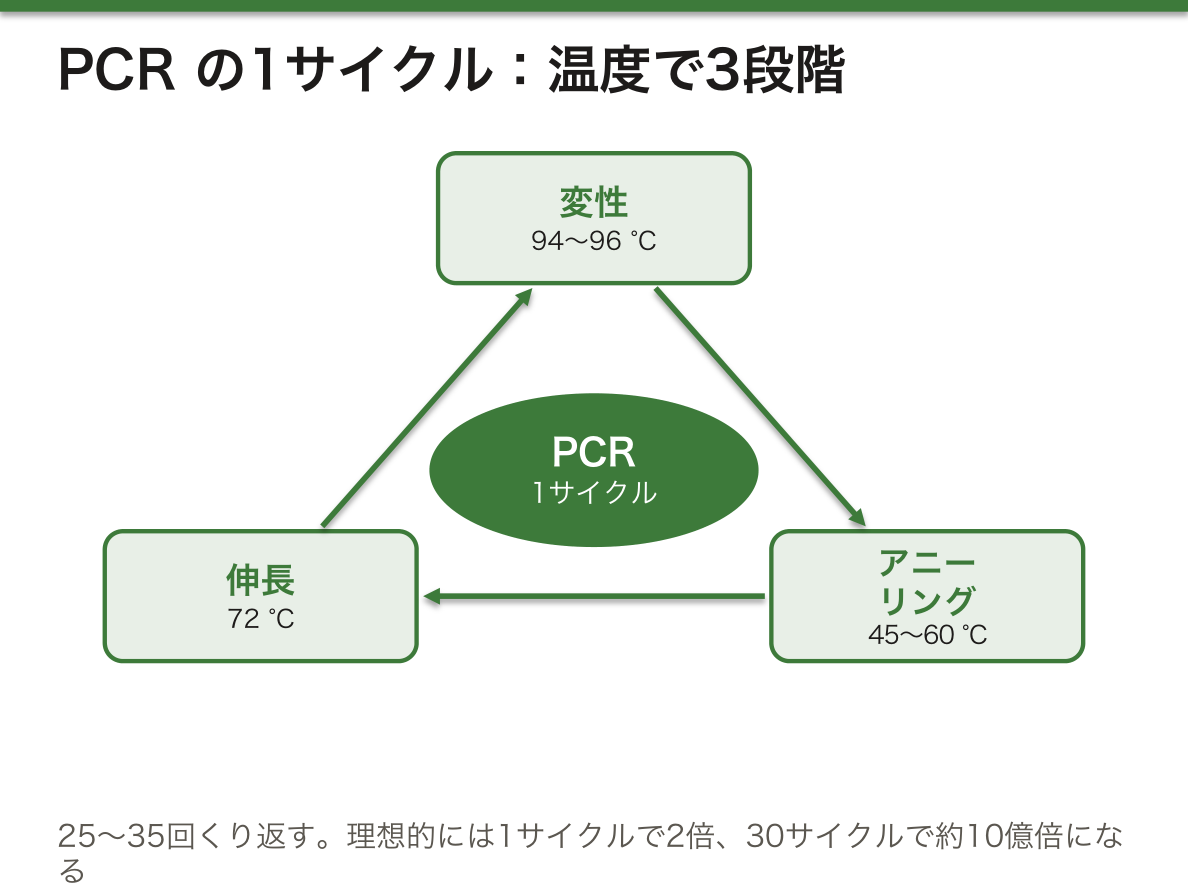
この3段階を1サイクルとして、25〜35回ほど繰り返す。2つのプライマーに挟まれた部分だけが増えるので、増えるのはゲノムの全体ではなく、狙った一部分だけである。
なぜヒトのDNAを増やすのに、ヒトのDNAポリメラーゼを使わないのか。毎サイクル94 °C以上に加熱するので、ふつうの酵素は最初の変性で壊れてしまう。そこで温泉に住む細菌Thermus aquaticusからとったTaq DNAポリメラーゼのような、熱で壊れない酵素を使う。
理想的には1サイクルで分子の数が2倍になるので、nサイクルで2n倍になる。30サイクルなら230≒109、つまり約10億倍で、ごく微量の試料からでも目に見える量にできる。この増え方の強さは、わずかな混入も増やしてしまうという弱点の裏返しでもある。そのため、鋳型DNAの代わりに水を入れた陰性対照と、確実に増える鋳型を入れた陽性対照を必ず一緒に流す。陰性対照にバンドが出れば汚染を、陽性対照にバンドが出なければ試薬やピペット操作の失敗を疑う。
PCRは遺伝子型の判定によく使われる。たとえば一部の人のゲノムにだけ入っている約300塩基のAlu配列の挿入を挟むようにプライマーを置くと、挿入のある対立遺伝子では長い産物、無い対立遺伝子では短い産物ができる。長いバンドだけなら挿入のホモ接合(+/+)、短いバンドだけなら挿入の無いホモ接合(−/−)、両方ならヘテロ接合(+/−)である。同じ考え方で、感染症の病原体の検出、遺伝性疾患の変異の検出、親子鑑定や法医学での個人識別などが行われている。
English version
Core
PCR (polymerase chain reaction) makes many copies of one part of a DNA whose sequence is known. It is the most basic method in molecular biology. It replaces the work of the enzymes in cellular replication with changes in temperature.
| Job | In the cell | In the tube (PCR) |
|---|---|---|
| Open the double strand | Helicase | Heat to about 94–96 °C (denaturation) |
| Starting point | Primase makes an RNA primer | Two synthetic DNA primers are added and bind at 45–60 °C (annealing) |
| Extend the strand | DNA polymerase and dNTPs | A heat-stable DNA polymerase extends at 72 °C (extension); dNTPs and Mg2+, needed by the enzyme, are added |
These three steps make one cycle, repeated about 25–35 times. Only the part between the two primers is copied, so what grows is not the whole genome but only the chosen region.
Going further
Why not use human DNA polymerase to copy human DNA? Each cycle heats the mix above 94 °C, and an ordinary enzyme is destroyed at the first denaturation. So a heat-stable enzyme is used, such as Taq DNA polymerase from Thermus aquaticus, a bacterium that lives in hot springs.
Ideally the number of molecules doubles each cycle, so after n cycles it is 2n times larger. After 30 cycles that is 230 ≈ 109, about a billion times, enough to see even from a tiny sample. This power has a downside: it also copies the smallest contamination. So a negative control (water instead of template DNA) and a positive control (a template that surely works) are always run together. A band in the negative control suggests contamination; no band in the positive control suggests a problem with reagents or pipetting.
In the clinic
PCR is often used for genotyping. For example, an Alu insertion of about 300 bases is present in the genome of only some people. If primers are placed on both sides of the insertion site, the allele with the insertion gives a long product and the allele without it gives a short product. Only the long band means homozygous for the insertion (+/+); only the short band means homozygous without it (−/−); both bands mean heterozygous (+/−). The same idea is used to detect pathogens in infections, find variants in inherited diseases, and identify individuals in paternity and forensic testing.
ゲル電気泳動 — 網目の中を、短いものほど速く走る
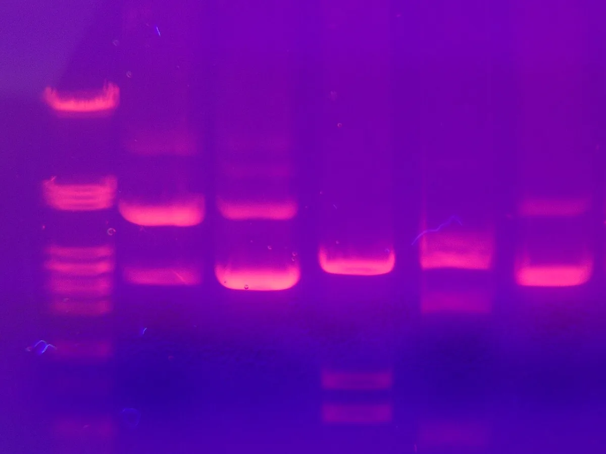
DNAはリン酸の骨格のため全体が負に荷電している。ゲルに電圧をかけると、DNAは陽極(+)に向かって移動する。ゲルは細かい網目なので、短い断片ほど抜けやすく速く進み、長い断片ほど遅れる。その結果、DNAは長さの順に並んだバンドに分かれる。長さのわかった断片を混ぜたサイズマーカーを隣のレーンに流し、それと比べて試料の長さを読む。
| アガロースゲル | ポリアクリルアミドゲル(PAG) | |
|---|---|---|
| 材料 | 海藻由来の多糖。冷やすと固まる | アクリルアミドとビスアクリルアミドを化学的に架橋 |
| 泳動の向き | 水平 | 垂直 |
| 分けられる長さ | 約50〜30,000塩基対(ゲルの濃さで調整) | 約5〜500塩基対 |
| 得意なこと | PCR産物の有無や大まかな長さの確認 | 短い断片の細かな差の区別(高い分解能) |
なぜ長さだけで分かれるのか。DNAは1塩基ごとにリン酸を1つもつので、長さが2倍になれば電荷も2倍になり、電荷と質量の比がほぼ一定になる。電気で引っぱる力の差が打ち消されるので、走る速さを決めるのは網目のくぐりやすさ、つまり長さだけになる。ゲルの濃度を上げると網目が細かくなり、短い断片どうしの差がよく分かれる。泳動後のDNAは見えないので、DNAに結合して蛍光を出す色素で染めて紫外線などで見る。
嚢胞性線維症で最も多い変異p.Phe508delは、3塩基だけの欠失である。この部分を挟んでPCRを行うと、正常な対立遺伝子からは92塩基対、変異のある対立遺伝子からは89塩基対の産物ができる。3塩基対の差はアガロースゲルではほとんど見分けられないので、分解能の高いポリアクリルアミドゲルが向いている。正常型のホモ接合では1本、変異のホモ接合では短い1本、ヘテロ接合では両方のバンドが出る(ヘテロ接合では、2種類の鎖が組み合わさった余分なバンドが見えることもある)。
English version
Core
Because of its phosphate backbone, DNA is negatively charged overall. When voltage is applied to a gel, DNA moves toward the anode (+). The gel is a fine mesh, so shorter fragments get through more easily and move faster, while longer ones fall behind. DNA separates into bands ordered by length. A size marker, a mix of fragments of known length, runs in the next lane, and the sample’s length is read by comparison.
| Agarose gel | Polyacrylamide gel (PAG) | |
|---|---|---|
| Material | A polysaccharide from seaweed; sets when cooled | Acrylamide and bis-acrylamide chemically cross-linked |
| Direction | Horizontal | Vertical |
| Separation range | About 50–30,000 bp (adjusted by gel concentration) | About 5–500 bp |
| Good for | Checking whether a PCR product is present and its rough size | Telling apart small differences between short fragments (high resolution) |
Going further
Why does DNA separate only by length? DNA has one phosphate per nucleotide, so doubling the length doubles the charge. The charge-to-mass ratio stays almost constant. Differences in electrical pull cancel out, so speed is decided only by how easily the fragment passes through the mesh, that is, by length. A more concentrated gel has a finer mesh and separates short fragments better. DNA cannot be seen after the run, so it is stained with a dye that binds DNA and glows, and viewed under UV or similar light.
In the clinic
The most common cystic fibrosis variant, p.Phe508del, is a deletion of only three bases. PCR across this region gives a 92 bp product from a normal allele and an 89 bp product from a variant allele. A 3 bp difference is hard to see on agarose, so high-resolution polyacrylamide gel is better. A normal homozygote shows one band, a variant homozygote one shorter band, and a heterozygote both bands (heterozygotes may also show extra bands from mixed pairs of strands).
シーケンシング — 1本を正確に、何百万本を同時に、長いまま読む
Sanger法(ジデオキシ法)は、合成の途中で鎖を止めて配列を読む方法である。DNAポリメラーゼに、ふつうのdNTPと少量のddNTP(ジデオキシヌクレオチド)を混ぜて与える。ddNTPは3′-OHを欠くので、取りこまれたところで鎖がそれ以上伸びない。4種類のddNTPを別々の蛍光色素で標識しておくと、あらゆる長さで止まった断片ができ、それをキャピラリー電気泳動で長さの順に並べて色を読めば、配列がそのまま並ぶ。読みとった配列は参照ゲノムと比べて変異を探す。
| Sanger法 | 次世代シーケンス(NGS) | ロングリード | |
|---|---|---|---|
| 1回に読む長さ | 約500〜900塩基 | 50〜300塩基(短い) | 10 kb超、100 kbを超えることも |
| 処理量 | 少ない(1断片ずつ) | 数百万〜数十億の断片を並行 | 中程度(本数は少ないが長い) |
| 正確さ | 非常に高い(99.9%超) | 高い(同じ場所を何度も読んで補う) | 改善中 |
| 主な用途 | 少数の変異の確認・検証 | エクソーム・全ゲノム・トランスクリプトーム | 構造変異・繰り返し配列・ゲノムの組み立て |
NGSは短い断片をばらばらに大量に読み、コンピューターで参照ゲノムに貼りつけていく。1か所の塩基を平均何回読んだかをカバレッジ(深さ)といい、深く読むほど読み間違いと本当の変異を区別しやすい。なぜロングリードが必要になるのか。短い断片は、同じ配列が何度も繰り返される場所や、大きな欠失・逆位などの構造変異では、どこに貼りつければよいか決められない。100塩基の断片で、数千塩基の繰り返しの長さを測ることはできない。1本を長いまま読めば、繰り返しや構造変異をまたいで読みきれる。
Sanger法の波形(エレクトロフェログラム)では、ヘテロ接合の位置に2色のピークが重なって現れる。嚢胞性線維症のp.Gly551Aspやp.Arg553Terのような1塩基の置換を確かめるには、この方法が今も標準である。一方、原因のわからない希少疾患では、NGSでエクソーム(すべてのエクソン)や全ゲノムを一度に調べ、見つかった候補の変異をSanger法で確認する、という組み合わせがよく使われる。見つかった変異は「病的・病的の可能性が高い・意義不明・良性の可能性が高い・良性」の段階で解釈される。
English version
Core
The Sanger (dideoxy) method reads a sequence by stopping chains during synthesis. DNA polymerase is given normal dNTPs mixed with a small amount of ddNTPs (dideoxynucleotides). A ddNTP lacks the 3′-OH, so the chain cannot grow once it is added. If each of the four ddNTPs carries a different fluorescent dye, fragments that stop at every length are made. Capillary electrophoresis lines them up by length, and reading the colours gives the sequence in order. The sequence is compared with the reference genome to look for variants.
| Sanger | Next-generation sequencing (NGS) | Long-read | |
|---|---|---|---|
| Read length | About 500–900 bases | 50–300 bases (short) | Over 10 kb, sometimes over 100 kb |
| Throughput | Low (one fragment at a time) | Millions to billions of fragments in parallel | Moderate (fewer reads, but long) |
| Accuracy | Very high (over 99.9%) | High (by reading the same place many times) | Improving |
| Main use | Checking and confirming a few variants | Exome, whole genome, transcriptome | Structural variants, repeats, genome assembly |
Going further
NGS reads huge numbers of short fragments separately, and a computer places them onto the reference genome. The average number of times each base is read is the coverage (depth); deeper reading makes it easier to tell reading errors from real variants. Why is long-read sequencing needed? Short fragments cannot be placed with certainty where the same sequence repeats many times, or across structural variants such as large deletions and inversions. A 100-base fragment cannot measure the length of a repeat thousands of bases long. Reading one long molecule can span repeats and structural variants completely.
In the clinic
In a Sanger trace (electropherogram), a heterozygous position shows two overlapping peaks of different colours. This method is still the standard for confirming single-base changes such as p.Gly551Asp and p.Arg553Ter in cystic fibrosis. In rare diseases of unknown cause, a common approach is to examine the exome (all exons) or whole genome by NGS, then confirm candidate variants by Sanger. Variants found are interpreted as pathogenic, likely pathogenic, of uncertain significance, likely benign, or benign.
オミクスとRT-PCR — 遺伝子から代謝物まで、層ごとに測る
体の分子をまとめて網羅的に調べる研究をオミクスと呼び、何を測るかで名前が変わる。ゲノミクスはDNA、トランスクリプトミクスはmRNA、プロテオミクスはタンパク質、メタボロミクスは代謝物(アミノ酸・有機酸・糖など)である。
RNAはPCRで直接増やせないので、まず逆転写酵素でRNAからDNA(cDNA)を写しとってからPCRにかける。これがRT-PCRである。増える様子を蛍光でサイクルごとに測るリアルタイム定量PCR(qPCR)では、蛍光が一定の線を越えたサイクル数(Ct値)を読む。最初の鋳型が多いほど早く線を越えるので、Ct値が小さいほど量が多い。試料を何万もの微小な滴に分けて、増えた滴の数を数えるデジタルPCRでは、基準の試料と比べなくても量を絶対値で出せる。
タンパク質や代謝物は、電気泳動や液体・ガスクロマトグラフィーで分けてから、質量分析(代謝物ではNMRも)で検出し、データベースと照らして同定する。
Ct値の差は2のべき乗で読む。効率が理想的なら1サイクルで2倍になるので、Ct値が1小さければ量は約2倍、約3.3小さければ約10倍である。
なぜゲノムだけでなく、タンパク質や代謝物まで測る必要があるのか。ヒトのタンパク質をつくる遺伝子は約2万個にすぎないが、選択的スプライシングと翻訳後修飾で、実際に働くタンパク質の種類ははるかに多い(100万種類にのぼるとも見積もられる)。さらに、遺伝子がどれだけ発現し、酵素が実際にどれだけ働いているかは、DNAの配列を見てもわからない。病気の「今の状態」に近いのは、下流の層である。全体を一度に測るRNA-seqは新しい転写産物やアイソフォームも見つけられ、1細胞ずつ測る方法では組織の中の細胞の違いまで見える。決まった遺伝子だけを正確に測るならqPCRが向いている。
臨床では層ごとに検査がある。SARS-CoV-2の診断はRT-PCRで、ウイルスのRNAをcDNAにして増やす。慢性骨髄性白血病では、BCR-ABL1の融合遺伝子のmRNAをqPCRで定期的に測り、治療の効き具合を追う。新生児のマススクリーニングは、乾燥ろ紙血のアミノ酸やアシルカルニチンをタンデム質量分析で測るメタボロミクスの応用である。原因不明の低血糖を起こした乳幼児では、尿の有機酸をガスクロマトグラフィー質量分析で調べて脂肪酸酸化の異常を疑い、関係する遺伝子(長鎖3-ヒドロキシアシルCoA脱水素酵素欠損症ならHADHA)の変異を確かめて診断を確定する、という流れになる。
English version
Core
Studying the body’s molecules all at once, comprehensively, is called omics, and the name changes with what is measured: genomics (DNA), transcriptomics (mRNA), proteomics (proteins) and metabolomics (metabolites such as amino acids, organic acids and sugars).
RNA cannot be copied directly by PCR, so reverse transcriptase first copies RNA into DNA (cDNA), which is then used for PCR. This is RT-PCR. In real-time quantitative PCR (qPCR), the increase is measured by fluorescence every cycle, and the cycle at which fluorescence crosses a set line (the Ct value) is read. More starting template crosses the line sooner, so a lower Ct means more template. In digital PCR, the sample is split into tens of thousands of tiny droplets and the droplets that amplify are counted, giving an absolute amount without a reference sample.
Proteins and metabolites are separated by electrophoresis or liquid or gas chromatography, detected by mass spectrometry (and NMR for metabolites), and identified using databases.
Going further
Read differences in Ct as powers of 2. With ideal efficiency the amount doubles each cycle, so a Ct one lower means about twice as much, and about 3.3 lower means about ten times as much.
Why measure proteins and metabolites, not just the genome? Humans have only about 20,000 protein-coding genes, but alternative splicing and post-translational modification create far more kinds of working protein (estimates reach a million). Also, the DNA sequence does not tell you how much a gene is expressed or how active an enzyme really is. The lower layers are closer to the “current state” of a disease. RNA-seq, which measures everything at once, can find new transcripts and isoforms, and single-cell methods show differences between cells within a tissue. For measuring chosen genes accurately, qPCR is best.
In the clinic
In the clinic, there are tests for each layer. SARS-CoV-2 is diagnosed by RT-PCR: viral RNA is turned into cDNA and amplified. In chronic myeloid leukaemia, BCR-ABL1 fusion mRNA is measured regularly by qPCR to follow the response to treatment. Newborn screening measures amino acids and acylcarnitines in dried blood spots by tandem mass spectrometry, an application of metabolomics. In an infant with unexplained hypoglycaemia, urine organic acids may be examined by gas chromatography–mass spectrometry to suspect a fatty acid oxidation disorder, and then a variant in the related gene (HADHA for long-chain 3-hydroxyacyl-CoA dehydrogenase deficiency) confirms the diagnosis.
精密医療と個別化医療 — 変異の種類から、効く薬が決まるMucoviscidosis
精密医療(precision medicine)は、遺伝子や分子の特徴で患者を小さな集団に分け、その集団に効く治療を選ぶ考え方である。個別化医療(personalized medicine)はもっと広く、遺伝的な特徴に加えて生活習慣・価値観・希望まで含めて、その人1人に合わせた医療を組み立てる。
| 精密医療 | 個別化医療 | |
|---|---|---|
| 焦点 | 生物学的・遺伝的な特徴 | 個人の全体 |
| 単位 | 患者の小集団 | 1人の患者 |
| 使うデータ | ゲノム・バイオマーカー | ゲノムに加え生活習慣・希望 |
| 主な分野 | 癌・希少疾患・薬理遺伝学 | 慢性疾患・精神疾患など |
精密医療のわかりやすい例が嚢胞性線維症(CF)である。CFTRタンパク質の変異は、どこが壊れるかでいくつかの型に分けられ、型ごとに使う薬が違う。
| 変異の例 | 何が起きるか | 効く薬 |
|---|---|---|
| p.Phe508del | 折りたたみに失敗し、小胞体にとどまってプロテアソームで分解される(膜に届かない) | コレクター(ルマカフトル・テザカフトル・エレクサカフトル)で膜まで届く量を増やす |
| p.Gly551Asp | 膜には届くが、チャネルが開かない(開閉の異常) | ポテンシエーター(アイバカフトル)で開く確率を上げる |
| p.Arg553Ter などのナンセンス変異 | 途中で終止し、タンパク質がほとんどできない | モジュレーターは働きかける相手が無く、効かない |
なぜ変異の型で薬が分かれるのか。コレクターは「形を整えて膜まで運ぶ」薬、ポテンシエーターは「膜にあるチャネルを開きやすくする」薬で、直す場所が違うからである。膜に届かないタンパク質をいくら開きやすくしても意味がなく、タンパク質そのものが無ければどちらの薬も手の出しようがない。
ヒトは対立遺伝子を2つもつので、2つの変異が違う場合(複合ヘテロ接合)はそれぞれの対立遺伝子が何を作るかを考える。片方がナンセンス変異でも、もう片方が開閉の異常なら、その対立遺伝子から作られる膜のタンパク質にポテンシエーターが効く。エレクサカフトル・テザカフトル・アイバカフトルの3剤併用は、少なくとも1つの対立遺伝子がp.Phe508delであれば使えるので、多くの患者に届くようになった。まれな変異では、患者の細胞から作ったオルガノイドなどで薬の効きを直接試すセラタイピングも行われる。
CFTRモジュレーターは肺機能を改善させた一方で、高価で、生涯飲み続ける必要があり、すべての変異には効かない。DNAの段階で直す遺伝子治療や、正常なCFTRのmRNAを気道に届ける治療は、どの変異にも使える可能性があるが、まだ臨床試験の段階である。栄養管理、感染対策、心理社会的な支援は、変異の種類にかかわらずすべての患者に必要であり、ここが個別化医療の領域になる。日本ではCFは非常にまれだが、欧州系の集団では最も多い単一遺伝子疾患の1つである。
English version
Core
Precision medicine sorts patients into small groups by genetic and molecular features and chooses the treatment that works for that group. Personalized medicine is broader: it builds care for one person, using genetic features plus lifestyle, values and wishes.
| Precision medicine | Personalized medicine | |
|---|---|---|
| Focus | Biological and genetic features | The whole person |
| Unit | Subgroups of patients | One patient |
| Data used | Genome, biomarkers | Genome plus lifestyle and preferences |
| Main fields | Cancer, rare diseases, pharmacogenomics | Chronic disease, mental health and others |
A clear example of precision medicine is cystic fibrosis (CF). CFTR variants are grouped by what part goes wrong, and each group needs a different drug.
| Example variant | What happens | Drug that works |
|---|---|---|
| p.Phe508del | Fails to fold, stays in the ER and is broken down by the proteasome (does not reach the membrane) | Correctors (lumacaftor, tezacaftor, elexacaftor) increase the amount that reaches the membrane |
| p.Gly551Asp | Reaches the membrane, but the channel does not open (gating defect) | A potentiator (ivacaftor) raises the chance of opening |
| Nonsense variants such as p.Arg553Ter | Translation stops early; almost no protein is made | Modulators have nothing to act on and do not work |
Going further
Why do drugs differ by variant type? Because they fix different places. A corrector “shapes the protein and gets it to the membrane”; a potentiator “helps the channel in the membrane open”. Making a protein easier to open is pointless if it never reaches the membrane, and if there is no protein at all, neither drug can help.
People have two alleles, so when the two variants differ (compound heterozygote), think about what each allele makes. Even if one allele has a nonsense variant, if the other has a gating defect, a potentiator works on the membrane protein made from that allele. The triple combination elexacaftor–tezacaftor–ivacaftor can be used if at least one allele is p.Phe508del, so it now reaches many patients. For rare variants, theratyping tests drug response directly in patient-derived cells such as organoids.
In the clinic
CFTR modulators have improved lung function, but they are expensive, must be taken for life, and do not work for every variant. Gene therapy that fixes the DNA, and therapy that delivers normal CFTR mRNA to the airways, could work for any variant, but they are still in clinical trials. Nutrition, infection control and psychosocial support are needed by every patient whatever the variant, and this is where personalized medicine comes in. CF is very rare in Japan, but it is one of the most common single-gene disorders in people of European origin.
癌の分子標的治療 — 大きさで、狙える場所が決まる
癌細胞では、体細胞に起きたDNAの変化が増殖の信号を出し続けていることが多い。1つは遺伝子の増幅や過剰発現で、HER2、EGFR、FGFR2、VEGF/VEGFRなどの受容体やリガンドが異常に多くなる。もう1つは活性化型の変異で、KRAS、BRAF、PIK3CAなど、シグナル伝達の途中の分子が「入」のまま固定される。
こうした分子を狙う薬は2種類に大別される。治療用モノクローナル抗体(名前の語尾が-mab)は細胞膜の受容体や細胞外の分子に結合する。低分子薬(キナーゼ阻害薬は語尾が-nib)は膜を通り抜けて細胞の中に入り、標的タンパク質の酵素活性を妨げる。
なぜ抗体は細胞の外しか狙えないのか。抗体は分子量約15万の大きなタンパク質で、細胞膜を通り抜けられない。だから標的は膜の外に出ている部分か、血液中の分子に限られる。低分子薬は小さく脂溶性も備えるので膜を通れ、細胞内のキナーゼのATP結合部位などに入りこんで働きを止める。標的がどこにあるかで、薬の形が決まる。
標的の下流が壊れていると、上流を止めても効かない。たとえば大腸癌でKRASに活性化型の変異があると、EGFRを抗体で止めても、その下のKRASが勝手に信号を出し続ける。抗EGFR抗体を使う前にKRASなどの変異を調べるのは、このためである。
分子標的薬は、標的があるかを調べるコンパニオン診断とセットで使われる。HER2陽性の乳癌や胃癌では、組織でHER2の過剰発現や遺伝子増幅を確かめてから、抗HER2抗体のトラスツズマブを使う。HER2が陰性なら効果は期待できない。また、治療を続けるうちに標的の分子に新たな変異が加わって薬が結合できなくなり、耐性が生じることがある(肺癌のEGFR阻害薬に対するT790M変異など)。耐性が出たら再び組織や血液の遺伝子を調べ、次の薬を選ぶ。
English version
Core
In cancer cells, DNA changes in somatic cells often keep growth signals switched on. One kind is gene amplification or overexpression: receptors and ligands such as HER2, EGFR, FGFR2 and VEGF/VEGFR become abnormally abundant. Another kind is activating variants: molecules in the middle of signalling pathways, such as KRAS, BRAF and PIK3CA, are locked “on”.
Drugs against these molecules fall into two groups. Therapeutic monoclonal antibodies (names end in -mab) bind receptors on the cell membrane or molecules outside the cell. Small-molecule drugs (kinase inhibitors end in -nib) pass through the membrane into the cell and block the enzyme activity of the target protein.
Going further
Why can antibodies only target things outside the cell? An antibody is a large protein of about 150,000 molecular weight and cannot cross the cell membrane. So its target must be the part of a receptor outside the membrane, or a molecule in the blood. Small-molecule drugs are small and fat-soluble enough to cross the membrane, and they fit into places such as the ATP-binding site of a kinase inside the cell to stop it. Where the target is decides what shape the drug must have.
If something below the target is broken, blocking above it does not work. For example, in colorectal cancer with an activating KRAS variant, blocking EGFR with an antibody does not help, because KRAS below it keeps sending the signal by itself. This is why KRAS and similar variants are tested before using anti-EGFR antibodies.
In the clinic
Targeted drugs are used together with companion diagnostics that test whether the target is present. In HER2-positive breast and gastric cancer, HER2 overexpression or gene amplification is confirmed in tissue before the anti-HER2 antibody trastuzumab is used. If HER2 is negative, no benefit is expected. During treatment, a new variant may appear in the target so the drug can no longer bind, causing resistance (such as the T790M variant against EGFR inhibitors in lung cancer). When resistance appears, tissue or blood is tested again and the next drug is chosen.
バイオテクノロジーと組換えインスリン — 細胞に、ヒトの薬を作らせる
バイオテクノロジーは、生物を使って製品(医薬品を含む)を作ったり改良したりする技術である。医療では、遺伝子工学・細胞培養・分子生物学を使って、薬・ワクチン・診断の道具が作られている。組換えインスリン、癌や自己免疫疾患に使うモノクローナル抗体、遺伝子治療、mRNAワクチンなどがその製品である。
ヒトのタンパク質を作らせる生物(発現系)には、細菌(大腸菌)、真菌(酵母)、植物、昆虫の細胞、哺乳類の培養細胞がある。遺伝子組換えで作られた最初の医薬品はヒトインスリンで、1982年に承認された。主に大腸菌と酵母で作られている。
膵β細胞の中では、インスリンは1本のプレプロインスリンとして作られる。先頭の24アミノ酸のシグナルペプチドが新しい鎖を小胞体へ導き、そこで切り取られてプロインスリンになる。小胞体で正しく折りたたまれ、3つのジスルフィド結合ができる。その後ゴルジ装置から分泌顆粒へ運ばれる途中で、真ん中のCペプチドが酵素で切り離され、A鎖とB鎖がジスルフィド結合でつながった活性のあるインスリンになる。
なぜ発現系をタンパク質ごとに選ぶのか。大腸菌は増えるのが速く安く大量に作れるが、小胞体もゴルジ装置も無いので、糖鎖の付加などの翻訳後修飾ができず、複雑なジスルフィド結合も正しく作りにくい。インスリンは糖鎖を必要とせず小さいので、大腸菌でA鎖とB鎖、あるいはプロインスリンを作らせ、あとから試験管の中で折りたたみと切断を行えば済む。一方、糖鎖が働きに欠かせない抗体などは、哺乳類の培養細胞で作る必要がある。そのタンパク質がどんな加工を必要とするかが、作らせる生物を決めている。
アミノ酸を少し入れ替えたインスリンアナログは、効き方の速さを変えてある。インスリンリスプロやアスパルトは皮下で集まりにくく速く効き、グラルギンは皮下で沈殿してゆっくり溶け出すので長く効く。
Cペプチドは、インスリンと同じ数だけβ細胞から分泌されるが、注射するインスリン製剤には含まれない。このため、低血糖の患者で血中インスリンが高いのにCペプチドが低ければ外から注射されたインスリンを、両方とも高ければインスリノーマなど体内での過剰分泌を考える。1型糖尿病で、残っているβ細胞の働きを評価するのにも使われる。
English version
Core
Biotechnology uses living organisms to make or improve products, including medicines. In medicine, genetic engineering, cell culture and molecular biology are used to make drugs, vaccines and diagnostic tools. Examples are recombinant insulin, monoclonal antibodies for cancer and autoimmune disease, gene therapy and mRNA vaccines.
Organisms used to make human proteins (expression systems) include bacteria (E. coli), fungi (yeast), plants, insect cells and cultured mammalian cells. The first medicine made by recombinant DNA technology was human insulin, approved in 1982. It is made mainly in E. coli and yeast.
In pancreatic β cells, insulin is made as a single chain, preproinsulin. A 24-amino-acid signal peptide at the front guides the new chain into the ER, where it is cut off to give proinsulin. In the ER it folds correctly and forms three disulfide bonds. Then, on the way from the Golgi apparatus to secretory granules, enzymes cut out the middle C-peptide, leaving active insulin: an A chain and a B chain joined by disulfide bonds.
Going further
Why choose the expression system for each protein? E. coli grows fast and can make large amounts cheaply, but it has no ER or Golgi apparatus, so it cannot do post-translational modifications such as adding sugar chains, and it has trouble forming complex disulfide bonds correctly. Insulin is small and needs no sugar chains, so E. coli can make the A and B chains, or proinsulin, and folding and cutting can be done afterwards in a test tube. Proteins whose sugar chains are essential, such as antibodies, must be made in cultured mammalian cells. What processing the protein needs decides which organism makes it.
In the clinic
Insulin analogues, with a few amino acids changed, act at different speeds. Insulin lispro and aspart do not cluster together under the skin, so they act fast. Glargine forms a deposit under the skin and dissolves slowly, so it acts for a long time.
C-peptide is released from β cells in the same number as insulin, but it is not in injected insulin products. So in a patient with hypoglycaemia, high blood insulin with low C-peptide suggests injected insulin, while high levels of both suggest overproduction in the body, such as an insulinoma. C-peptide is also used to assess remaining β-cell function in type 1 diabetes.
まとめ
| 光学顕微鏡と染色 — 低倍から始め、油浸で解像度を稼ぐ | 光学顕微鏡では、必ず低倍率の対物レンズ(10×)で標本を探して焦点を合わせてから、40×、さらに100×の油浸レンズへ進む。 |
|---|---|
| PCR — DNA複製を、温度で操って試験管で繰り返す | PCR(ポリメラーゼ連鎖反応)は、配列のわかっているDNAの一部分だけを大量に増やす方法で、分子生物学の最も基本的な手技である。 |
| ゲル電気泳動 — 網目の中を、短いものほど速く走る | DNAはリン酸の骨格のため全体が負に荷電している。 |
| シーケンシング — 1本を正確に、何百万本を同時に、長いまま読む | Sanger法(ジデオキシ法)は、合成の途中で鎖を止めて配列を読む方法である。 |
| オミクスとRT-PCR — 遺伝子から代謝物まで、層ごとに測る | 体の分子をまとめて網羅的に調べる研究をオミクスと呼び、何を測るかで名前が変わる。 |
| 精密医療と個別化医療 — 変異の種類から、効く薬が決まる | 精密医療(precision medicine)は、遺伝子や分子の特徴で患者を小さな集団に分け、その集団に効く治療を選ぶ考え方である。 |
| 癌の分子標的治療 — 大きさで、狙える場所が決まる | 癌細胞では、体細胞に起きたDNAの変化が増殖の信号を出し続けていることが多い。 |
| バイオテクノロジーと組換えインスリン — 細胞に、ヒトの薬を作らせる | バイオテクノロジーは、生物を使って製品(医薬品を含む)を作ったり改良したりする技術である。 |
このノートは医学生が授業の内容を自分の言葉でまとめたもので、診療の判断に使うものではありません。誤りに気づいたらお知らせください。