軟骨は基質が硬いゲルになった結合組織である。線維(II型コラーゲン)が骨組みを作り、そこにアグリカンが水を抱え込む。押されると水が逃げ、離すと戻る——この粘弾性が軟骨の本体で、骨のような剛性とは別種の強さである。
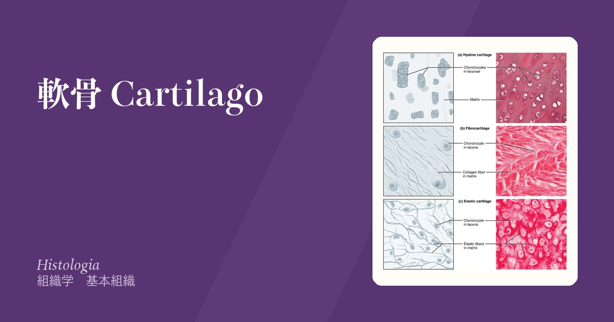
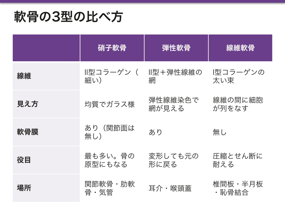
3つの型は入っている線維で決まる。硝子軟骨(II型のみ、基質が均質でガラス様)、弾性軟骨(+弾性線維)、線維軟骨(+I型コラーゲンが束で走る)。どこに何があるかは要求される力の種類から導ける。
軟骨には血管・リンパ管・神経が無い。この一点が、軟骨のほぼ全ての臨床的性質——治らない、痛みを感じない、免疫にさらされにくい——を説明する。変形性関節症も、半月板損傷も、成長軟骨の障害も、すべてここに戻ってくる。
- 軟骨
- 硝子軟骨
- 弾性軟骨
- 線維軟骨
- 軟骨細胞
- 骨端軟骨(成長板)
- 関節軟骨 — 治らない組織を、どう守るか
- 軟骨膜 — 2つの成長のしかたと、治るかどうかを分けるもの
- 関節の分類と滑膜関節
軟骨Cartilago
軟骨細胞が軟骨小腔 lacuna に収まり、そのまわりを硬い基質が固める組織。表面は軟骨膜 perichondrium に覆われる(関節軟骨と線維軟骨には無い)。
血管・リンパ管・神経がいずれも無い。栄養は軟骨膜または滑液から拡散で届く。
無血管であることの帰結を並べると、軟骨の臨床像がそのまま出てくる。
- 治癒しない——修復細胞も栄養も届かない。損傷は瘢痕(線維軟骨)で埋まり、元の硝子軟骨には戻らない
- 痛みを感じない——神経が無い。変形性関節症の痛みは軟骨下骨・滑膜・関節包から来ており、軟骨自体は痛まない。「軟骨がすり減っている=痛い」ではないので、画像所見と症状が乖離する
- 免疫特権に近い——血流が無いため免疫細胞が届きにくく、同種軟骨移植の拒絶が起きにくい
- 厚さに上限がある——拡散で届く距離が限られるため、軟骨は厚くなれない
栄養が拡散頼みなので、関節を動かすこと自体が栄養供給になる。荷重と除荷で滑液が軟骨内へ出入りするポンプ作用があり、長期の不動が軟骨を萎縮させる。ギプス固定後のリハビリが早期に始まる根拠のひとつである。
血管が無いから、治らない。軟骨は基質からの拡散だけで栄養されるため、代謝が遅く、損傷しても修復されにくい。
この性質は移植では利点になる。血管が無く免疫細胞が届きにくいので拒絶されにくい——肋軟骨や耳介軟骨を自家移植して鼻・気管・耳を再建するのはこのためである。
逆に感染すると治りにくい。耳介軟骨膜炎・気管軟骨の感染は難治になる。ピアスの軟骨部への穿孔が問題になるのも同じ理由である。
再発性多発軟骨炎 — II 型コラーゲンに対する自己免疫。耳介(耳垂を除く)・鼻(鞍鼻)・気道軟骨が侵される。耳垂に軟骨が無いので腫れない——組織学を知っていれば診断できる数少ない所見である。
English version
Core
Chondrocytes sit in lacunae surrounded by a firm matrix. The surface is covered by the perichondrium — except on articular cartilage and fibrocartilage.
There are no blood vessels, no lymphatics and no nerves. Nutrition arrives by diffusion from the perichondrium or the synovial fluid.
Going further
List the consequences of being avascular and the clinical picture of cartilage falls out directly.
- It does not heal — neither repair cells nor nutrients arrive. A defect fills with scar (fibrocartilage) and never returns to hyaline cartilage.
- It does not feel pain — there are no nerves. The pain of osteoarthritis comes from the subchondral bone, synovium and capsule; the cartilage itself does not hurt. So "worn cartilage" does not equal pain, and imaging and symptoms diverge.
- It is close to immunologically privileged — without blood flow, immune cells scarcely reach it, so allografts of cartilage are rarely rejected.
- Its thickness has a ceiling — diffusion reaches only so far, so cartilage cannot become thick.
Because nutrition depends on diffusion, moving the joint is itself the supply of nutrition. Loading and unloading pump synovial fluid in and out, and prolonged immobility atrophies the cartilage. This is one of the reasons rehabilitation begins early after a cast is removed.
In the clinic
It does not heal because there are no vessels. Nourished only by diffusion, cartilage is metabolically slow and repairs poorly.
This property becomes an advantage in transplantation. Without vessels, immune cells scarcely reach it and rejection is unlikely — which is why costal or auricular cartilage is autografted to reconstruct the nose, trachea and ear.
Conversely, infection is hard to clear. Perichondritis of the auricle and infection of the tracheal cartilage are refractory — the same reason piercing through the cartilage of the ear causes trouble.
Relapsing polychondritis — autoimmunity against type II collagen. It attacks the auricle (sparing the lobule), the nose (saddle nose) and the airway cartilages. The lobule contains no cartilage and therefore does not swell — one of the few findings that can be diagnosed from a knowledge of histology alone.
硝子軟骨Cartilago hyalina
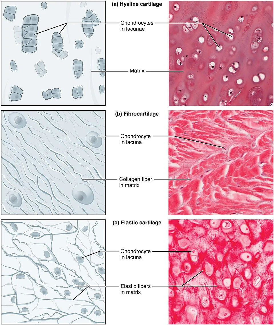
最も多い軟骨。基質はII型コラーゲンとアグリカンで、線維が細く基質と屈折率が近いため、光学顕微鏡では均質でガラス様に見える(hyalos=ガラス)。
場所:関節軟骨・肋軟骨・鼻軟骨・気管と気管支の軟骨・骨端軟骨(成長板)・胎児の骨の原型。
基質は好塩基性(GAGの負電荷)で青紫に染まり、軟骨小腔の周囲がとくに濃い。
関節軟骨は4層構造で、層ごとにコラーゲンの向きが変わる。表層は関節面と平行(せん断力に耐える)、中間層は斜め、深層は垂直、その下に石灰化層があって軟骨下骨に固定される。この配列をarcade 構造と呼ぶ。
変形性関節症はこの構造が表層から壊れていく過程である。表層の線維化・毛羽立ち(fibrillation)→ 亀裂 → 全層欠損 → 軟骨下骨の露出(eburnation)。骨が露出すると初めて神経のある組織にたどり着くので痛みが出る。
X線で見える所見も組織像の裏返しである。関節裂隙狭小化(軟骨の喪失)・骨棘(辺縁の軟骨内骨化)・軟骨下骨硬化(荷重集中への反応)・骨嚢胞。関節リウマチの骨びらん+周囲の骨萎縮とは所見の組み合わせが逆になる点が鑑別の軸になる。
関節軟骨は再生しない——これが治療戦略を決める。若年の限局した欠損には骨髄刺激法(微小骨折術)で骨髄から間葉系細胞を呼び込むが、埋まるのは線維軟骨で耐久性が劣る。より本格的には自家骨軟骨移植や自家培養軟骨細胞移植、進行例では人工関節置換術となる。
English version
Core
The commonest cartilage. The matrix is type II collagen and aggrecan; the fibres are fine and close to the matrix in refractive index, so by light microscopy it appears homogeneous and glassy (hyalos = glass).
Sites: articular cartilage, costal cartilage, nasal cartilage, the cartilages of trachea and bronchi, the epiphysial (growth) plate, and the fetal model of bone.
The matrix is basophilic (from the negative charge of GAGs), staining blue-purple, and is especially dense immediately around the lacunae.
Going further
Articular cartilage has four zones, and the direction of the collagen changes with each: the superficial zone runs parallel to the surface (resisting shear), the intermediate zone obliquely, the deep zone perpendicular, and beneath them a calcified zone anchors it to subchondral bone. This arrangement is the arcade structure.
Osteoarthritis is the process of that structure failing from the surface downward: fibrillation of the superficial zone → fissures → full-thickness defect → exposure of subchondral bone (eburnation). Pain appears only when the bone is exposed, because that is the first tissue reached that has nerves.
The radiographic findings are the histology turned inside out: joint space narrowing (loss of cartilage), osteophytes (endochondral ossification at the margin), subchondral sclerosis (a response to concentrated load) and subchondral cysts. The combination is the reverse of the erosions with periarticular osteopenia of rheumatoid arthritis, which is the axis of the differential.
In the clinic
Articular cartilage does not regenerate — and this determines the strategy of treatment.
For a localised defect in a young patient, marrow stimulation (microfracture) draws mesenchymal cells from the marrow, but what fills the defect is fibrocartilage, of inferior durability. More definitive options are osteochondral autograft and autologous chondrocyte implantation; in advanced disease, arthroplasty.
弾性軟骨Cartilago elastica
硝子軟骨の基質に弾性線維の網が加わったもの。変形しても正確に元の形へ戻るのが役目。
場所:耳介・外耳道・耳管・喉頭蓋・喉頭の小軟骨(喉頭蓋の形を保つ)。
弾性線維は HE で見えないので、弾性線維染色で確認する。
分布に共通するのは「繰り返し曲げられるが、形が変わっては困る場所」である。耳介は日常的に押しつぶされ、喉頭蓋は嚥下のたびに折れ曲がる。硝子軟骨では割れ、線維軟骨では戻らない。
弾性軟骨は加齢による石灰化が起きにくい点も特徴で、肋軟骨(硝子軟骨)が高齢で石灰化して胸部X線に写るのとは対照的である。
再発性多発軟骨炎——II型コラーゲンに対する自己免疫で、軟骨膜のある軟骨が繰り返し炎症を起こす。耳介軟骨炎(耳垂を避けて耳介だけが腫れるのが特徴的。耳垂には軟骨が無い)、鼻軟骨の破壊による鞍鼻、気道軟骨の虚脱による気道閉塞。「軟骨のある場所だけが腫れる」という所見の分布そのものが診断の手がかりになる。
English version
Core
Hyaline matrix with the addition of a network of elastic fibres. Its role is to deform and return precisely to its original shape.
Sites: auricle, external acoustic meatus, auditory tube, epiglottis, and the small laryngeal cartilages.
Elastic fibres are invisible on H&E; confirm with an elastic stain.
Going further
What the sites have in common is "repeatedly bent, but must not change shape". The auricle is crushed daily and the epiglottis folds with every swallow. Hyaline cartilage would crack and fibrocartilage would not spring back.
Elastic cartilage is also resistant to calcification with age — in contrast to costal cartilage (hyaline), which calcifies in the elderly and becomes visible on the chest radiograph.
In the clinic
Relapsing polychondritis — autoimmunity against type II collagen, causing repeated inflammation of cartilages that possess a perichondrium.
- Auricular chondritis — characteristically the auricle swells while the lobule is spared, because the lobule contains no cartilage.
- Destruction of the nasal cartilage giving a saddle nose.
- Collapse of the airway cartilages causing obstruction.
The distribution of the findings — only the places that have cartilage swell — is itself the clue to the diagnosis.
線維軟骨Cartilago fibrosa
I型コラーゲンの太い束が層状に走り、そのあいだに軟骨細胞が列をなす。軟骨膜が無い。密性結合組織と硝子軟骨の中間の性質で、圧縮とせん断の両方に耐える。
場所:椎間板の線維輪・膝の半月板・恥骨結合・関節唇・腱の骨付着部。
分布は「強い力が向きを変えながらかかる継ぎ目」に一致する。骨と骨のあいだ(恥骨結合・椎間板)、骨と腱のあいだ(付着部)、荷重を分散する場所(半月板)。
半月板は血流の分布が治療方針を決める好例である。外周1/3(red zone)には関節包からの血管が入るため縫合で治癒しうるが、内側1/3(white zone)は無血管で治らず切除の対象になる。同じ組織でも、血が来るかどうかで手術が変わる。
椎間板も同様に無血管で、拡散に頼るため加齢で最も早く変性する組織のひとつである。線維輪が裂けて髄核が脱出するのが椎間板ヘルニアで、後外側は後縦靭帯が薄く弱いため後外側へ脱出しやすく、神経根を圧迫する。
脱出した髄核は自然に縮小しうる。無血管の椎間板内にあった髄核が硬膜外腔へ出ると、そこで初めて血管とマクロファージに曝され貪食されるためで、大きく脱出したものほど吸収されやすいという逆説的な現象が起きる。「まず保存療法」という原則の組織学的な根拠である。
English version
Core
Thick bundles of type I collagen run in layers, with chondrocytes in rows between them. There is no perichondrium. It is intermediate between dense connective tissue and hyaline cartilage, resisting both compression and shear.
Sites: annulus fibrosus of the intervertebral disc, the menisci of the knee, the pubic symphysis, the glenoid and acetabular labra, and the insertions of tendons into bone.
Going further
The distribution matches the junctions where strong forces act while changing direction: between bone and bone (pubic symphysis, intervertebral disc), between bone and tendon (the enthesis), and where load must be distributed (the menisci).
The meniscus is the clearest example of blood supply dictating treatment. The outer third (red zone) receives vessels from the capsule and can heal after repair; the inner third (white zone) is avascular, does not heal, and is resected. The same tissue, and the operation changes according to whether blood reaches it.
The intervertebral disc is likewise avascular and dependent on diffusion, making it one of the earliest tissues to degenerate with age. When the annulus tears and the nucleus pulposus herniates, it is a disc herniation; because the posterior longitudinal ligament is thin and weak posterolaterally, herniation tends to occur there and compress the nerve root.
In the clinic
A herniated nucleus pulposus can shrink spontaneously. Material that was inside the avascular disc, once it enters the epidural space, is exposed for the first time to vessels and macrophages and is phagocytosed — producing the paradox that the larger the herniation the more readily it resorbs. This is the histological basis of the principle of treating conservatively first.
軟骨細胞Chondrocytus
基質に囲まれた小腔(lacuna)に1〜数個ずつ収まる細胞。基質を作り、維持する。
1つの母細胞が分裂してできた集まりを同源細胞群(isogenous group)と呼び、数個が固まって見える。これは内部から増える=介在性成長の証拠である。
低酸素環境にいるため、代謝はほぼ嫌気性解糖に依存する。
軟骨の成長には2通りある。
- 介在性成長 interstitial——軟骨細胞が基質の中で分裂し、内側から膨らむ。成長板での骨の伸長はこの様式
- 付加性成長 appositional——軟骨膜の内層の細胞が軟骨芽細胞に分化し、表面に積み増す
成人ではどちらもほぼ止まる。軟骨が治らない理由は無血管であることに加え、この増殖能の低さにもある。
低酸素に適応しているため、軟骨細胞はHIF-1α を恒常的に使って生存している。この性質が、軟骨が血管の無い環境で生き延びられる仕組みそのものである。
軟骨細胞が増えると腫瘍になり、減ると変形性関節症になる。
内軟骨腫 — 手足の短管骨に多い良性腫瘍。多発すると Ollier 病、血管腫を伴うと Maffucci 症候群で悪性化のリスクが上がる。
軟骨肉腫 — 骨盤・大腿骨近位など体幹に近い骨に多い。化学療法と放射線が効きにくく、広範切除が原則——血管が乏しく代謝が遅いという軟骨の性質がそのまま治療の制約になる。
成長板の軟骨細胞は増殖して背を伸ばす。軟骨無形成症(FGFR3 の機能獲得)ではこの増殖が抑えられて四肢が短くなる。膜性骨化する頭蓋冠は正常なので頭だけが相対的に大きい。
同源細胞群が目立つ=間質性成長が盛んな若い軟骨である。
English version
Core
A cell enclosed singly or in small groups within a lacuna, which makes and maintains the matrix.
A cluster derived from one parent cell by division is an isogenous group, seen as several cells together — evidence of growth from within, that is, interstitial growth.
Living in a hypoxic environment, its metabolism depends almost entirely on anaerobic glycolysis.
Going further
Cartilage grows in two ways.
- Interstitial growth — chondrocytes divide within the matrix and the cartilage expands from inside. This is how bone lengthens at the growth plate.
- Appositional growth — cells of the inner perichondrium become chondroblasts and add matrix to the surface.
In the adult both largely cease. Cartilage fails to heal not only because it is avascular but also because of this low proliferative capacity.
Adapted to hypoxia, the chondrocyte survives by constitutive use of HIF-1α — the very mechanism by which cartilage can live in an environment without vessels.
In the clinic
When chondrocytes proliferate the result is a tumour; when they are lost the result is osteoarthritis.
Enchondroma — a benign tumour, common in the short tubular bones of the hand and foot. Multiple lesions constitute Ollier disease, and with haemangiomas Maffucci syndrome, both carrying an increased risk of malignant change.
Chondrosarcoma — common in bones near the trunk, such as the pelvis and proximal femur. Chemotherapy and radiotherapy work poorly and wide excision is the rule — the poor vascularity and slow metabolism of cartilage becoming directly a constraint on treatment.
The chondrocytes of the growth plate proliferate and lengthen the body. In achondroplasia (gain of function in FGFR3) this proliferation is suppressed and the limbs are short. The cranial vault, which ossifies in membrane, is normal, so the head appears relatively large.
Prominent isogenous groups indicate young cartilage growing interstitially.
骨端軟骨(成長板)Lamina epiphysialis
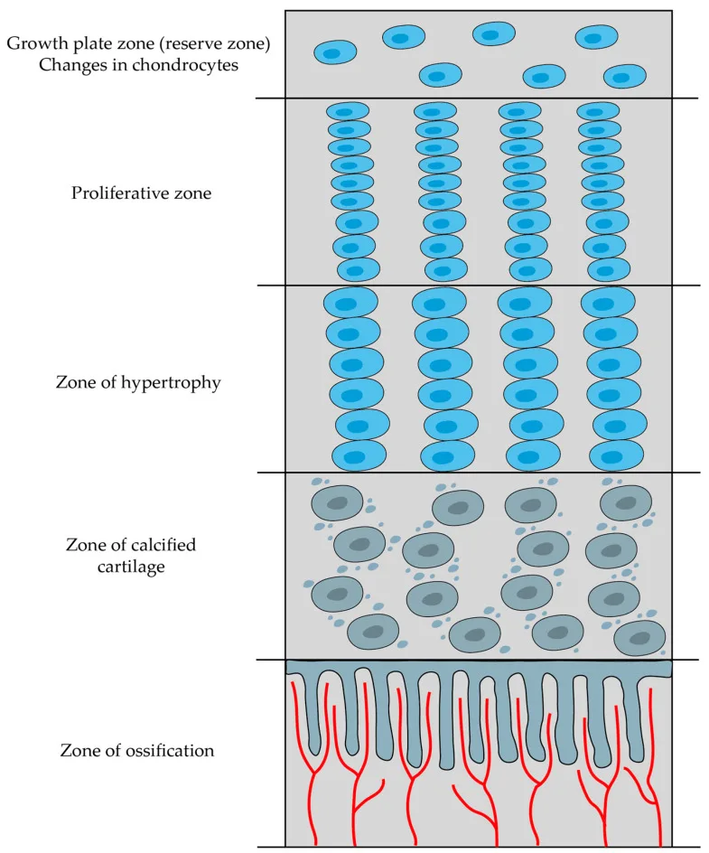
骨端と骨幹端のあいだに残る硝子軟骨の板。ここで軟骨が作られては骨に置き換わることで、骨が長さ方向に伸びる。
骨端側から骨幹側へ、5つの帯が順に並ぶ。
- 静止帯——予備の軟骨細胞
- 増殖帯——細胞が分裂し縦に積み重なる(銭を積んだように見える)
- 肥大帯——細胞が大きく膨らむ。ここが最も脆い
- 石灰化帯——基質が石灰化し軟骨細胞が死ぬ
- 骨化帯——血管が侵入し、骨芽細胞が骨を置く
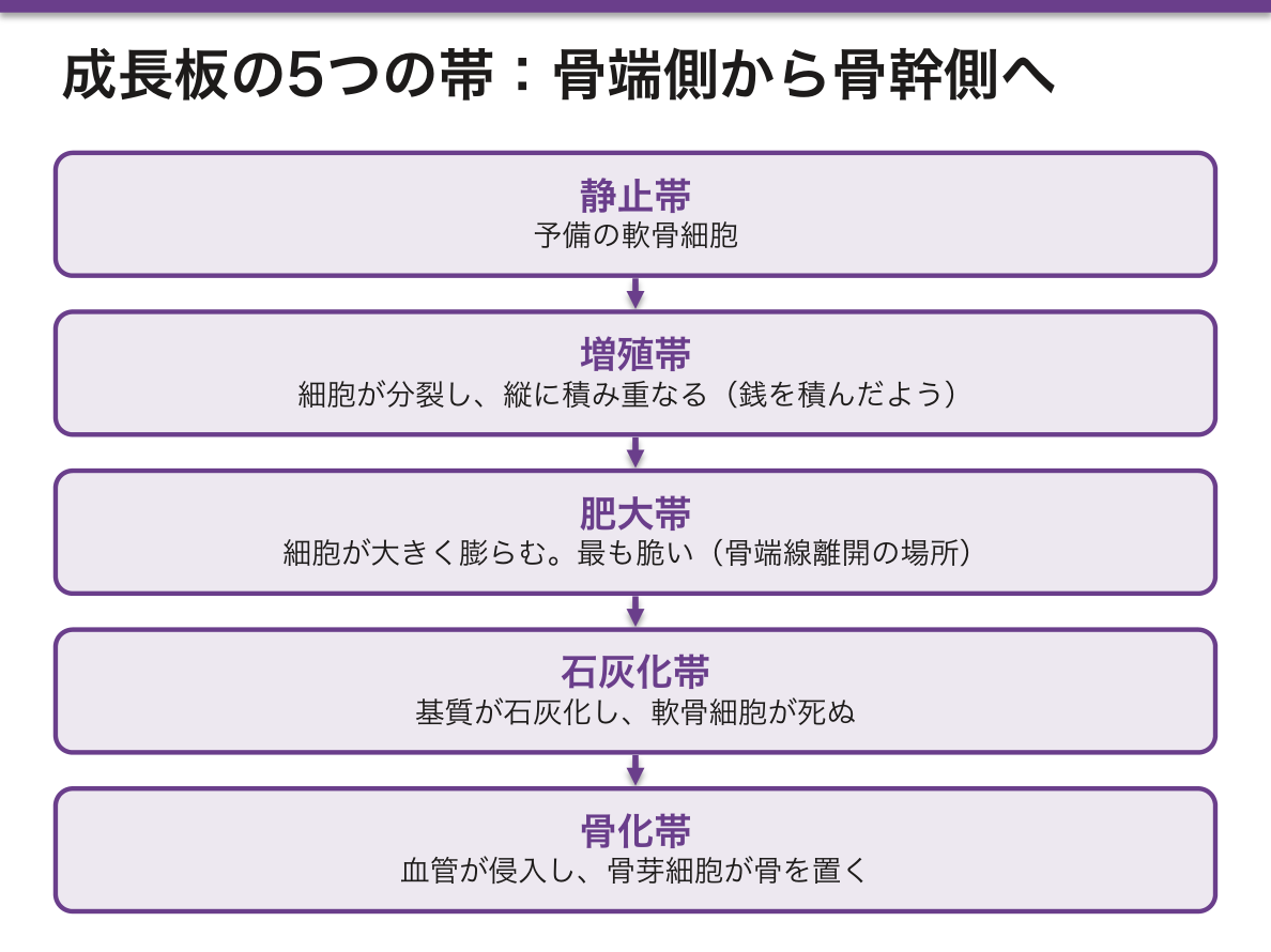
肥大帯が力学的に最弱という事実が、小児の外傷学をほぼ決めている。小児では靭帯より成長板のほうが弱いため、成人なら靭帯損傷になる外力が成長板の骨折(骨端線離開)になる。分類は Salter-Harris の I〜V型で、III・IV型は関節面と成長板の両方を横切るため整復精度が要求され、V型(圧挫)は最も予後が悪い。
成長板が障害されると成長障害・変形が残る。部分的に閉鎖すれば片側だけ伸びなくなり角状変形を生じる。小児の骨折を「成人の小型版」として扱ってはいけない理由がここにある。
閉鎖の時期はホルモンで決まる。成長ホルモン・IGF-1 が成長を促し、エストロゲンが成長板を閉鎖させる。思春期の急伸のあと成長が止まるのはこのためで、男女で身長差が生じるのも閉鎖時期の差による。
軟骨無形成症——FGFR3 の機能獲得型変異(常染色体顕性、多くは新生突然変異で高齢父性が関連)。FGFR3 は軟骨細胞の増殖を抑制する受容体なので、活性化すると成長板の増殖帯が働かない。
結果として軟骨内骨化だけが障害され、膜内骨化は正常という特徴的な体型になる——四肢(長管骨)が短く、体幹と頭蓋(膜内骨化)は保たれる。前額部突出・鞍鼻・三尖手。知能と寿命は原則正常。「どちらの骨化様式か」で侵される骨が決まるのがこの疾患の要点である。
English version
Core
A plate of hyaline cartilage remaining between epiphysis and metaphysis. Bone lengthens because cartilage is made here and replaced by bone.
Five zones, from the epiphysial towards the diaphysial side:
- Resting zone — the reserve of chondrocytes.
- Proliferative zone — cells divide and stack vertically, like a pile of coins.
- Hypertrophic zone — the cells swell. This is the weakest point.
- Calcification zone — the matrix calcifies and the chondrocytes die.
- Ossification zone — vessels invade and osteoblasts lay down bone.
Going further
That the hypertrophic zone is mechanically weakest determines almost the whole of paediatric trauma. In a child the growth plate is weaker than the ligament, so a force that would cause a ligamentous injury in an adult produces a physeal fracture instead.
They are classified as Salter–Harris types I–V: types III and IV cross both the articular surface and the physis and demand accurate reduction; type V (crush) has the worst prognosis.
Damage to the growth plate leaves growth disturbance and deformity. Partial closure stops growth on one side only and produces angular deformity. This is why a fracture in a child must not be treated as a small version of an adult fracture.
The timing of closure is hormonal: growth hormone and IGF-1 promote growth, and oestrogen closes the plate. This is why growth stops after the pubertal spurt, and why the difference in adult height between the sexes arises from a difference in the timing of closure.
In the clinic
Achondroplasia — a gain-of-function mutation in FGFR3 (autosomal dominant, mostly new mutations, associated with advanced paternal age).
FGFR3 is a receptor that suppresses chondrocyte proliferation, so when it is activated the proliferative zone of the growth plate does not work. The result is that endochondral ossification alone is impaired while intramembranous ossification is normal, giving the characteristic body form: short limbs with a preserved trunk and cranial vault. Frontal bossing, saddle nose, trident hand. Intelligence and lifespan are normally unaffected.
The point of this disease is that which bones are affected is decided by which mode of ossification they use.
関節軟骨 — 治らない組織を、どう守るかCartilago articularis
硝子軟骨だが、関節面には軟骨膜が無い——ここが再生できない最大の理由になる。
4層に分かれ、コラーゲン線維の向きが層ごとに変わる。
| 層 | 線維の向き | 細胞 |
|---|---|---|
| 表層 | 関節面と平行 | 扁平・密 |
| 移行層 | 斜め | 円形 |
| 深層 | 関節面と垂直 | 柱状に並ぶ |
| 石灰化層 | — | tidemark で非石灰化層と分かれる |
血管・リンパ管・神経が無い——栄養は滑液からの拡散に頼り、荷重による出し入れ(ポンプ作用)が要る。
滑膜は2層——A細胞(マクロファージ性)とB細胞(線維芽細胞性、ヒアルロン酸を作る)。基底膜が無い。
「神経が無い」ことが、変形性関節症の症状のずれを説明する。軟骨がすり減っても痛みは出ない。痛むのは滑膜・骨・靱帯になる(変形性関節症の章)——「画像の変化と痛みが一致しない理由が、ここにある」。
「血管が無い」ことが、治らない理由になる。損傷しても炎症細胞も幹細胞も来ないので、修復の過程が始まらない(修復と創傷治癒の章)——「軟骨下骨まで届く損傷だけが、骨髄から細胞を呼べる」。
だから微小骨折術(マイクロフラクチャー)という手技が成り立つ。わざと軟骨下骨に穴を開け、骨髄の細胞を呼び込む。ただしできるのは線維軟骨になる(スポーツ外傷の章)——「元と同じ硝子軟骨は戻らない」。
栄養が荷重に依存することは、リハビリテーションの根拠になる。動かさなければ軟骨は痩せる。免荷の期間が長いほど、軟骨は薄くなる(運動処方の章)——「安静そのものが、関節にとっては害になりうる」。
滑膜に基底膜が無いことは、関節液の性質を決めている。血漿成分が容易に出入りするので、炎症のときは関節液に細胞と蛋白が増える(関節液の章)——「関節穿刺の白血球数が、炎症性か非炎症性かを分ける」。
軟骨膜が無いから、関節軟骨は再生しない。これが変形性関節症が治らない理由であり、人工関節が要る理由でもある。
変形性関節症の X 線4徴 — 関節裂隙の狭小化・骨硬化・骨棘・軟骨下嚢胞。軟骨が減った結果、下の骨に負荷が移ったことを映している。炎症は主体ではないので朝のこわばりは短い(30分以内)——関節リウマチとの鑑別点になる。
骨に達する損傷(全層欠損)だけが治る。骨髄から間葉系細胞が出てくるからで、微小骨折術(microfracture)はこれを人為的に起こす手技である。ただしできるのは線維軟骨で、硝子軟骨には及ばない。
自家培養軟骨細胞移植が行われるのも、自然には埋まらないという前提があるからである。
English version
Core
Hyaline cartilage, but the articular surface has no perichondrium — the principal reason it cannot regenerate.
Four zones, with the collagen changing direction in each:
| Zone | Fibre direction | Cells |
|---|---|---|
| Superficial | Parallel to the surface | Flattened, densely packed |
| Transitional | Oblique | Round |
| Deep | Perpendicular to the surface | Arranged in columns |
| Calcified | — | Separated from the uncalcified zone by the tidemark |
No vessels, lymphatics or nerves — nutrition depends on diffusion from the synovial fluid, and requires the pumping action of loading.
The synovium has two cell types — A cells (macrophage-like) and B cells (fibroblast-like, making hyaluronic acid) — and no basal lamina.
Going further
Having no nerves explains the mismatch of symptoms in osteoarthritis. Wearing away the cartilage does not itself cause pain; the pain comes from synovium, bone and ligament — here is why the imaging and the pain do not correspond.
Having no vessels is why it does not heal. After injury neither inflammatory cells nor stem cells arrive, so the process of repair never begins — only a lesion reaching the subchondral bone can summon cells from the marrow.
This is what makes microfracture possible: holes are deliberately made in the subchondral bone to bring marrow cells in. But what forms is fibrocartilage — the original hyaline cartilage does not return.
That nutrition depends on loading is the rationale of rehabilitation: cartilage wastes if it is not moved, and the longer the period of non-weight-bearing the thinner it becomes — rest itself can be harmful to a joint.
That the synovium has no basal lamina determines the nature of the joint fluid: plasma constituents pass freely, so in inflammation cells and protein increase — the white cell count on joint aspiration is what separates inflammatory from non-inflammatory disease.
In the clinic
Because there is no perichondrium, articular cartilage does not regenerate. This is why osteoarthritis does not resolve and why joint replacement is needed.
The four radiographic signs of osteoarthritis — joint space narrowing, sclerosis, osteophytes and subchondral cysts — reflect the transfer of load to the bone beneath after the cartilage has gone. Inflammation is not the primary process, so morning stiffness is brief (under 30 minutes) — a point of distinction from rheumatoid arthritis.
Only a lesion reaching bone (a full-thickness defect) heals, because mesenchymal cells emerge from the marrow; microfracture induces this artificially, though what forms is fibrocartilage rather than hyaline. Autologous chondrocyte implantation exists on the same premise: the defect will not fill on its own.
軟骨膜 — 2つの成長のしかたと、治るかどうかを分けるものPerichondrium
軟骨は2 通りの伸び方をする。どちらの様式で伸びるかは、軟骨膜があるかどうかで決まる。
| 付加成長 | 軟骨膜の内層の細胞が軟骨芽細胞になり、表面に新しい基質を足す | 太くなる方向。軟骨膜が要る |
| 間質成長 | 軟骨細胞が基質の中で分裂し、同壁性細胞群を作る | 内側から膨らむ。成長期に盛んで、成人ではほぼ止まる |
軟骨膜は 2 層からなる。外層は線維性(血管と神経を含む)、内層は細胞性(軟骨をつくる細胞の供給源)。軟骨そのものには血管が無いので、栄養は軟骨膜から拡散で届く。
軟骨膜が無い軟骨が2つあり、どちらも臨床的に問題になる。
- 関節軟骨——関節腔に面する側に軟骨膜が無い。供給源も血管も無いので、傷が治らない。変形性関節症が不可逆に進む土台がここにある
- 線維軟骨——そもそも軟骨膜を持たない
だから軟骨の修復を狙う治療は、「外から細胞を持ち込む」か「骨髄と繋げる」かのどちらかになる——骨髄刺激法(軟骨下骨に穴を開けて骨髄の細胞を呼び込む)、自家骨軟骨移植、培養軟骨細胞移植。いずれも本来の硝子軟骨ではなく線維軟骨で埋まることが多く、耐久性が劣る。
⚠️ 軟骨膜の炎症と損傷も、それ自体が疾患になる。
- 耳介血腫——軟骨膜が剥がれて血が溜まると、栄養が届かず軟骨が壊死する。放置すると変形(カリフラワー耳)が残るので、早期に血腫を除去して圧迫する
- 再発性多発軟骨炎——軟骨への自己免疫。耳介(耳垂を除く)・鼻・気道の軟骨を侵し、鞍鼻と気道の虚脱を起こす。耳垂には軟骨が無いので腫れない——この所見が診断の手がかりになる
耳介の外傷では、腫れが軟骨膜の下かどうかを見る。血腫を残すと、数週間で不可逆の変形になる。
English version
Core
Cartilage grows in two ways, and which one operates depends on whether a perichondrium is present.
| Mode | Mechanism | Character |
|---|---|---|
| Appositional | Cells of the inner perichondrium become chondroblasts and add new matrix to the surface | Growth in thickness. Requires a perichondrium |
| Interstitial | Chondrocytes divide within the matrix, forming isogenous groups | Expansion from within. Active during growth, largely ceasing in the adult |
The perichondrium has two layers: an outer fibrous layer (containing vessels and nerves) and an inner cellular layer (the source of cartilage-forming cells). Since cartilage itself has no vessels, nutrition diffuses from the perichondrium.
Going further
Two cartilages have no perichondrium, and both are clinically troublesome.
- Articular cartilage — none on the surface facing the joint cavity. With neither a source of cells nor a blood supply, injury does not heal. Here is the foundation of the irreversible progression of osteoarthritis.
- Fibrocartilage — has none at all.
So treatments aimed at repairing cartilage must either bring cells in from outside or connect the defect to the marrow: marrow stimulation (drilling the subchondral bone to bring marrow cells in), osteochondral autograft, autologous chondrocyte implantation. All tend to fill with fibrocartilage rather than true hyaline cartilage, and are less durable.
⚠️ Inflammation and injury of the perichondrium are themselves diseases.
- Auricular haematoma — when the perichondrium is stripped and blood collects beneath it, nutrition no longer reaches the cartilage and it necroses. Left alone it leaves deformity (cauliflower ear), so the haematoma must be evacuated early and compressed.
- Relapsing polychondritis — autoimmunity against cartilage, attacking the auricle (sparing the lobule), nose and airway. The lobule has no cartilage and does not swell — a clue to the diagnosis.
In the clinic
In trauma to the auricle, establish whether the swelling lies beneath the perichondrium. A haematoma left in place becomes an irreversible deformity within weeks.
関節の分類と滑膜関節Articulatio synovialis
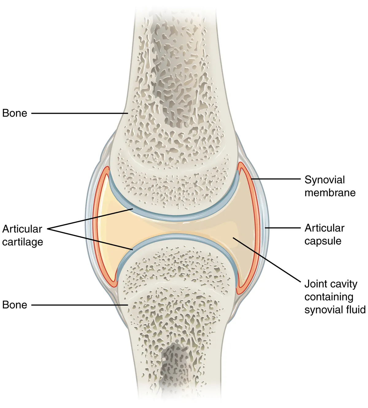
関節は「何で繋がっているか」で3つ——線維性(結合組織。縫合・釘植・靭帯結合)、軟骨性(軟骨。硝子軟骨結合=骨端軟骨、線維軟骨結合=恥骨結合や椎間円板)、滑膜性(関節腔がある。可動性が高い)。
滑膜関節の6要素——①関節軟骨(硝子軟骨、軟骨膜が無い)②関節包(外の線維膜と内の滑膜)③関節腔④滑液⑤靭帯⑥補助構造(関節円板・半月・滑液包)。
滑膜 membrana synovialis——上皮ではない。1〜3層の滑膜細胞が並ぶが、基底膜が無く、細胞間に隙間があるのが決定的な特徴。A 細胞(マクロファージ系。異物を掃除する)とB 細胞(線維芽細胞系。ヒアルロン酸を作る)の2種。滑膜は関節軟骨の表面は覆わない。
滑液——血漿の濾液にヒアルロン酸とルブリシンを加えたもの。潤滑と、血管の無い関節軟骨への栄養供給を担う。
関節軟骨に血管・神経・リンパ管が無いことが、痛みが出にくく、しかも治らない理由。関節リウマチでは滑膜が増殖してパンヌスを作り、軟骨と骨を破壊する——滑膜の病気が軟骨を壊す、という順序が要点。
English version
Core
Joints are classified by what joins the bones.
- Fibrous — connective tissue: suture, gomphosis, syndesmosis.
- Cartilaginous — cartilage: synchondrosis (the growth plate), symphysis (pubic symphysis, intervertebral disc).
- Synovial — with a joint cavity; freely mobile.
Six components of a synovial joint — ① articular cartilage (hyaline, without perichondrium) ② capsule (outer fibrous membrane, inner synovium) ③ joint cavity ④ synovial fluid ⑤ ligaments ⑥ accessory structures (discs, menisci, bursae).
Synovium — not an epithelium. One to three layers of synoviocytes, but with no basal lamina and with gaps between the cells, which is the decisive feature. A cells (macrophage lineage, clearing debris) and B cells (fibroblast lineage, making hyaluronic acid). The synovium does not cover the surface of the articular cartilage.
Synovial fluid — a filtrate of plasma with hyaluronic acid and lubricin added; it lubricates and nourishes the avascular articular cartilage.
Going further
That articular cartilage has no vessels, nerves or lymphatics is why pain appears late and why it does not heal.
In rheumatoid arthritis the synovium proliferates into a pannus and destroys cartilage and bone — the essential point is the order: a disease of the synovium destroys the cartilage.
まとめ
| 軟骨 | 軟骨細胞が軟骨小腔 lacuna に収まり、そのまわりを硬い基質が固める組織。 |
|---|---|
| 硝子軟骨 | 最も多い軟骨。基質はII型コラーゲンとアグリカンで、線維が細く基質と屈折率が近いため、光学顕微鏡では均質でガラス様に見える(hyalos=ガラス)。 |
| 弾性軟骨 | 硝子軟骨の基質に弾性線維の網が加わったもの。 |
| 線維軟骨 | I型コラーゲンの太い束が層状に走り、そのあいだに軟骨細胞が列をなす。 |
| 軟骨細胞 | 基質に囲まれた小腔(lacuna)に1〜数個ずつ収まる細胞。 |
| 骨端軟骨(成長板) | 骨端と骨幹端のあいだに残る硝子軟骨の板。 |
| 関節軟骨 — 治らない組織を、どう守るか | 硝子軟骨だが、関節面には軟骨膜が無い——ここが再生できない最大の理由になる。 |
| 軟骨膜 — 2つの成長のしかたと、治るかどうかを分けるもの | 軟骨は2 通りの伸び方をする。どちらの様式で伸びるかは、軟骨膜があるかどうかで決まる。 |
| 関節の分類と滑膜関節 | 関節は「何で繋がっているか」で3つ——線維性(結合組織。 |
このノートは医学生が授業の内容を自分の言葉でまとめたもので、診療の判断に使うものではありません。誤りに気づいたらお知らせください。