血液は基質が液体になった結合組織である。細胞(血球)が基質(血漿)に浮かび、線維は普段は溶けている——必要なときだけフィブリンとして析出する。この見方をすると、凝固は「結合組織が必要に応じて線維を作る」現象として理解できる。
血球は1個の造血幹細胞から分かれる。骨髄系とリンパ系に分かれ、そこから先は分化の樹をたどるだけ。白血病の分類も、血算の読み方も、この樹のどこで止まったかで決まるので、樹の形を先に入れておくと後がすべて楽になる。
血球の寿命(赤血球120日・血小板約10日・好中球わずか数時間〜1日)を覚えておくと、異常が現れるまでの時間が読める。抗癌剤で好中球が先に落ち、貧血が最後に来るのはこの差による。
- 血液
- 赤血球
- 好中球
- 白血球の残り4種 — 好酸球・好塩基球・リンパ球・単球
- 血小板
- 造血
- 造血の階層 — 幹細胞・前駆細胞・CFU を区別する
- 赤血球造血 — 青から赤へ、そして核を捨てるまで
- 顆粒球造血と単球造血 — 特殊顆粒が出た段階で、系統が決まる
- 赤血球膜 — 形を保つ骨組みが壊れると、脾で壊される
- 血液塗抹 — 赤血球の形が、そのまま診断名になる
血液Sanguis
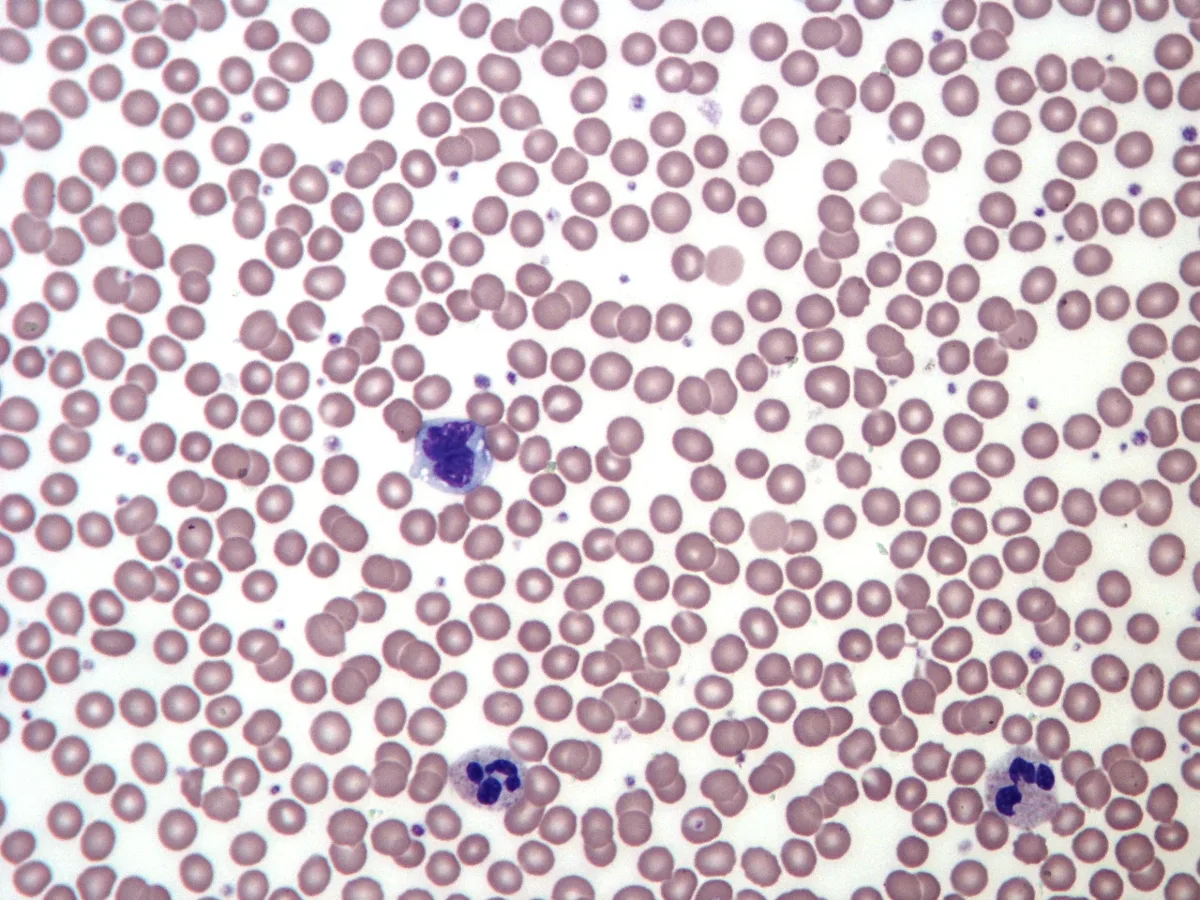
液体の基質(血漿)に血球が浮かぶ、特殊な結合組織。成人で約5L、体重の約8%。
- 血漿 約55%——水91%、蛋白7%(アルブミン・グロブリン・フィブリノゲン)、電解質・栄養素
- 血球 約45%(=ヘマトクリット)——赤血球がほぼ全部を占める
凝固させたあとの上澄みが血清で、フィブリノゲンと凝固因子が消費されて含まれない点が血漿との違い。
血漿蛋白の役割を分けて覚えると臨床が読める。
- アルブミン——膠質浸透圧の約80%を担い、薬物・ビリルビン・Ca を運ぶ。肝で作られるので肝硬変・ネフローゼ・低栄養で低下 → 浮腫
- グロブリン——免疫(γ)と輸送(α・β)
- フィブリノゲン——凝固の最終基質であり、急性期蛋白。炎症で上がり赤沈亢進の主因になる
血清Caの約40%はアルブミンと結合しているため、低アルブミン血症では総Caが低く出るが、生理活性のあるイオン化Caは正常なことが多い。補正Ca=実測Ca+(4−Alb) を使う理由である。
血算の3系統を分けて読むのが診療の出発点である。1系統だけの異常か、汎血球減少かで考える範囲が変わる。汎血球減少=骨髄の問題(再生不良性貧血・白血病・骨髄異形成症候群)か、脾機能亢進か、ビタミン B12/葉酸欠乏を疑う。
ヘマトクリットは相対値である。脱水では血液が濃縮して見かけ上高くなり、輸液で下がる——出血直後は Hb が下がらないのは、血漿と血球が同じ割合で失われるからである。数時間〜1日かけて血管内に水が戻って初めて貧血として現れる。
アルブミンは膠質浸透圧を担う。低アルブミン血症では浮腫と腹水になり、薬の遊離型が増えて効きすぎる(ワルファリン・フェニトイン)。
フィブリノゲンは炎症でも上がる急性期蛋白で、赤沈亢進の主因である。
English version
Core
A special connective tissue in which cells float in a liquid matrix. About 5 litres in the adult, some 8 per cent of body weight.
- Plasma, about 55 per cent — 91 per cent water, 7 per cent protein (albumin, globulin, fibrinogen), electrolytes and nutrients.
- Cells, about 45 per cent (the haematocrit) — almost entirely red cells.
The supernatant after clotting is serum, which differs from plasma in that fibrinogen and the clotting factors have been consumed.
Going further
Separate the roles of the plasma proteins and the clinical picture becomes readable.
- Albumin — carries about 80 per cent of the colloid osmotic pressure and transports drugs, bilirubin and calcium. Made in the liver, so it falls in cirrhosis, nephrotic syndrome and malnutrition → oedema.
- Globulins — immunity (γ) and transport (α, β).
- Fibrinogen — the final substrate of coagulation and an acute phase protein; it rises in inflammation and is the main reason the ESR is raised.
About 40 per cent of serum calcium is bound to albumin, so in hypoalbuminaemia the total calcium reads low while the physiologically active ionised calcium is often normal. This is why the corrected calcium (measured Ca + (4 − albumin)) is used.
In the clinic
Reading the three lineages of the blood count separately is the starting point of practice. Whether one lineage is abnormal or all three changes the range of what to consider.
Pancytopenia = a problem of the marrow (aplastic anaemia, leukaemia, myelodysplastic syndrome), hypersplenism, or deficiency of vitamin B12 or folate.
The haematocrit is a relative value. In dehydration the blood is concentrated and it reads high, falling with fluid. Immediately after haemorrhage the haemoglobin does not fall, because plasma and cells are lost in the same proportion; anaemia appears only after hours to a day, once water has returned to the circulation.
Albumin carries the colloid osmotic pressure: hypoalbuminaemia gives oedema and ascites, and increases the free fraction of drugs so that they act too strongly (warfarin, phenytoin).
Fibrinogen is an acute phase protein that also rises in inflammation and is the main cause of a raised ESR.
赤血球Erythrocytus
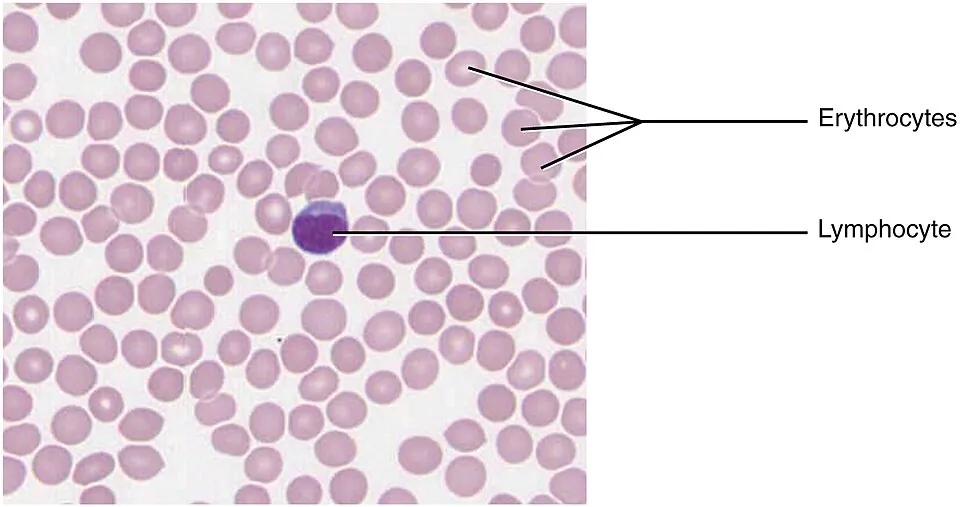
直径約7.5μm の両凹円板。成熟すると核もミトコンドリアも失う——中身をヘモグロビンで満たすためである。寿命約120日、脾臓で処理される。
両凹の形が効いている——表面積が大きく拡散に有利、変形しやすく自分より細い毛細血管を通れる。
ミトコンドリアが無いので、代謝は嫌気性解糖のみ。運ぶ酸素を自分では使わない。
「核もミトコンドリアも無い」ことの帰結が国試の頻出事項になる。
- 自分でタンパクを作れない——酵素が失活しても補充できず、寿命が尽きる
- ATPは解糖系だけ——ピルビン酸キナーゼ欠損症でATPが不足すると、Na⁺/K⁺-ATPase が止まり細胞が膨らんで溶血
- 酸化ストレスに弱い——グルコース6リン酸脱水素酵素(G6PD)欠損症ではNADPHが作れずグルタチオンを還元できない。ソラマメ・抗マラリア薬・感染で急性溶血、Heinz 小体が出る
赤血球のサイズ(MCV)で貧血を分ける習慣も、作られ方の違いを見ている。小球性=ヘモグロビンが作れない(鉄欠乏・サラセミア・慢性炎症)、大球性=DNAが作れない(ビタミンB12・葉酸欠乏。核の成熟だけが遅れ細胞質が先に育つ=巨赤芽球性)、正球性=出血・溶血・骨髄不全。
脾摘後や無脾症では、通常なら脾臓が除去するHowell-Jolly 小体(核の残骸)が末梢血に見える。同時に莢膜を持つ細菌(肺炎球菌・インフルエンザ桿菌・髄膜炎菌)への抵抗が落ち、電撃的な敗血症(OPSI)のリスクがあるためワクチンが必須になる。血液像の1つの所見が、予防接種の適応まで繋がる。
English version
Core
A biconcave disc about 7.5 µm across. On maturing it loses both nucleus and mitochondria — in order to fill itself with haemoglobin. Lifespan about 120 days; removed in the spleen.
The biconcave shape works: a large surface area favouring diffusion, and deformability allowing it to pass through capillaries narrower than itself.
Without mitochondria its metabolism is anaerobic glycolysis alone. It does not use the oxygen it carries.
Going further
The consequences of having neither nucleus nor mitochondria are frequently examined.
- It cannot make protein for itself — an enzyme that is inactivated cannot be replaced, and the cell simply reaches the end of its life.
- ATP comes from glycolysis alone — in pyruvate kinase deficiency, ATP runs short, the Na⁺/K⁺-ATPase stops, the cell swells and haemolyses.
- It is vulnerable to oxidative stress — in G6PD deficiency NADPH cannot be made and glutathione cannot be reduced. Broad beans, antimalarials and infection cause acute haemolysis, with Heinz bodies.
The habit of classifying anaemia by size (MCV) is also looking at how the cell was made: microcytic = haemoglobin cannot be made (iron deficiency, thalassaemia, chronic inflammation); macrocytic = DNA cannot be made (B12 or folate deficiency, where only nuclear maturation lags while the cytoplasm develops — megaloblastic); normocytic = haemorrhage, haemolysis or marrow failure.
In the clinic
After splenectomy or in asplenia, Howell–Jolly bodies (nuclear remnants) that the spleen would normally remove appear in the peripheral blood.
At the same time resistance to encapsulated organisms (pneumococcus, Haemophilus influenzae, meningococcus) falls, with a risk of overwhelming post-splenectomy infection, so vaccination is mandatory.
A single finding on the blood film leads all the way to an indication for immunisation.
好中球Granulocytus neutrophilus
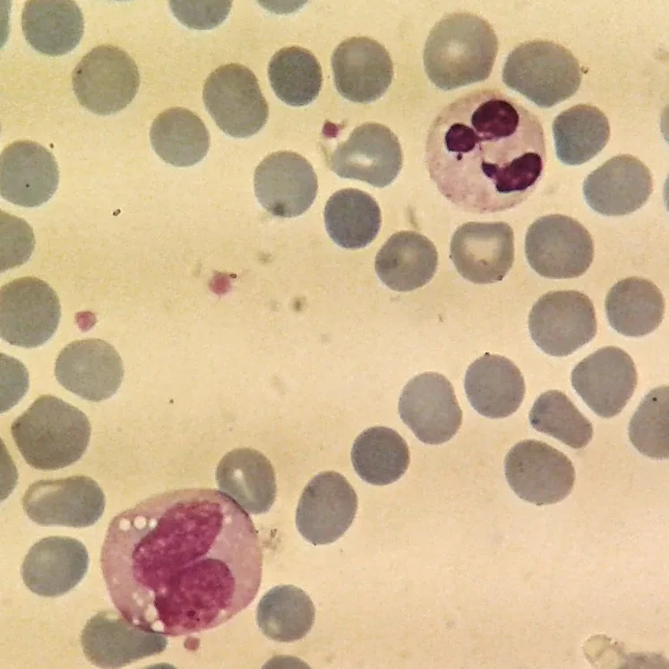
白血球の約60%を占める。核が2〜5葉に分節し(分葉核)、細胞質の顆粒は HE でほとんど染まらない(=中性=neutro)。
急性細菌感染の主役。血管外へ出て貪食し、活性酸素とリソソーム酵素で殺菌する。寿命は数時間〜1日と極端に短い。
未熟な桿状核球が増えることを核左方移動といい、急性感染の指標になる。
好中球の数と形の読み方を3つ。
- 核左方移動——需要が供給を上回り、未熟なまま出てきた。急性細菌感染・炎症
- 過分節(5葉以上)——巨赤芽球性貧血の所見。DNA合成障害で核の成熟がずれる。貧血なのに白血球の形で診断がつく
- 好中球減少——500/μL未満で発熱性好中球減少症。感染源がはっきりしなくても広域抗菌薬を直ちに開始する
好中球は死ぬときにNETs(クロマチンの網)を放出して細菌を絡め取る。膿は死んだ好中球と組織の残骸であり、好中球が居なければ膿は作れない。だから好中球減少患者の感染は膿瘍を作らず所見に乏しいまま重症化する——「熱だけ」で動く根拠である。
核の分葉が、そのまま病態を教える。
左方移動 — 桿状核好中球や幼若な細胞が増える。急性細菌感染で骨髄が急いで出荷している状態。類白血病反応では著明な白血球増加になるが、好中球アルカリホスファターゼ(NAP)スコアが高いことが慢性骨髄性白血病(低値)との鑑別になる。
過分節好中球(5分葉以上) — ビタミン B12/葉酸欠乏。巨赤芽球性貧血より先に現れることがあり、末梢血塗抹の1枚で気づける。
好中球減少(500/μL 未満)は発熱性好中球減少症のリスク。抗癌薬投与後の発熱は緊急事態として直ちに広域抗菌薬を始める。好中球が少ないと膿が作れず、所見が乏しい——「症状が軽く見える」ことが危険である。
English version
Core
About 60 per cent of the white cells. The nucleus is segmented into 2–5 lobes and the granules scarcely stain with H&E (neutral = neutro).
The principal cell of acute bacterial infection. It leaves the vessel, phagocytoses, and kills using reactive oxygen species and lysosomal enzymes.
Lifespan is only hours to a day — extremely short. An increase in immature band forms is a left shift, an index of acute infection.
Going further
Three ways to read the number and the form.
- Left shift — demand has outstripped supply and immature cells have been released. Acute bacterial infection and inflammation.
- Hypersegmentation (five lobes or more) — a finding of megaloblastic anaemia; impaired DNA synthesis puts nuclear maturation out of step. The diagnosis of an anaemia made from the shape of a white cell.
- Neutropenia — below 500/µL is febrile neutropenia. Start broad-spectrum antibiotics immediately even without an identified source.
Neutrophils release NETs (nets of chromatin) as they die, entangling bacteria. Pus is dead neutrophils and tissue debris, so without neutrophils there can be no pus. Hence infection in a neutropenic patient forms no abscess and becomes severe with few signs — the justification for acting on fever alone.
In the clinic
The lobation of the nucleus tells you the process directly.
Left shift — band forms and immature cells increase, the marrow shipping urgently in acute bacterial infection. In a leukaemoid reaction the white count is markedly raised, but a high neutrophil alkaline phosphatase score distinguishes it from chronic myeloid leukaemia, where it is low.
Hypersegmented neutrophils (five lobes or more) — vitamin B12 or folate deficiency. They can appear before the megaloblastic anaemia, and can be noticed on a single blood film.
Neutropenia below 500/µL carries the risk of febrile neutropenia. Fever after chemotherapy is an emergency and broad-spectrum antibiotics are started at once.
Without neutrophils no pus can form and the signs are sparse — and it is dangerous precisely because it looks mild.
白血球の残り4種 — 好酸球・好塩基球・リンパ球・単球
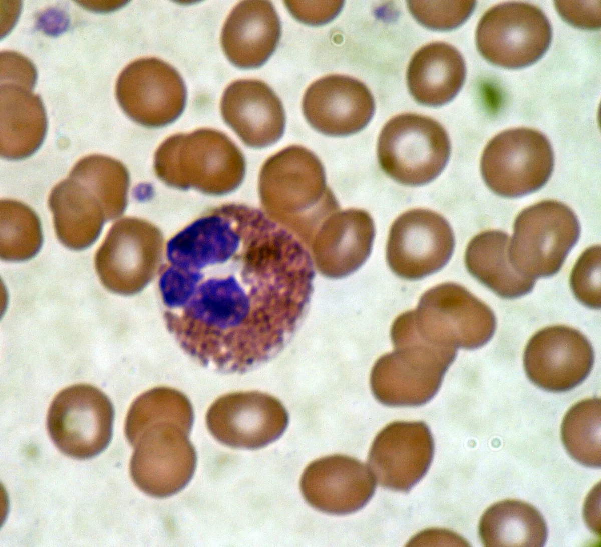
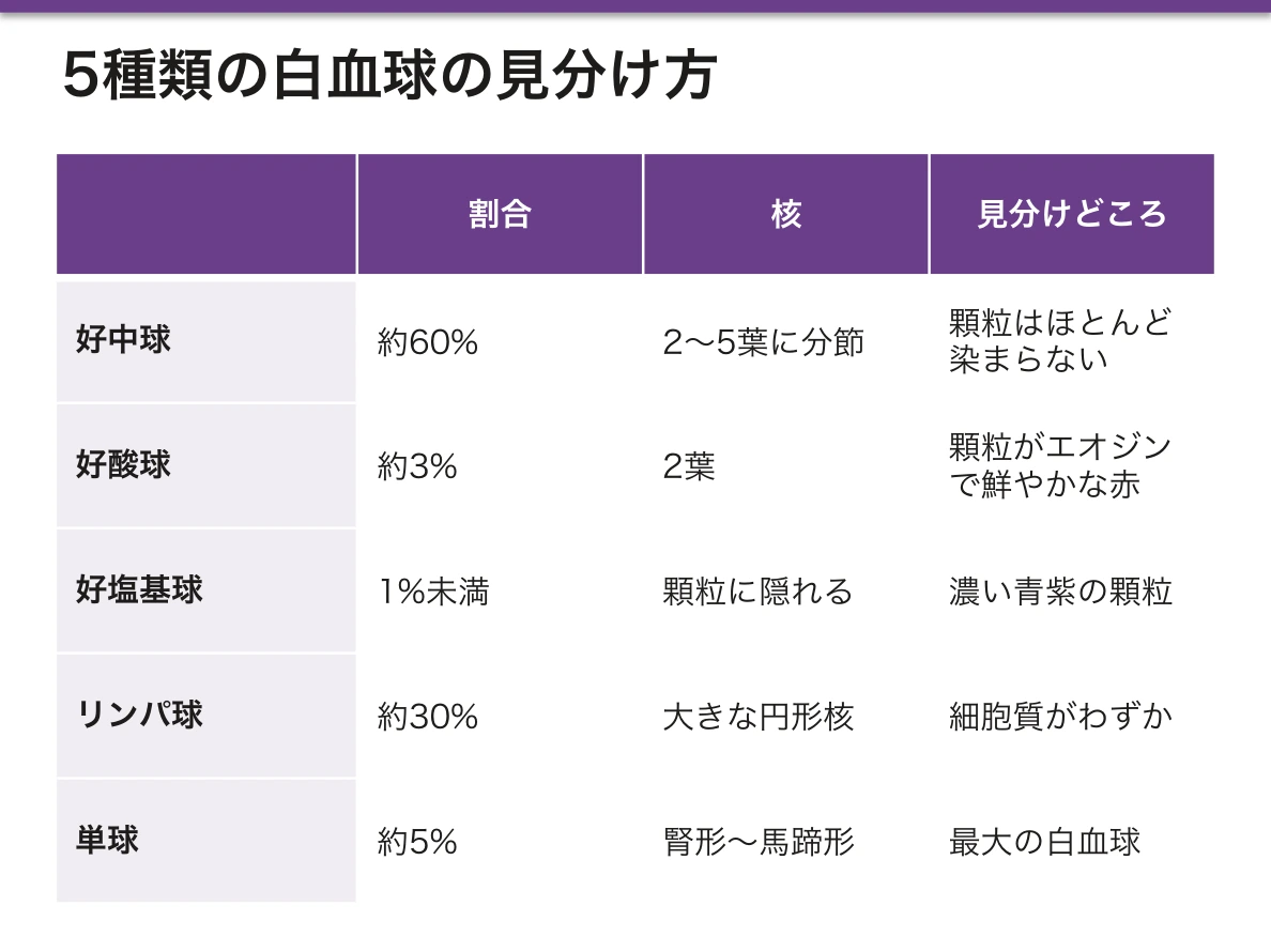
- 好酸球(約3%)——顆粒がエオジンで鮮やかな赤、核は2葉。寄生虫・アレルギーで増える
- 好塩基球(<1%)——濃い青紫の顆粒が核を隠す。ヒスタミン・ヘパリンを持ち、肥満細胞に似るが別の細胞(好塩基球は血中、肥満細胞は組織)
- リンパ球(約30%)——大きな円形核が細胞質をほぼ占める。T細胞・B細胞・NK細胞。形では区別できない
- 単球(約5%)——最大の白血球。腎形〜馬蹄形の核。組織へ出てマクロファージになる
好酸球増多の鑑別は語呂で押さえられる——NAACP:Neoplasm(腫瘍)/Allergy/Asthma/Connective tissue disease(膠原病・血管炎)/Parasite(寄生虫)。ただし組織内に留まる寄生虫でこそ増えるのであって、腸管内にいるだけの蟯虫では増えない点が問われる。
好酸球の顆粒には主要塩基性蛋白(MBP)が入っており、寄生虫を殺すと同時に自分の気道上皮も傷つける。喘息の気道リモデリングに好酸球が関わるのはこのためで、抗IL-5抗体が重症喘息に効く根拠になる。
ステロイドは好中球を増やし、リンパ球・好酸球を減らす。好中球が増えるのは骨髄からの動員と血管壁からの脱離(marginated pool の遊離)であって、感染ではない——ステロイド投与中の白血球増多を感染と誤読しない。
どれが増えているかで、原因の見当がつく。
好酸球増多 — アレルギー・寄生虫・薬剤・膠原病(好酸球性多発血管炎性肉芽腫症)・副腎不全・血液腫瘍。「アレルギーと寄生虫」からまず考える。ステロイドで急速に減る。
リンパ球増多 — ウイルス感染・百日咳・結核・慢性リンパ性白血病。異型リンパ球が目立てば伝染性単核球症(EBV)を疑う。アモキシシリンを投与すると高率に皮疹が出るので、咽頭炎の抗菌薬選択で問題になる。
単球増多 — 結核・感染性心内膜炎・慢性炎症。
好塩基球増多 — 慢性骨髄性白血病を強く示唆する。数少ない特異性の高い所見である。
リンパ球減少 — HIV・ステロイド・放射線・重症感染。
English version
Core
- Eosinophil (about 3 per cent) — granules stain brilliant red with eosin; the nucleus is bilobed. Increased in parasitic infestation and allergy.
- Basophil (under 1 per cent) — deep blue-purple granules obscuring the nucleus. Contains histamine and heparin; resembles the mast cell but is a different cell (basophil in blood, mast cell in tissue).
- Lymphocyte (about 30 per cent) — a large round nucleus filling almost the whole cell. T, B and NK cells, indistinguishable by morphology.
- Monocyte (about 5 per cent) — the largest white cell, with a kidney-shaped or horseshoe nucleus. Becomes a macrophage on entering tissue.
Going further
The differential of eosinophilia can be held with the mnemonic NAACP: Neoplasm, Allergy, Asthma, Connective tissue disease (including vasculitis), Parasite. Note that it is parasites within tissue that raise the count; a threadworm living in the lumen alone does not.
The eosinophil granules contain major basic protein, which kills parasites and also damages the patient’s own airway epithelium. This is why eosinophils contribute to airway remodelling in asthma, and the basis for anti-IL-5 antibodies in severe asthma.
Corticosteroids raise the neutrophil count and lower lymphocytes and eosinophils. The neutrophils rise through release from the marrow and detachment from the vessel wall (the marginated pool), not because of infection — do not misread the leucocytosis of a patient on steroids as infection.
In the clinic
Which cell is raised suggests the cause.
Eosinophilia — allergy, parasites, drugs, connective tissue disease (eosinophilic granulomatosis with polyangiitis), adrenal insufficiency, haematological malignancy. Think first of allergy and parasites. It falls rapidly with corticosteroids.
Lymphocytosis — viral infection, pertussis, tuberculosis, chronic lymphocytic leukaemia. Prominent atypical lymphocytes suggest infectious mononucleosis (EBV); amoxicillin causes a rash in a high proportion, which matters when choosing an antibiotic for pharyngitis.
Monocytosis — tuberculosis, infective endocarditis, chronic inflammation.
Basophilia — strongly suggests chronic myeloid leukaemia; one of the few highly specific findings.
Lymphopenia — HIV, corticosteroids, radiation, severe infection.
血小板Thrombocytus
細胞ではなく、巨核球の細胞質がちぎれた断片。核が無い。直径2〜3μm、寿命約10日。
一次止血を担う。血管が破れるとvon Willebrand 因子(vWF)を介して露出したコラーゲンに粘着し、活性化して凝集し血小板血栓を作る。
顆粒には ADP・セロトニン・トロンボキサンA₂・第V因子などが入り、放出してさらに血小板を呼ぶ。
止血を一次と二次に分けると、出血の様子から障害部位が読める。
- 一次止血の障害(血小板・vWF)——皮膚粘膜出血:点状出血・紫斑・鼻出血・歯肉出血・過多月経。出血時間延長
- 二次止血の障害(凝固因子)——深部出血:関節内出血・筋肉内血腫。血友病が典型。APTT/PT延長
点状出血なら血小板、関節出血なら凝固因子——この対応は国試で繰り返し問われる。
薬も作用点で分かれる。アスピリンはCOX-1を不可逆的に阻害してトロンボキサンA₂を止める。血小板は核が無く酵素を作り直せないため、効果が血小板の寿命(約10日)続く。手術前に休薬期間が要るのはこのためで、「核が無い」という組織学的事実が休薬日数を決めている。
血小板は数だけでなく、働きも見る。
von Willebrand 病 — 最も多い遺伝性出血性疾患。血小板数は正常だが粘着できない。粘膜出血・鼻出血・過多月経。vWF は第 VIII 因子の担体でもあるのでAPTT が延長することがある。
特発性血小板減少性紫斑病 — 抗血小板抗体で脾で破壊される。骨髄では巨核球が増えていることが産生低下(再生不良性貧血)との鑑別点である。
血栓性血小板減少性紫斑病 — ADAMTS13 の欠損でvWF の巨大分子が切れず、微小血栓ができる。血小板減少・溶血性貧血(破砕赤血球)・腎障害・発熱・神経症状の5徴。血小板輸血は禁忌で、血漿交換が治療になる。
アスピリンはシクロオキシゲナーゼを不可逆に阻害するので、核の無い血小板は回復できず、効果が寿命(約10日)続く。
English version
Core
Not a cell but a fragment of megakaryocyte cytoplasm. It has no nucleus; 2–3 µm across, lifespan about 10 days. It performs primary haemostasis.
When a vessel breaks it adheres to exposed collagen through von Willebrand factor, activates, aggregates and forms a platelet plug. Its granules contain ADP, serotonin, thromboxane A₂ and factor V, which are released to recruit further platelets.
Going further
Divide haemostasis into primary and secondary and the site of the defect can be read from the pattern of bleeding.
- Defect of primary haemostasis (platelets, vWF) — mucocutaneous bleeding: petechiae, purpura, epistaxis, gingival bleeding, menorrhagia. Prolonged bleeding time.
- Defect of secondary haemostasis (clotting factors) — deep bleeding: haemarthrosis, muscle haematoma. Haemophilia is typical. Prolonged APTT or PT.
Petechiae mean platelets; joint bleeding means clotting factors — a correspondence asked repeatedly.
The drugs divide by their point of action. Aspirin irreversibly inhibits COX-1 and stops thromboxane A₂. Because the platelet has no nucleus and cannot remake the enzyme, the effect lasts the lifespan of the platelet (about 10 days). This is why a washout period is needed before surgery — a histological fact, the absence of a nucleus, determines the number of days.
In the clinic
Look at platelet function as well as number.
von Willebrand disease — the commonest inherited bleeding disorder. The platelet count is normal but the platelets cannot adhere. Mucosal bleeding, epistaxis, menorrhagia. vWF also carries factor VIII, so the APTT may be prolonged.
Immune thrombocytopenic purpura — antiplatelet antibodies cause destruction in the spleen. Increased megakaryocytes in the marrow distinguish it from failure of production (aplastic anaemia).
Thrombotic thrombocytopenic purpura — deficiency of ADAMTS13 leaves ultra-large vWF multimers uncleaved and microthrombi form. The pentad of thrombocytopenia, haemolytic anaemia (schistocytes), renal impairment, fever and neurological signs. Platelet transfusion is contraindicated; plasma exchange is the treatment.
Aspirin inhibits cyclooxygenase irreversibly, and the anucleate platelet cannot recover, so the effect lasts the lifespan of about 10 days.
造血Haemopoësis
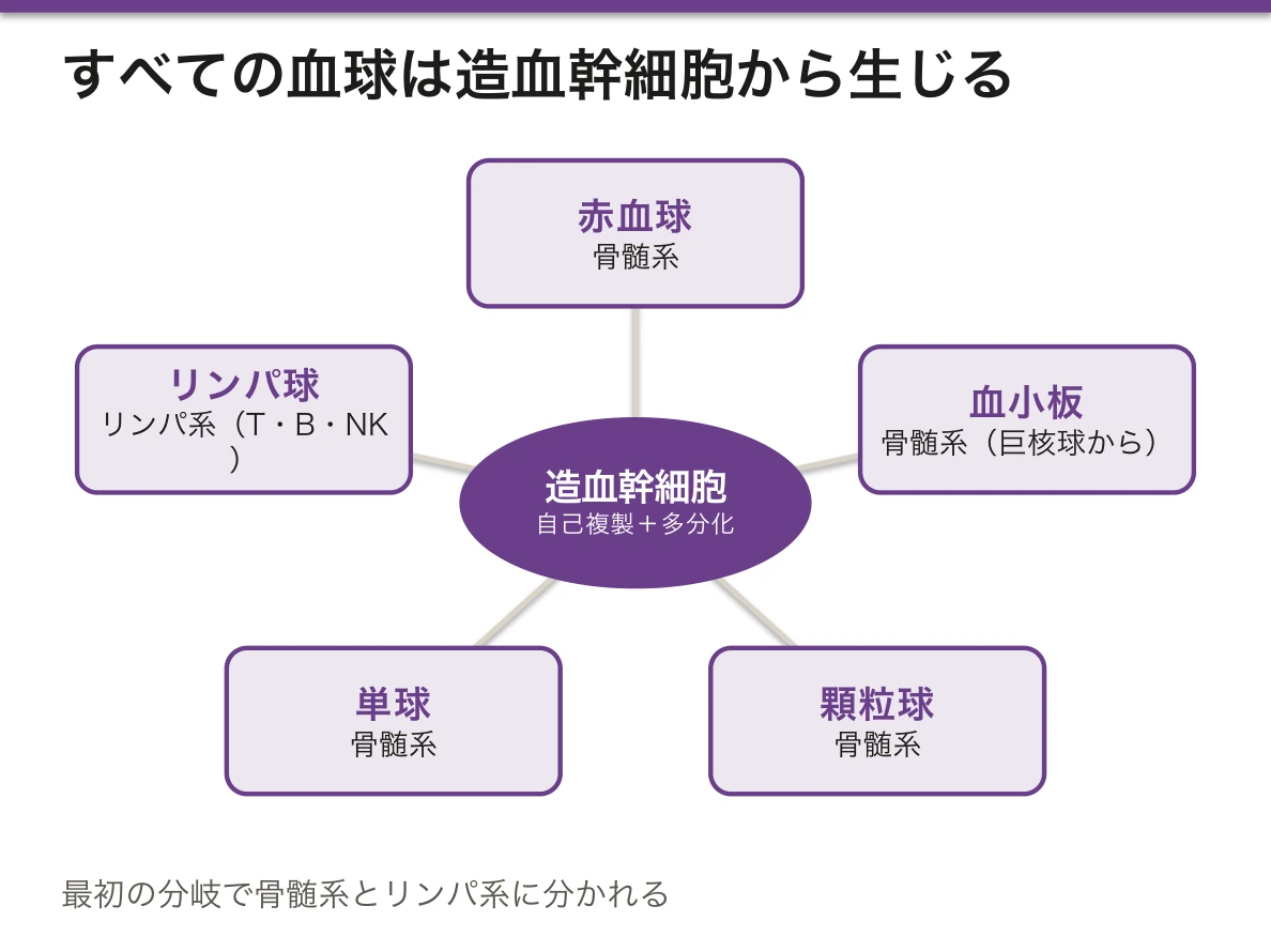
すべての血球は1種類の造血幹細胞(HSC)から生じる。HSC は自己複製能と多分化能を併せ持つ。
最初の分岐で骨髄系(赤血球・血小板・顆粒球・単球)とリンパ系(T・B・NK)に分かれる。
造血の場は年齢で移る——卵黄嚢(〜2か月)→ 肝・脾(2〜7か月)→ 骨髄(5か月〜生涯)。
成人では造血する骨髄(赤色骨髄)が体幹に限られる——椎体・胸骨・肋骨・腸骨・頭蓋。四肢の長管骨は黄色骨髄(脂肪)に置き換わる。骨髄穿刺を腸骨や胸骨で行うのはこのためである。
強い造血需要があると、黄色骨髄が赤色に戻り、それでも足りなければ髄外造血が起きる。胎生期に造血していた肝・脾が再び動員されるので、肝脾腫として現れる。サラセミア・骨髄線維症・大理石骨病でみられる。
幹細胞は骨髄のニッチ(骨内膜側と血管周囲)で維持される。骨芽細胞と細網細胞が作る微小環境であり、造血は骨と不可分——ここで骨の章と繋がる。G-CSF が幹細胞をニッチから引き剥がして末梢血へ動員する仕組みが、末梢血幹細胞移植の基礎になっている。
どの段階で止まるかが、病名を決める。
造血幹細胞そのものが減る — 再生不良性貧血。汎血球減少で、骨髄は脂肪髄になる。
分化が途中で止まって未熟な細胞が増える — 急性白血病。骨髄の芽球 20%以上。
分化はするが質が悪い — 骨髄異形成症候群。血球減少があるのに骨髄は過形成という無効造血が特徴である。
成人の造血は扁平骨(腸骨・胸骨・椎体・肋骨)に限られる。だから骨髄穿刺は後腸骨稜で行う。小児では長管骨にも造血がある。
髄外造血——骨髄が使えなくなると肝・脾で造血が再開する。骨髄線維症・重症溶血性貧血・サラセミアで肝脾腫になる理由である。
G-CSF は顆粒球系を、エリスロポエチンは赤芽球系を刺激する。
English version
Core
All blood cells arise from a single kind of haemopoietic stem cell, which combines self-renewal with multipotency.
The first division separates the myeloid line (red cells, platelets, granulocytes, monocytes) from the lymphoid line (T, B, NK).
The site of haemopoiesis moves with age — yolk sac (to 2 months) → liver and spleen (2–7 months) → bone marrow (5 months onwards, for life).
Going further
In the adult, haemopoietic (red) marrow is confined to the trunk — vertebrae, sternum, ribs, ilium, skull. The long bones of the limbs are replaced by yellow (fatty) marrow. This is why marrow is aspirated from the ilium or sternum.
Under strong demand yellow marrow reverts to red, and if that is still not enough extramedullary haemopoiesis begins: the liver and spleen, which produced blood in fetal life, are recruited again, presenting as hepatosplenomegaly. Seen in thalassaemia, myelofibrosis and osteopetrosis.
Stem cells are maintained in niches in the marrow (endosteal and perivascular), a microenvironment created by osteoblasts and reticular cells. Haemopoiesis is inseparable from bone — and here this chapter joins the chapter on bone. That G-CSF prises stem cells out of the niche and mobilises them into the blood is the basis of peripheral blood stem cell transplantation.
In the clinic
Where the process stops determines the name of the disease.
- The stem cells themselves are depleted — aplastic anaemia: pancytopenia with a fatty marrow.
- Differentiation halts and immature cells accumulate — acute leukaemia: 20 per cent or more blasts in the marrow.
- Differentiation occurs but the product is defective — myelodysplastic syndrome, characterised by ineffective haemopoiesis: cytopenia despite a hypercellular marrow.
Adult haemopoiesis is confined to flat bones (ilium, sternum, vertebrae, ribs), which is why marrow is aspirated from the posterior iliac crest. In children the long bones are still haemopoietic.
Extramedullary haemopoiesis — when the marrow can no longer function, the liver and spleen resume production, which is why myelofibrosis, severe haemolytic anaemia and thalassaemia cause hepatosplenomegaly.
造血の階層 — 幹細胞・前駆細胞・CFU を区別する
造血は3つの段に分けて考えると整理しやすい。
| 段 | 例 | 自己複製 | 分化できる範囲 | 塗抹で名前が付くか |
|---|---|---|---|---|
| 造血幹細胞(HSC) | — | 生涯つづく | すべての血球 | 付かない(小リンパ球に似た目立たない細胞) |
| 前駆細胞(CFU で呼ぶ) | CFU-GEMM、CFU-GM、BFU-E→CFU-E、CFU-Meg、リンパ系前駆細胞(CFU-L) | ほとんど無い | 段を下るごとに狭くなる | 付かない |
| 前駆体(芽球から成熟途中まで) | 前赤芽球・骨髄芽球・単芽球・巨核芽球 | 無い(分裂はする) | 1系統に決まっている | 付く |
CFU(colony-forming unit、コロニー形成単位)はもともと実験から生まれた名前である。骨髄の細胞を半固形の培地にまき、サイトカインを加えると、1個の細胞が分裂して1つの細胞の集団(コロニー)を作る。そのコロニーにどの血球が混ざっているかを見て、元の細胞の能力を後から名付けた。顆粒球(G)・赤血球(E)・単球(M)・巨核球(M)がすべて出れば CFU-GEMM、顆粒球と単球だけなら CFU-GM である。
骨髄系の枝分かれは、CFU-GEMM(共通骨髄系前駆細胞)から顆粒球・単球の枝(CFU-GM → CFU-G・CFU-M、別に好酸球系・好塩基球系)と赤血球・巨核球の枝(BFU-E → CFU-E、CFU-Meg)に分かれる。リンパ系は CFU-L から T・B・NK に分かれる。
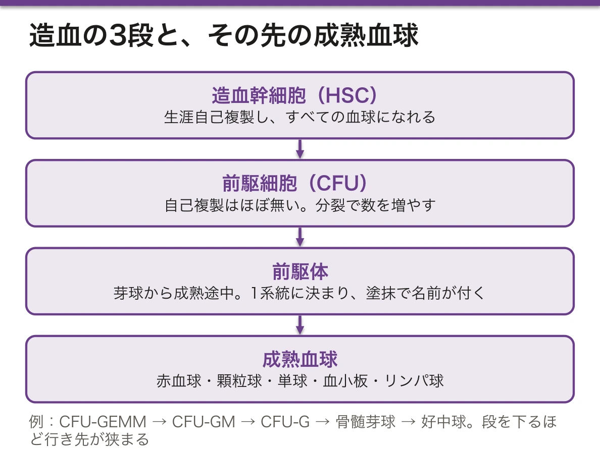
なぜ段を分けるのか。幹細胞はめったに分裂しない。分裂のたびに DNA の複製の誤りがたまり、数も使い減るからである。そこで数を増やす仕事は前駆細胞が引き受ける。前駆細胞は自己複製をほぼ捨てる代わりに、何回も分裂して数を何桁も増やす。こうして、少数の幹細胞を守りながら、成人で毎日およそ 2,000 億個の赤血球を作ることができる。
コロニーを作らせるときに加えた因子が、そのまま薬の名前になっている。G-CSF(顆粒球コロニー刺激因子)、GM-CSF、M-CSF の「コロニー刺激」とは、この培養でコロニーを増やした働きのことである。
胎生期の造血も、場所で型を分ける。卵黄嚢の血島(受精後3週ころから)→ 大動脈周囲の領域(AGM 領域)→ 肝(6週ころから出生まで)→ 脾 → 骨髄(最初は鎖骨で、2か月半ころ)。卵黄嚢で作られる最初の赤血球は核を持ち、胎芽型のヘモグロビンを含む。
どの段が侵されるかで、減る血球・増える血球の組み合わせが決まる。
パルボウイルス B19 は赤血球系の前駆細胞(BFU-E・CFU-E から前赤芽球)にだけ感染して壊す。赤血球の寿命は約120日あるので健康な人では目立たないが、遺伝性球状赤血球症のように寿命が短い人では急に重い貧血になる(無形成発作)。白血球と血小板は保たれる——前駆細胞の段で1系統だけが止まったからである。
反対に幹細胞の段の病気では、複数の系統が同時に動く。慢性骨髄性白血病(BCR-ABL)では顆粒球だけでなく好塩基球・好酸球・血小板も増え、真性多血症(JAK2 変異)では赤血球に加えて白血球と血小板も増える。異常が幹細胞にあるので、その子孫すべてに異常が受け継がれるからである。
English version
Core
It helps to think of haemopoiesis in three levels.
| Level | Examples | Self-renewal | Range of fates | Named on a smear? |
|---|---|---|---|---|
| Haemopoietic stem cell (HSC) | — | lifelong | all blood cells | no (looks like a small lymphocyte) |
| Progenitor (named as CFU) | CFU-GEMM, CFU-GM, BFU-E→CFU-E, CFU-Meg, lymphoid progenitor (CFU-L) | almost none | narrows at each step | no |
| Precursor (blast to maturing cell) | proerythroblast, myeloblast, monoblast, megakaryoblast | none (but it divides) | one lineage only | yes |
CFU (colony-forming unit) is originally a name from an experiment. Marrow cells are put in a semi-solid medium with cytokines. One cell divides and forms one colony. The cell types inside the colony tell us, afterwards, what the first cell could make. If granulocytes, erythrocytes, monocytes and megakaryocytes all appear, it was a CFU-GEMM; if only granulocytes and monocytes, a CFU-GM.
In the myeloid line, CFU-GEMM (common myeloid progenitor) splits into a granulocyte–monocyte branch (CFU-GM → CFU-G and CFU-M, plus separate eosinophil and basophil lines) and an erythrocyte–megakaryocyte branch (BFU-E → CFU-E, and CFU-Meg). The lymphoid line starts from CFU-L and gives T, B and NK cells.
Going further
Why have levels? Stem cells divide rarely. Each division adds DNA copying errors and uses the cell up. So the job of making numbers goes to the progenitors. They give up self-renewal, but divide many times and multiply the number by several orders of magnitude. In this way a small, protected pool of stem cells can supply about 200 billion red cells every day in an adult.
The factors added to the culture became the names of drugs. The "colony-stimulating" in G-CSF, GM-CSF and M-CSF means exactly this: they increased colonies in the dish.
Fetal haemopoiesis is also classified by site: yolk sac blood islands (from about week 3), the AGM region around the aorta, the liver (from about week 6 until birth), the spleen, and the bone marrow (first in the clavicle, at about 2.5 months). The first red cells from the yolk sac keep their nucleus and carry embryonic haemoglobin.
In the clinic
Which level is hit decides which combination of blood cells falls or rises.
Parvovirus B19 infects and kills only erythroid progenitors (BFU-E/CFU-E to proerythroblast). Red cells live about 120 days, so a healthy person hardly notices. But in a person with short red-cell survival, such as hereditary spherocytosis, severe anaemia appears suddenly (aplastic crisis). White cells and platelets stay normal, because only one line stopped at the progenitor level.
In diseases of the stem cell, several lines move together. In chronic myeloid leukaemia (BCR-ABL), basophils, eosinophils and platelets rise as well as neutrophils. In polycythaemia vera (JAK2 mutation), white cells and platelets rise as well as red cells. The defect is in the stem cell, so all its descendants inherit it.
赤血球造血 — 青から赤へ、そして核を捨てるまでErythropoësis
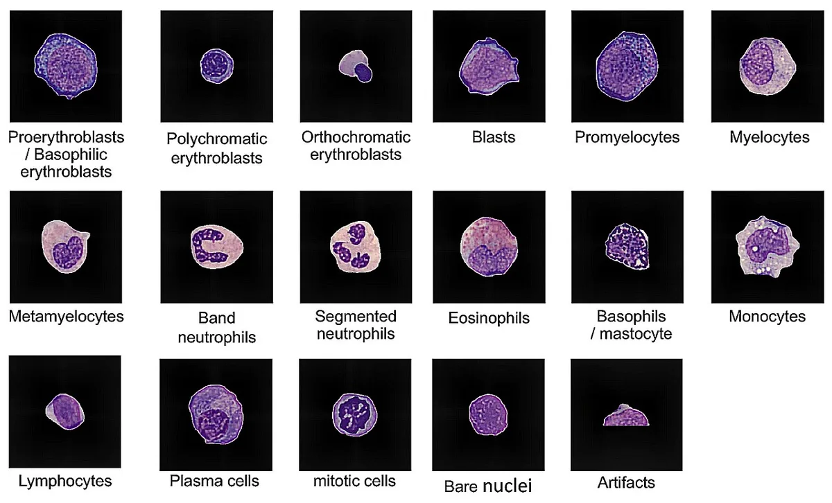
赤血球は前赤芽球 → 好塩基性赤芽球 → 多染性赤芽球 → 正染性赤芽球 → 網赤血球 → 赤血球の順に成熟する。前赤芽球から網赤血球まで約1週間かかる。
| 段階 | 大きさ・核 | 細胞質の色(May-Giemsa) | 起きていること |
|---|---|---|---|
| 前赤芽球 | 最も大きい。核小体のある大きな核 | 濃い青 | リボソームを大量に作る |
| 好塩基性赤芽球 | やや小さい。クロマチンが粗くなる | 濃い青 | ヘモグロビン合成が始まる |
| 多染性赤芽球 | さらに小さい。核が濃くなる | 青灰色〜紫 | ヘモグロビンが増え、リボソームが減る。分裂はこの段階まで |
| 正染性赤芽球 | 小さい。核は濃縮して小さく黒い | ほぼピンク | 核を細胞の外へ押し出す(脱核) |
| 網赤血球 | 核なし。成熟赤血球よりやや大きい | ピンク(超生体染色で網状の RNA が見える) | 骨髄に1〜2日、血中で約1日のあいだ RNA が残る |
| 赤血球 | 直径約 7.5 µm、核なし | ピンク・中央が淡い | 寿命約120日 |
骨髄では赤芽球が1個のマクロファージを囲んで島(赤芽球島)を作る。中心のマクロファージは鉄を渡し、捨てられた核を食べる。
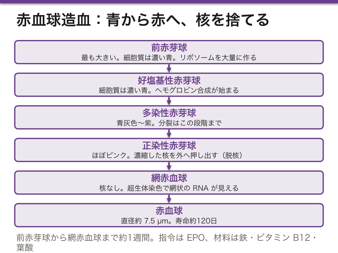
色の変化には理由がある。リボソーム(RNA)は塩基性色素で青く、ヘモグロビンは酸性色素でピンクに染まる。成熟が進むと、蛋白を作る道具(リボソーム)が減り、作った製品(ヘモグロビン)が増える。だから青 → 灰紫 → ピンクと色が移る。色を読めば、成熟の段階が読める。
調節する因子は2種類に分けると覚えやすい。指令はエリスロポエチン(EPO)で、腎皮質の尿細管周囲の間質細胞が低酸素を感じて作る(HIF を介する)。EPO は主に CFU-E から早い時期の赤芽球に働き、細胞が死なずに分裂を続けられるようにする。材料は、ヘモグロビンのための鉄と、DNA を合成するためのビタミン B12 と葉酸である。
網赤血球の数は、貧血を2つに分ける。多ければ骨髄は働いていて、赤血球が壊れるか失われている(溶血・出血)。少なければ作る側(骨髄・EPO・材料)に問題がある。塗抹で青みのある大きな赤血球(多染性)が多いのは、網赤血球が増えている印である。
材料が足りないと、核と細胞質の成熟がずれる。B12・葉酸の欠乏では DNA 合成だけが遅れ、ヘモグロビンは作られ続ける。核は若いのに細胞質は成熟している大きな細胞(巨赤芽球)ができ、多くが骨髄の中で壊れる(無効造血。LDH と間接ビリルビンが上がる)。鉄欠乏では反対にヘモグロビンが足りず、細胞は分裂を余分に重ねて小さく淡くなる。
慢性腎臓病では EPO が作れないので貧血になり、赤血球造血刺激因子製剤で治療する。
English version
Core
Red cells mature in this order: proerythroblast → basophilic erythroblast → polychromatophilic erythroblast → orthochromatophilic erythroblast → reticulocyte → erythrocyte. From proerythroblast to reticulocyte takes about one week.
| Stage | Size and nucleus | Cytoplasm (May-Giemsa) | What happens |
|---|---|---|---|
| Proerythroblast | largest; big nucleus with nucleoli | deep blue | makes many ribosomes |
| Basophilic erythroblast | a little smaller; coarser chromatin | deep blue | haemoglobin synthesis starts |
| Polychromatophilic erythroblast | smaller; darker nucleus | blue-grey to purple | more haemoglobin, fewer ribosomes; last stage that divides |
| Orthochromatophilic erythroblast | small; condensed dark nucleus | almost pink | pushes the nucleus out (enucleation) |
| Reticulocyte | no nucleus; slightly larger than a mature cell | pink (supravital stain shows an RNA network) | keeps RNA for 1–2 days in marrow and about 1 day in blood |
| Erythrocyte | about 7.5 µm, no nucleus | pink, pale centre | lives about 120 days |
In the marrow, erythroblasts form an island around one macrophage. The central macrophage hands over iron and eats the discarded nuclei.
Going further
The colour change has a reason. Ribosomes (RNA) take basic dye and look blue; haemoglobin takes acidic dye and looks pink. As the cell matures, the tools (ribosomes) decrease and the product (haemoglobin) increases. So the colour moves blue → grey-purple → pink. Reading the colour means reading the stage.
Split the regulating factors into two groups. The order is erythropoietin (EPO), made by peritubular interstitial cells of the renal cortex when they sense hypoxia (through HIF). EPO acts mainly on CFU-E and early erythroblasts and lets them survive and keep dividing. The materials are iron for haemoglobin and vitamin B12 and folate for DNA synthesis.
In the clinic
The reticulocyte count splits anaemia in two. If it is high, the marrow is working and red cells are destroyed or lost (haemolysis, bleeding). If it is low, the problem is on the production side (marrow, EPO, materials). Many large bluish red cells on a smear (polychromasia) mean more reticulocytes.
When materials are missing, nuclear and cytoplasmic maturation separate. In B12 or folate deficiency only DNA synthesis is slow, while haemoglobin is still made. Large cells with a young nucleus and a mature cytoplasm (megaloblasts) appear, and many die inside the marrow (ineffective erythropoiesis; LDH and indirect bilirubin rise). In iron deficiency it is the reverse: haemoglobin is short, the cell divides extra times, and becomes small and pale.
In chronic kidney disease EPO is not made, so anaemia develops; it is treated with erythropoiesis-stimulating agents.
顆粒球造血と単球造血 — 特殊顆粒が出た段階で、系統が決まるGranulocytopoësis
顆粒球は骨髄芽球から始まり、前骨髄球・骨髄球・後骨髄球・桿状核球を経て分葉核球になる。骨髄芽球から成熟まで約2週間である。
| 段階 | 核 | 顆粒 |
|---|---|---|
| 骨髄芽球 | 大きく丸い。核小体が数個 | 無い。細胞質は青い |
| 前骨髄球 | 丸〜楕円。最も大きい細胞 | アズール顆粒(一次顆粒=リソソーム、ミエロペルオキシダーゼを含む)が現れる |
| 骨髄球 | 丸く、やや偏る | 特殊顆粒(二次顆粒)が初めて現れ、好中・好酸・好塩基の3系統に分かれる。分裂はここまで |
| 後骨髄球 | 腎臓形にくぼむ | 特殊顆粒が主体 |
| 桿状核球 | 馬蹄形・棒状 | 同上(三次顆粒も加わる) |
| 分葉核球 | 2〜5葉 | 成熟 |
単球は CFU-GM → CFU-M → 単芽球 → 前単球 → 単球と成熟する。単球は血中に1〜3日いたあと組織へ出て、マクロファージになる。破骨細胞と多くの樹状細胞も同じ系統から生じる。
因子は、IL-3 と GM-CSF が早い段階を広く支え、G-CSF が好中球、IL-5 が好酸球、M-CSF が単球を進める。
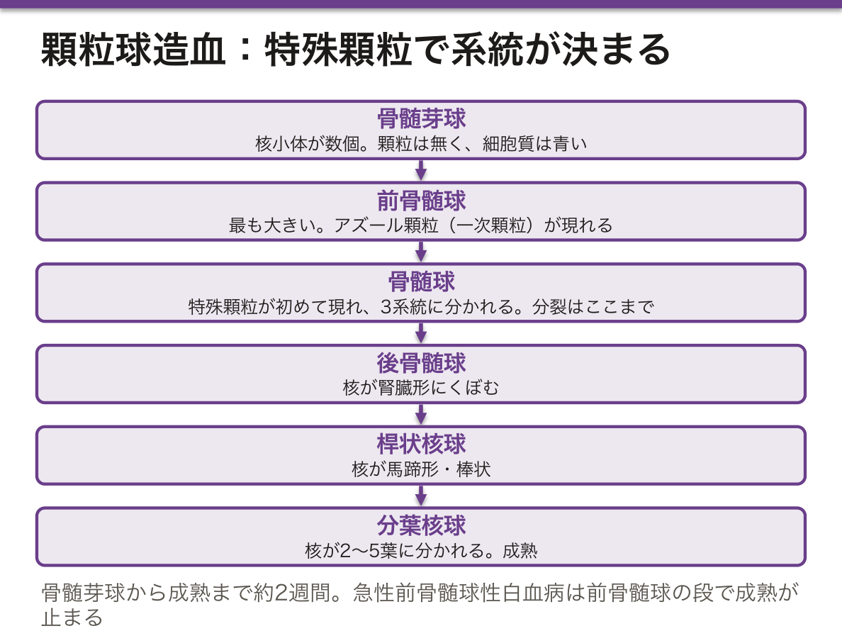
系統の見分けは特殊顆粒が出る骨髄球の段階で初めてできる。それより前の骨髄芽球と前骨髄球は、どの顆粒球になるかを形では言えない。アズール顆粒は前骨髄球の段階でしか作られないので、その後の分裂で娘細胞に分けられて薄まり、成熟好中球では少なく見える。
骨髄の顆粒球は2つの池に分かれている。骨髄芽球から骨髄球までの分裂する池と、後骨髄球から分葉核球までの成熟・貯蔵の池である。貯蔵の池には血中の好中球の何倍もの細胞が控えていて、感染があるとまずここから放出される。血中の好中球は数時間で組織へ出ていく。
急性前骨髄球性白血病では前骨髄球の段で成熟が止まる。アズール顆粒が集まった針状のAuer 小体を持ち、顆粒の中身が凝固を起こすのでDICを合併しやすい。全トランス型レチノイン酸で止まった成熟を先へ進めると、細胞は好中球へ分化して治る——段階で止まった病気を、段階を進めて治す例である。
慢性骨髄性白血病では骨髄芽球から分葉核球まですべての段階が末梢血に並ぶ。ミエロペルオキシダーゼ染色が芽球に陽性なら骨髄系、陰性ならリンパ系を考える。
抗癌薬のあと好中球が最も減るのは投与後おおよそ1〜2週である。貯蔵の池が使い切られるまで時間がかかるからで、この時期の発熱に G-CSF と抗菌薬で備える。
English version
Core
Granulocytes mature as myeloblast → promyelocyte → myelocyte → metamyelocyte → band → segmented cell. From myeloblast to maturity takes about two weeks.
| Stage | Nucleus | Granules |
|---|---|---|
| Myeloblast | large, round; several nucleoli | none; blue cytoplasm |
| Promyelocyte | round to oval; the largest cell | azurophilic granules (primary granules = lysosomes with myeloperoxidase) appear |
| Myelocyte | round, slightly eccentric | specific (secondary) granules appear for the first time and the neutrophil, eosinophil and basophil lines separate; last stage that divides |
| Metamyelocyte | kidney-shaped | mainly specific granules |
| Band cell | horseshoe or rod | same (tertiary granules added) |
| Segmented cell | 2–5 lobes | mature |
Monocytes mature as CFU-GM → CFU-M → monoblast → promonocyte → monocyte. A monocyte stays in blood for 1–3 days, then enters tissue and becomes a macrophage. Osteoclasts and many dendritic cells come from the same line.
Factors: IL-3 and GM-CSF support the early stages broadly; G-CSF drives neutrophils, IL-5 eosinophils and M-CSF monocytes.
Going further
The lineage can be told only from the myelocyte stage, when specific granules appear. Before that, the myeloblast and promyelocyte do not show which granulocyte they will become. Azurophilic granules are made only in the promyelocyte, so later divisions share them between daughter cells and they look sparse in the mature neutrophil.
Marrow granulocytes sit in two pools: a dividing pool (myeloblast to myelocyte) and a maturation–storage pool (metamyelocyte to segmented cell). The storage pool holds several times the number of neutrophils in blood, and in infection it is released first. Neutrophils leave the blood for the tissues within hours.
In the clinic
In acute promyelocytic leukaemia maturation stops at the promyelocyte. The cells carry needle-like Auer rods (fused azurophilic granules), and the granule contents trigger clotting, so DIC is common. All-trans retinoic acid pushes the blocked maturation forward and the cells become neutrophils — a disease stuck at one stage is treated by moving it to the next.
In chronic myeloid leukaemia every stage from myeloblast to segmented cell appears in the blood. If blasts are positive for myeloperoxidase, think myeloid; if negative, think lymphoid.
After chemotherapy, neutrophils fall lowest roughly 1–2 weeks after the dose, because the storage pool takes that long to run out. Fever in this period is covered with G-CSF and antibiotics.
赤血球膜 — 形を保つ骨組みが壊れると、脾で壊されるMembrana erythrocytica
両凹円板の形は、膜の裏に張られた蛋白の網(膜骨格)が作っている。
- スペクトリン(αとβ)が網を作る
- アンキリン・バンド3・プロテイン4.2 が、網を脂質二重層につなぎ止める(垂直方向)
- プロテイン4.1・アクチンが、網どうしをつなぐ(水平方向)
両凹円板であることの意味——表面積に余裕があるので変形でき、直径 8 μm の細胞が 3 μm の毛細血管を通れる。
膜蛋白が血液型抗原にもなる——ABO は糖鎖、Rh は膜貫通蛋白。
グリコホリンが表面の負電荷(シアル酸)を作り、赤血球どうしが反発する。
「壊れる方向」で病名が分かれるのが、この項の要点になる。
| 壊れる向き | 起こること | 病名 |
|---|---|---|
| 垂直(膜と骨格の連結) | 膜が小胞として失われ、球状になる | 遺伝性球状赤血球症 |
| 水平(骨格どうしの連結) | 形が保てず楕円になる | 遺伝性楕円赤血球症 |
(溶血の章)——「連結の方向が、そのまま細胞の形になる」。
球状になると、表面積の余裕が失われる。変形できなくなり、脾の索を通り抜けられずに捕まって壊される(血管外溶血)(脾臓の章)——「脾摘が治療になるのは、壊す場所を取り除いているから」。
浸透圧抵抗試験が診断に使われるのも、同じ理屈になる——「余裕が無い細胞は、少し水を吸っただけで破裂する」。
グリコホリンが作る負電荷は、赤沈の速さを決めている。炎症でフィブリノゲンや免疫グロブリンが増えると反発が打ち消され、連銭形成が起きて沈むのが速くなる(血算を読む章)——「赤沈は、蛋白の変化を赤血球の動きで見ている検査になる」。
マラリア原虫は、この膜蛋白を入口として使う(寄生虫の章)——「Duffy 抗原を持たない人が、三日熱マラリアに感染しにくい」。
膜骨格が壊れると、脾で壊される。変形能を失った赤血球が脾洞の細い隙間を通れないためである。
遺伝性球状赤血球症 — スペクトリン・アンキリンの異常。球状赤血球・MCHC 高値・浸透圧脆弱性亢進・網赤血球増加。脾摘で溶血が止まる——壊す場所を除くのが治療になる。胆石(ビリルビン結石)を合併しやすい。
発作性夜間ヘモグロビン尿症 — GPI アンカーの欠損で CD55・CD59 が膜に留まらず、補体で壊れる。血管内溶血・血栓症・血球減少。抗補体薬(エクリズマブ)が効くが髄膜炎菌ワクチンが必須である。
末梢血塗抹の赤血球の形が診断そのものになる。破砕赤血球=微小血管障害、標的赤血球=サラセミア・肝疾患、涙滴赤血球=骨髄線維症、連銭形成(rouleaux)=多発性骨髄腫。
English version
Core
The biconcave shape is created by a network of proteins beneath the membrane (the membrane skeleton).
- Spectrin (α and β) forms the network.
- Ankyrin, band 3 and protein 4.2 tie the network to the lipid bilayer (vertical connections).
- Protein 4.1 and actin tie the network to itself (horizontal connections).
The point of being a biconcave disc is that there is surplus surface area, so the cell can deform: an 8 µm cell passes through a 3 µm capillary.
Membrane proteins also carry the blood group antigens — ABO is carbohydrate, Rh is a transmembrane protein. Glycophorin provides the surface negative charge (sialic acid) so that red cells repel one another.
Going further
The point of this section is that the direction in which it breaks determines the disease.
| Direction of failure | What happens | Disease |
|---|---|---|
| Vertical (skeleton to membrane) | Membrane is lost as vesicles and the cell becomes spherical | Hereditary spherocytosis |
| Horizontal (skeleton to skeleton) | The shape cannot be held and becomes elliptical | Hereditary elliptocytosis |
The direction of the connection is directly the shape of the cell.
Once spherical, the surplus surface area is gone. The cell can no longer deform, is trapped in the cords of the spleen and destroyed (extravascular haemolysis) — splenectomy is a treatment because it removes the place of destruction. The osmotic fragility test works on the same logic — a cell with no surplus bursts after taking up only a little water.
The negative charge from glycophorin sets the rate of the ESR: when fibrinogen and immunoglobulin rise in inflammation the repulsion is cancelled, rouleaux form and sedimentation accelerates — the ESR sees a change in protein through the movement of red cells.
The malaria parasite uses these membrane proteins as its entry — people lacking the Duffy antigen are resistant to Plasmodium vivax.
In the clinic
When the membrane skeleton fails, the cell is destroyed in the spleen, because a cell that has lost deformability cannot pass the narrow gaps of the splenic sinuses.
Hereditary spherocytosis — abnormal spectrin or ankyrin. Spherocytes, a high MCHC, increased osmotic fragility and reticulocytosis. Splenectomy stops the haemolysis — removing the place of destruction is the treatment. Pigment gallstones are a common complication.
Paroxysmal nocturnal haemoglobinuria — a defect of the GPI anchor leaves CD55 and CD59 off the membrane and complement destroys the cell. Intravascular haemolysis, thrombosis and cytopenia. Anti-complement therapy (eculizumab) works, but meningococcal vaccination is mandatory.
The shape of the red cell on a film is itself the diagnosis: schistocytes = microangiopathy; target cells = thalassaemia or liver disease; teardrop cells = myelofibrosis; rouleaux = multiple myeloma.
血液塗抹 — 赤血球の形が、そのまま診断名になる
自動分析器は数と大きさを測るが、形は塗抹標本でしか分からない。形が特定の疾患を名指しするので、貧血の精査では必ず見る。
| 破砕赤血球 | 切れて角ばる | 血栓性微小血管症(TTP・HUS)・DIC・機械弁。見つけたら緊急の対応を考える |
| 球状赤血球 | 小型で中心の淡明部が消える | 遺伝性球状赤血球症・自己免疫性溶血性貧血 |
| 標的赤血球 | 的のような同心円 | サラセミア・肝疾患・脾摘後。膜が余っている状態 |
| 涙滴赤血球 | 涙のしずく型 | 骨髄線維症・骨髄への浸潤。白赤芽球症を伴う |
| 連銭形成 | コインを積んだように重なる | 多発性骨髄腫・高γグロブリン血症。血沈も亢進する |
| 鎌状 | 三日月型 | 鎌状赤血球症 |
赤血球の中に見えるものにも意味がある。
- Howell-Jolly 小体——核の残骸。脾臓が取り除くはずのものなので、これが見えたら脾摘後・無脾症・機能的無脾を意味する。莢膜を持つ菌への感染に弱いことに直結する所見である
- 好塩基性斑点——リボソームの残り。鉛中毒・サラセミア
- Heinz 小体——変性したヘモグロビン。特殊染色が要る(通常の染色では見えない)。G6PD 欠損症
- 咬合赤血球——Heinz 小体を脾が噛み取った跡。同じく G6PD 欠損症
白血球の形も同時に見る。好中球の過分葉(5 分葉以上)は、ビタミン B12・葉酸欠乏を示し、大球性貧血の原因を絞る。幼若な細胞の出現(左方移動)は感染や骨髄の異常を示す。
⚠️ 塗抹は「作り方」で像が変わる。厚く塗った場所では赤血球が重なり、薄すぎる縁では変形する。見るのは、赤血球が接しつつ重ならない領域である。EDTA による偽の変化(時間が経つと形が崩れる)もあるので、採血後は速やかに標本を作る。
原因のはっきりしない貧血や血小板減少では、塗抹を自分で見るか、検査室に「形態を見てほしい」と依頼する。数字だけでは分からない。
English version
Core
An automated analyser measures number and size, but shape can only be seen on a film. Because certain shapes name a disease, the film is always examined when investigating anaemia.
| Cell | Appearance | Meaning |
|---|---|---|
| Schistocyte | Fragmented and angular | Thrombotic microangiopathy (TTP, HUS), DIC, mechanical valve. Finding them calls for urgent action |
| Spherocyte | Small, with loss of central pallor | Hereditary spherocytosis, autoimmune haemolytic anaemia |
| Target cell | Concentric, like a target | Thalassaemia, liver disease, post-splenectomy. Surplus membrane |
| Teardrop cell | Tear-shaped | Myelofibrosis, marrow infiltration; with a leucoerythroblastic picture |
| Rouleaux | Stacked like coins | Multiple myeloma, hypergammaglobulinaemia; the ESR is also raised |
| Sickle cell | Crescent-shaped | Sickle cell disease |
Going further
What is seen inside the red cell also has meaning.
- Howell–Jolly body — a nuclear remnant. The spleen ought to have removed it, so its presence means splenectomy, asplenia or functional hyposplenism — a finding that leads directly to vulnerability to encapsulated organisms.
- Basophilic stippling — residual ribosomes. Lead poisoning, thalassaemia.
- Heinz body — denatured haemoglobin, requiring a special stain (invisible on routine staining). G6PD deficiency.
- Bite cell — where the spleen has bitten out a Heinz body; also G6PD deficiency.
Look at the white cells at the same time. Hypersegmented neutrophils (five lobes or more) indicate B12 or folate deficiency and narrow the cause of a macrocytic anaemia. Immature forms (a left shift) indicate infection or marrow disease.
⚠️ The picture changes with how the film is made. Where it is thick the cells overlap; at the thin edge they distort. Examine the zone where the cells touch but do not overlap. EDTA also causes artefact with time, so make the film promptly after venepuncture.
In the clinic
In an anaemia or thrombocytopenia of unclear cause, look at the film yourself, or ask the laboratory to examine the morphology. The numbers alone will not tell you.
まとめ
| 血液 | 液体の基質(血漿)に血球が浮かぶ、特殊な結合組織。 |
|---|---|
| 赤血球 | 直径約7.5μm の両凹円板。成熟すると核もミトコンドリアも失う——中身をヘモグロビンで満たすためである。 |
| 好中球 | 白血球の約60%を占める。核が2〜5葉に分節し(分葉核)、細胞質の顆粒は HE でほとんど染まらない(=中性=neutro)。 |
| 白血球の残り4種 — 好酸球・好塩基球・リンパ球・単球 | 好酸球(約3%)——顆粒がエオジンで鮮やかな赤、核は2葉。 |
| 血小板 | 細胞ではなく、巨核球の細胞質がちぎれた断片。 |
| 造血 | すべての血球は1種類の造血幹細胞(HSC)から生じる。 |
| 造血の階層 — 幹細胞・前駆細胞・CFU を区別する | 造血は3つの段に分けて考えると整理しやすい。 |
| 赤血球造血 — 青から赤へ、そして核を捨てるまで | 赤血球は前赤芽球 → 好塩基性赤芽球 → 多染性赤芽球 → 正染性赤芽球 → 網赤血球 → 赤血球の順に成熟する。 |
| 顆粒球造血と単球造血 — 特殊顆粒が出た段階で、系統が決まる | 顆粒球は骨髄芽球から始まり、前骨髄球・骨髄球・後骨髄球・桿状核球を経て分葉核球になる。 |
| 赤血球膜 — 形を保つ骨組みが壊れると、脾で壊される | 両凹円板の形は、膜の裏に張られた蛋白の網(膜骨格)が作っている。 |
| 血液塗抹 — 赤血球の形が、そのまま診断名になる | 自動分析器は数と大きさを測るが、形は塗抹標本でしか分からない。 |
このノートは医学生が授業の内容を自分の言葉でまとめたもので、診療の判断に使うものではありません。誤りに気づいたらお知らせください。