この章の実験は、目に見えない反応を見える変化に置き換える工夫でできている。脂肪の分解は、できた脂肪酸の量を水酸化ナトリウムの滴定で数えて見る。胆汁が表面張力を下げることは、水に浮く硫黄の粉が沈むかで見る。脱水素酵素が水素を外したことは、それを受け取ったメチレンブルーの青が消えることで見る。過酸化水素を壊す酵素は、酸素の泡や、酸化されて色づくヒドロキノンで見る。
どの実験でも、まず「何を何に置き換えて見ているのか」を押さえ、次に対照(1つだけ条件を抜いた試験管)がそれぞれ何を証明しているかを考えるとよい。胆汁酸の化学、クエン酸回路と電子伝達系、活性酸素の理論そのものは脂質・TCA 回路・活性酸素の章にあるので、ここでは実験の組み立てと結果の読み方を中心に書く。
- 胆汁の pH と成分 — 酵素を含まない消化液
- 胆汁はリパーゼを助ける — 牛乳の脂肪から出た脂肪酸を滴定で数える
- 表面張力 — 硫黄の粉が沈むのは、胆汁酸塩が水面に並ぶから
- コハク酸脱水素酵素とメチレンブルー — 人工の水素受容体で、脱水素を色に変える
- 消える・戻る・また消える — 3相の観察と、足りない対照
- カタラーゼ — 溶血させた血液に過酸化水素を注ぐと、泡があふれる
- ペルオキシダーゼ — 西洋わさびとヒドロキノン、4本の対照
胆汁の pH と成分 — 酵素を含まない消化液Bilis
胆汁は肝細胞が休みなく作る黄緑〜褐色の液で、1日に 0.5〜1 L ほど分泌される。食事のあいだは胆嚢にためられて濃縮され、脂肪を含む食物が十二指腸に入るとコレシストキニンの刺激で胆嚢が収縮して送り出される。胆汁には消化酵素が1つも入っていない。それでも脂肪の消化と吸収に欠かせないのは、中の胆汁酸塩が洗剤として働くからである。
万能 pH 試験紙を浸すと、胆汁は弱アルカリ性を示す(肝臓から出たばかりの胆汁でおよそ pH 7.5〜8)。成分ごとに pH への効き方を整理すると次のようになる。
| 成分 | 中身 | pH への影響 |
|---|---|---|
| 胆汁酸塩 | グリシンやタウリンと抱合したコール酸などのナトリウム塩 | ほぼ中性〜わずかにアルカリ側。抱合で酸が強くなり、腸の pH ではほぼ全部が陰イオン(塩)になっている |
| 炭酸水素イオン HCO3− | 胆管の上皮細胞が分泌 | pH を上げる主役 |
| リン脂質 | 主にレシチン(ホスファチジルコリン) | ほぼ影響しない |
| コレステロール | 胆汁酸に変えきれなかった分 | 影響しない(中性) |
| ビリルビン抱合体 | ビリルビンジグルクロニド | 弱い酸だが量が少なく、影響は小さい。胆汁の色のもと |
| 電解質と水 | Na+・K+・Cl− など | 影響しない |
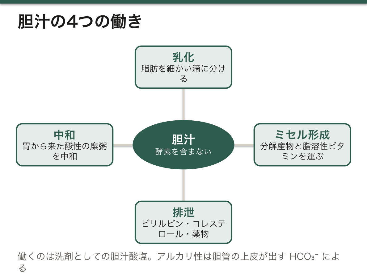
胆汁の働きは大きく4つにまとめられる。① 脂肪を細かい滴に分ける乳化、② 脂肪の分解産物と脂溶性ビタミン(A・D・E・K)を包んで小腸の上皮へ運ぶミセル形成、③ ビリルビン・余ったコレステロール・一部の薬物などを捨てる排泄路、④ 胃から来た酸性の糜粥を中和すること。さらに最近は、胆汁酸が核内受容体などを介して代謝を調節するシグナル分子でもあることが分かっている。
なぜアルカリ性なのか。胆管の上皮細胞は、膵管の細胞と同じようにセクレチンに反応して HCO3− を分泌する。酸性の糜粥が十二指腸に入るとセクレチンが出て、膵液と胆汁の両方から HCO3− が送られ、膵酵素の働ける pH に整える。胆嚢の中では水と電解質が吸収されて胆汁は何倍にも濃縮され、胆汁酸塩・コレステロール・ビリルビンの濃度が上がる一方で HCO3− は減るので、胆嚢胆汁は肝胆汁より pH が低めになる。実験に使う胆汁がどちらかで、読める pH は少し変わる。
コレステロールは水にほとんど溶けず、胆汁の中では胆汁酸塩とレシチンが作る混合ミセルに入ってはじめて溶けていられる。コレステロールが多すぎるか、胆汁酸塩やレシチンが足りないか、胆嚢の中で胆汁がよどむと、コレステロールが結晶になってコレステロール胆石ができる。胆石や腫瘍で胆汁の流れが止まると(閉塞性黄疸)、便は灰白色になり、尿は抱合型ビリルビンで濃く色づく。脂肪と脂溶性ビタミンが吸収できなくなり、ビタミン K の不足からプロトロンビン時間が延びるのも、胆汁の働きから説明できる。
English version
Core
Bile is a yellow-green to brown fluid made continuously by hepatocytes, about 0.5–1 L a day. Between meals it is stored and concentrated in the gallbladder. When fatty food enters the duodenum, cholecystokinin makes the gallbladder contract and push it out. Bile contains no digestive enzymes at all. It is still essential for digesting and absorbing fat because its bile salts act as a detergent.
Universal indicator paper shows that bile is weakly alkaline (about pH 7.5–8 for bile fresh from the liver). The effect of each component on pH is as follows.
| Component | What it is | Effect on pH |
|---|---|---|
| Bile salts | Sodium salts of cholic acid and others, conjugated with glycine or taurine | Almost neutral to slightly alkaline. Conjugation makes them stronger acids, so at intestinal pH they are almost all anions (salts) |
| Bicarbonate HCO3− | Secreted by bile duct epithelial cells | The main factor raising pH |
| Phospholipids | Mainly lecithin (phosphatidylcholine) | Almost no effect |
| Cholesterol | The part not turned into bile acids | No effect (neutral) |
| Conjugated bilirubin | Bilirubin diglucuronide | A weak acid, but present in small amounts, so little effect. Gives bile its colour |
| Electrolytes and water | Na+, K+, Cl− and others | No effect |
Bile has four main functions: (1) emulsification, breaking fat into fine droplets; (2) micelle formation, carrying fat digestion products and fat-soluble vitamins (A, D, E, K) to the intestinal epithelium; (3) an excretion route for bilirubin, excess cholesterol and some drugs; and (4) neutralising acidic chyme from the stomach. More recently, bile acids have also been shown to be signalling molecules that regulate metabolism through nuclear receptors and other receptors.
Going further
Why is bile alkaline? Bile duct epithelial cells, like pancreatic duct cells, secrete HCO3− in response to secretin. When acidic chyme enters the duodenum, secretin is released, and HCO3− arrives from both pancreatic juice and bile, bringing the pH to a level where pancreatic enzymes can work. In the gallbladder, water and electrolytes are absorbed and bile becomes several times more concentrated: bile salts, cholesterol and bilirubin rise, while HCO3− falls, so gallbladder bile has a lower pH than liver bile. The pH you read depends a little on which bile is used.
In the clinic
Cholesterol barely dissolves in water; in bile it stays dissolved only inside mixed micelles made of bile salts and lecithin. If there is too much cholesterol, too little bile salt or lecithin, or bile stagnates in the gallbladder, cholesterol crystallises and cholesterol gallstones form. When a stone or tumour blocks bile flow (obstructive jaundice), stools turn pale grey and urine darkens with conjugated bilirubin. Fat and fat-soluble vitamins are no longer absorbed, and lack of vitamin K prolongs the prothrombin time — all explained by the functions of bile.
胆汁はリパーゼを助ける — 牛乳の脂肪から出た脂肪酸を滴定で数える
基質は牛乳の脂肪(トリアシルグリセロール)、リパーゼの源はパンクレアチン(膵臓から取った消化酵素の混合物)である。4つのフラスコに牛乳 4 mL ずつを入れ、次のように組み合わせる。水で全体の量をそろえるのは、どのフラスコでも濃度を同じにして比べるためである。指示薬にフェノールフタレインを1滴ずつ加え、38 ℃ で20分置いてから、0.1 mol/L の NaOH を1滴ずつ加えて、薄いピンクが消えずに残るところまで滴定する。
| フラスコ | パンクレアチン | 胆汁 | 見ているもの | NaOH の量(予想) |
|---|---|---|---|---|
| I | − | − | 牛乳そのものの酸(基準) | 中 |
| II | + | − | リパーゼだけの働き | 多い |
| III | − | + | 胆汁そのもののアルカリ性 | 最も少ない |
| IV | + | + | リパーゼ+胆汁 | 最も多い |
膵リパーゼはトリアシルグリセロールの1位と3位のエステル結合を加水分解し、2-モノアシルグリセロール1分子と遊離脂肪酸2分子を作る。脂肪酸はカルボン酸なので H+ を出して液を酸性にし、それを中和するのに NaOH が使われる。フェノールフタレインは pH 約 8.2 より下では無色、上ではピンクなので、ピンクになるまでに要った NaOH の量=フラスコの中の酸の量になる。pH の低い順に並べると IV → II → I → III となる。胆汁の入ったフラスコは胆汁の色素のせいでピンクが橙がかるので、色の強さをそろえて終点を決める。
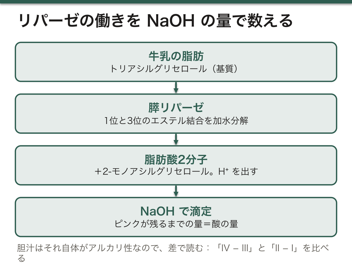
読むときは差で比べる。胆汁はそれ自体がアルカリ性なので、胆汁を入れたフラスコは最初から NaOH が少なくて済む。そこで「IV − III」(胆汁があるときにリパーゼが増やした酸)と「II − I」(胆汁がないときにリパーゼが増やした酸)を比べる。前者が大きければ、胆汁がリパーゼの働きを強めたと言える。I と III は、牛乳そのものの酸(リン酸塩・クエン酸・カゼインなど)と胆汁そのもののアルカリ性という背景を差し引くための対照である。
なぜ胆汁で速くなるのか。リパーゼは水に溶けた酵素で、油と水の境目でしか働けない。胆汁酸塩が脂肪の滴を細かく割ると、同じ量の脂肪でも境目の面積が大きくなり、酵素が取りつける場所が増える。ただし胆汁酸塩が滴の表面を覆うと、リパーゼはかえって表面からはがされやすくなる。これを防ぐのが膵臓から一緒に出るコリパーゼで、胆汁酸塩に覆われた滴の上にリパーゼをつなぎとめる。パンクレアチンにはコリパーゼも入っている。
脂肪の消化には胆汁と膵リパーゼの両方が要る。胆汁が来ない(閉塞性黄疸、回腸の切除で胆汁酸が再吸収されず失われる)場合も、リパーゼが足りない(慢性膵炎、嚢胞性線維症)場合も、脂肪が吸収されずに便に出る脂肪便になり、脂溶性ビタミンも不足する。膵外分泌不全の治療に使う消化酵素の製剤(パンクレリパーゼ)は、この実験のパンクレアチンと同じ種類のものである。逆に、肥満の治療に使われるオルリスタットはリパーゼを阻害して脂肪の吸収を減らし、油のような便が副作用になる。
English version
Core
The substrate is milk fat (triacylglycerol) and the source of lipase is pancreatin (a mixture of digestive enzymes from the pancreas). Four flasks each get 4 mL of milk and are combined as below. Water is used to make the total volume equal, so that concentrations are the same in every flask. One drop of phenolphthalein is added to each, the flasks stay at 38 °C for 20 minutes, and then 0.1 mol/L NaOH is added drop by drop until a faint pink stays.
| Flask | Pancreatin | Bile | What it shows | NaOH needed (expected) |
|---|---|---|---|---|
| I | − | − | Acid of milk itself (baseline) | Medium |
| II | + | − | Lipase alone | More |
| III | − | + | Alkalinity of bile itself | Least |
| IV | + | + | Lipase + bile | Most |
Pancreatic lipase hydrolyses the ester bonds at positions 1 and 3 of triacylglycerol and makes one 2-monoacylglycerol and two free fatty acids. Fatty acids are carboxylic acids; they release H+ and make the liquid acidic, and NaOH is used to neutralise them. Phenolphthalein is colourless below about pH 8.2 and pink above it, so the NaOH needed to reach pink = the amount of acid in the flask. From lowest to highest pH the order is IV → II → I → III. Bile pigments make the pink look orange in flasks with bile, so the end point is set by matching the intensity of the colour.
Going further
Compare differences, not raw numbers. Bile is itself alkaline, so flasks with bile need less NaOH from the start. So compare “IV − III” (acid added by lipase with bile) with “II − I” (acid added by lipase without bile). If the first is larger, bile boosted lipase. Flasks I and III are controls that let you subtract the background: the acid already in milk (phosphates, citrate, casein and others) and the alkalinity of bile itself.
Why does bile speed things up? Lipase is a water-soluble enzyme and works only at the boundary between oil and water. When bile salts break fat into fine droplets, the same amount of fat has a much larger boundary, so the enzyme has more places to bind. But bile salts covering the droplet surface can actually push lipase off. Colipase, released from the pancreas together with lipase, prevents this by anchoring lipase onto bile-salt-coated droplets. Pancreatin contains colipase too.
In the clinic
Fat digestion needs both bile and pancreatic lipase. If bile does not arrive (obstructive jaundice, or loss of bile acids after ileal resection) or lipase is lacking (chronic pancreatitis, cystic fibrosis), fat is not absorbed and appears in the stool as steatorrhoea, and fat-soluble vitamins run short. The enzyme preparation used to treat pancreatic exocrine insufficiency (pancrelipase) is the same kind of material as the pancreatin in this experiment. In the other direction, orlistat, used for obesity, inhibits lipase to reduce fat absorption, and oily stools are its side effect.
表面張力 — 硫黄の粉が沈むのは、胆汁酸塩が水面に並ぶから
2本の試験管に水 3 mL ずつを入れ、片方にだけ胆汁を3滴加える。両方に硫黄の粉(黄色の細かい粒)を少量ずつ落として振る。水だけの試験管では、硫黄は水面に浮いたままか、壁にはりつく。胆汁を入れた試験管では、硫黄は水にぬれて沈んでいく。
なぜ水だけでは浮くのか。硫黄は水をはじく(疎水性の)物質で、粒が軽い。水の表面の分子は、内側の分子とは水素結合で引き合うが外側(空気)には相手がいないので、内側へ引き込まれ、表面がぴんと張った膜のようにふるまう。これが表面張力で、水は表面張力の大きい液体である。疎水性の軽い粒は、この膜を破れずに上に乗ってしまう。
胆汁の中で効いているのは胆汁酸塩である。胆汁酸塩は1つの分子の中に水になじむ部分と水をはじく部分をもつ両親媒性の分子で、水と空気の境目に集まり、水をはじく側を空気に、水になじむ側を水に向けて並ぶ。すると表面の分子どうしの引き合いが弱まり、表面張力が下がる。同時に硫黄の粒の表面にも胆汁酸塩が付いて、粒が水になじむ「ぬれる」状態になり、沈む。
胆汁酸の形は、石けんとは少し違う。石けん(脂肪酸の塩)は「頭」と「しっぽ」をもつ細長い分子だが、胆汁酸はステロイドの平たい板で、OH 基がすべて板の片面に、メチル基が反対の面に並んでいる。片面が水になじみ、もう片面が水をはじく「表と裏のある板」なので、境目に平たく寝そべって並ぶ。グリシンやタウリンが結合(抱合)すると酸が強くなり、腸の pH でもイオンのまま水に溶けていられる。一定の濃度(臨界ミセル濃度)を超えるとミセルを作る。
硫黄の粉を沈めたのと同じ性質が、十二指腸で大きな脂肪の滴を細かい滴に割り、割れた滴がまたくっつかないように表面を覆っている。つまりこの試験管は、乳化を水面の上で見せている実験である。
この現象は、尿の中の胆汁酸塩を調べる古い検査(Hay 試験)そのものである。尿の上に硫黄の粉をまき、沈めば尿に胆汁酸塩が出ていると判断した。胆汁の流れが止まって胆汁酸が血中にたまり、尿に漏れ出る胆汁うっ滞の手がかりになった。いまは血液の胆汁酸の測定や画像検査に置き換わっているが、「胆汁酸塩は表面張力を下げる」という性質を検査に変えた例として理解しておくとよい。
English version
Core
Two tubes each get 3 mL of water, and only one gets 3 drops of bile. A little sulphur powder (fine yellow grains) is dropped into both, and they are shaken. In water alone, the sulphur stays floating on the surface or sticks to the wall. In the tube with bile, the sulphur becomes wet and sinks.
Why does it float in water alone? Sulphur is water-repelling (hydrophobic), and the grains are light. Water molecules at the surface are pulled by hydrogen bonds toward the molecules inside, but have no partners on the outside (air). So they are pulled inward, and the surface acts like a tight film. This is surface tension, and water has a high surface tension. Light hydrophobic grains cannot break through this film and sit on top of it.
The active component of bile is the bile salts. A bile salt is an amphipathic molecule, with a water-loving part and a water-repelling part in one molecule. It gathers at the water–air boundary, with the water-repelling side toward the air and the water-loving side toward the water. This weakens the pull between surface molecules and lowers the surface tension. At the same time, bile salts coat the sulphur grains so that they become “wettable”, and they sink.
Going further
The shape of a bile acid is a little different from soap. Soap (a fatty acid salt) is a long molecule with a “head” and a “tail”. A bile acid is a flat steroid plate: all its OH groups are on one face and the methyl groups on the other. It is a plate with a water-loving face and a water-repelling face, so it lies flat at the boundary. Joining with glycine or taurine (conjugation) makes it a stronger acid, so it stays ionised and dissolved at intestinal pH. Above a certain concentration (the critical micelle concentration) it forms micelles.
The same property that sank the sulphur also breaks large fat droplets into small ones in the duodenum, and coats them so they do not join again. In other words, this tube shows emulsification at a water surface.
In the clinic
This is exactly an old test for bile salts in urine (Hay’s test). Sulphur powder was sprinkled on urine; if it sank, bile salts were present. It was a clue to cholestasis, where bile flow stops, bile acids build up in the blood and leak into urine. Today it has been replaced by blood bile acid measurement and imaging, but it is a good example of turning the property “bile salts lower surface tension” into a test.
コハク酸脱水素酵素とメチレンブルー — 人工の水素受容体で、脱水素を色に変える
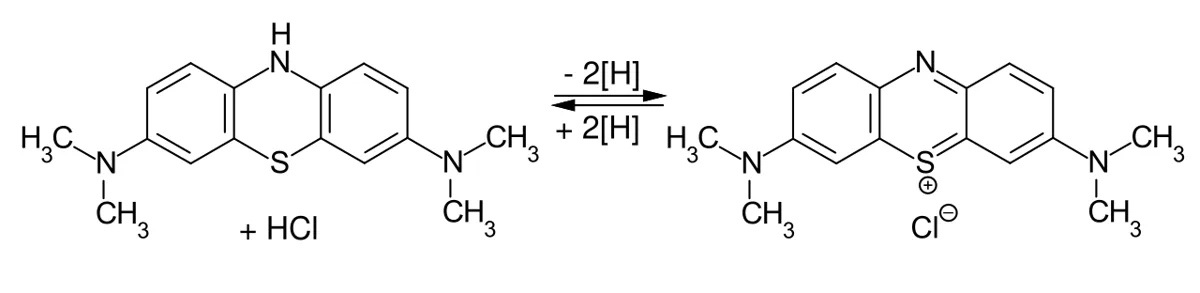
コハク酸脱水素酵素(SDH)は、コハク酸から水素原子2個を外してフマル酸(トランス形の二重結合をもつ)に変える酸化還元酵素で、外した水素は酵素に結合したFAD が受け取って FADH2 になる。ミトコンドリアの中では、この電子はそのままユビキノンへ渡される。SDH はクエン酸回路の第6段階の酵素であり、同時に電子伝達系の複合体 II そのものでもある。両方の経路に属し、回路の酵素で唯一ミトコンドリア内膜に埋まっている。
実験では、刻んだ肝臓2〜3片を乳鉢ですりつぶし、水を少しずつ(合計 5 mL まで)加えてどろどろの液(ホモジネート)にする。試験管に移し、基質のコハク酸アンモニウム液 2〜3 mL とメチレンブルーを2滴加えて混ぜる。液は肝臓の黄褐色とメチレンブルーの青が重なって緑がかって見える。38 ℃ に30分置き、振らずにそっと取り出して色を見る。
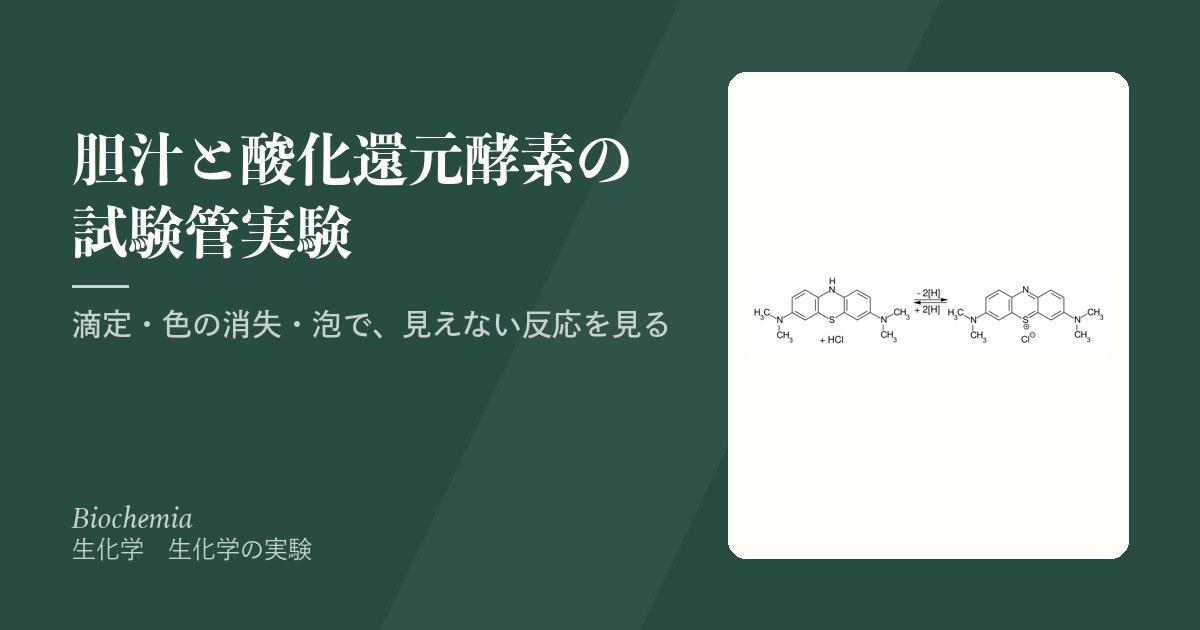
メチレンブルーは、3つの環に電子が広がった(共役した)色素で、正の電荷も分子全体に広がっている。この形(酸化型)は赤い光を吸うので青い。水素2個ぶん(電子2個とプロトン)を受け取ると、中央の環の窒素に H が付いて共役が切れ、光を吸わなくなって無色のロイコメチレンブルー(還元型)になる。試験管の中では、すりつぶしで電子伝達系の並びがこわれ、酸素も少ないので、メチレンブルーがユビキノンの代わりに人工の水素受容体として電子を受け取る。青が消えること自体が、コハク酸から水素が外された証拠になる。
なぜ肝臓を使うのか。肝細胞は1個に千を超えるミトコンドリアをもち、SDH は内膜に結合しているので、少しの組織から多くの酵素が取れる。肝臓はやわらかく、すりつぶしやすいのも利点である。ミトコンドリアの多い心筋や骨格筋も同じ理由で使え、古典的な実験ではハトの胸筋などの筋肉が使われた。
手順にも理由がある。メチレンブルーを入れすぎない(2滴)のは、酵素が還元しきれずに青が残ると、酵素が働いていても陰性に見えるからである。取り出すときに振らないのは、空気の酸素が混ざるとロイコ体がすぐ青に戻り、観察が台なしになるからである。この実験が証明するのは脱水素であって、ATP の合成ではない。すりつぶした組織には膜の内外の H+ の勾配も ADP も用意されていない。
SDH の遺伝子(SDHA・SDHB・SDHC・SDHD)に生まれつき変異があると、遺伝性の傍神経節腫・褐色細胞腫が起こりやすくなる。SDH が働かないとコハク酸がたまり、それが酸素の不足を感じる仕組み(HIF を分解する酵素)を止めてしまうため、細胞は酸素が十分あっても「低酸素だ」と受け取って増殖の信号を出し続ける(偽低酸素)。代謝物そのものが腫瘍を起こす例として知られている。また FAD はビタミン B2(リボフラビン)から作られるので、SDH は B2 が補酵素として働く代表的な酵素でもある。
English version
Core
Succinate dehydrogenase (SDH) is an oxidoreductase that removes two hydrogen atoms from succinate and turns it into fumarate (which has a trans double bond). The hydrogens are taken by FAD bound to the enzyme, which becomes FADH2. In mitochondria the electrons go straight on to ubiquinone. SDH is the enzyme of step 6 of the citric acid cycle and, at the same time, is complex II of the electron transport chain itself. It belongs to both pathways, and it is the only cycle enzyme embedded in the inner mitochondrial membrane.
In the experiment, 2–3 pieces of chopped liver are ground in a mortar while water is added little by little (up to 5 mL in total), to make a thick liquid (homogenate). It is poured into a tube, and 2–3 mL of ammonium succinate (the substrate) and 2 drops of methylene blue are added and mixed. The liquid looks greenish, because the yellow-brown of liver and the blue of the dye overlap. The tube stays at 38 °C for 30 minutes and is then taken out gently, without shaking, and its colour is noted.
Methylene blue is a dye whose electrons are spread (conjugated) over three rings, with its positive charge also spread over the molecule. This oxidised form absorbs red light and so looks blue. When it accepts two hydrogens (two electrons and a proton), an H is added to the nitrogen of the central ring, the conjugation breaks, it no longer absorbs light, and it becomes colourless leucomethylene blue (the reduced form). In the tube, grinding has disrupted the electron transport chain and there is little oxygen, so methylene blue takes the electrons instead of ubiquinone, as an artificial hydrogen acceptor. The loss of blue is itself the proof that hydrogen was removed from succinate.
Going further
Why liver? A hepatocyte has more than a thousand mitochondria, and SDH is bound to the inner membrane, so a small piece of tissue gives a lot of enzyme. Liver is also soft and easy to grind. Heart and skeletal muscle, also rich in mitochondria, work for the same reason; classic experiments used muscle such as pigeon breast muscle.
The steps have reasons too. Not too much methylene blue (2 drops) is used, because if the enzyme cannot reduce all of it, blue remains and the tube looks negative even though the enzyme worked. The tube is not shaken when taken out, because oxygen from the air quickly turns the leuco form blue again and spoils the observation. What this experiment proves is dehydrogenation, not ATP synthesis: ground tissue has no H+ gradient across a membrane and no ADP supplied.
In the clinic
Inherited variants in the SDH genes (SDHA, SDHB, SDHC, SDHD) cause hereditary paraganglioma and phaeochromocytoma. When SDH does not work, succinate builds up and blocks the system that senses low oxygen (the enzymes that break down HIF). The cell then “thinks” it is hypoxic even when oxygen is plentiful and keeps sending growth signals (pseudohypoxia). It is a known example of a metabolite itself causing a tumour. Also, FAD is made from vitamin B2 (riboflavin), so SDH is a typical enzyme that uses B2 as its coenzyme.
消える・戻る・また消える — 3相の観察と、足りない対照
恒温器から出した試験管は、時間とともに3つの相を見せる。
| 相 | いつ | 見える色 | 起きていること |
|---|---|---|---|
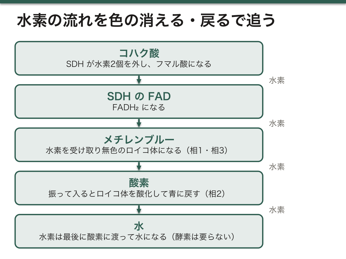
| 1 | 恒温器から出した直後(振らない) | 無色(肝臓の淡い黄褐色)。液面のごく上だけ青い輪が残ることがある | SDH がコハク酸から外した水素を、メチレンブルーが受け取ってロイコ体になった。液面は空気に触れているので青い |
| 2 | 振った直後 | 青が戻る | 振って入った空気の酸素が、ロイコ体を酸化して青に戻す。水素は酸素に渡って水になる(酵素は要らない) |
| 3 | さらに10分置いた後 | また無色 | 溶けた酸素を使い切ると、まだ働いている酵素が、残っているコハク酸から水素を外してメチレンブルーを再び還元する |
まとめると、コハク酸 →(SDH・FAD)→ メチレンブルー → 酸素 → 水という水素の流れを、色の消える・戻るで追いかけていることになる。メチレンブルーは、本物の電子伝達系の代わりにコハク酸と酸素の間を行き来する運び屋として働いている。
この試験管1本だけでは、厳密には「SDH が働いた」とまでは言い切れない。きちんと言うには、次の対照を並べるとよい。
| 対照 | 中身 | 予想 | 証明すること |
|---|---|---|---|
| 基質なし | 肝ホモジネート+水+メチレンブルー | ゆっくり、または弱く色が消える | 組織にもともとあるコハク酸や、NADH を使う別の脱水素酵素の分を差し引ける。コハク酸の試験管がこれより速いことが SDH の証拠 |
| 煮沸 | 煮沸したホモジネート+コハク酸+メチレンブルー | 青のまま | 色が消えるのが酵素によることを示す |
| 阻害剤 | ホモジネート+コハク酸+マロン酸+メチレンブルー | 色が消えるのが遅れる | SDH を狙って止めると色の変化も止まる。コハク酸を増やすと阻害が弱まる |
マロン酸(−OOC–CH2–COO−)は、コハク酸(−OOC–CH2–CH2–COO−)より炭素が1つ少ないだけで、両端のカルボキシ基で活性部位にはまる。しかし水素を外せる CH2–CH2 の部分がないので反応できず、席をふさぐだけになる。基質と同じ場所を奪い合う競合阻害の代表例で、基質を増やせば打ち勝てる。クエン酸回路が回路であることを示した歴史的な研究でも、マロン酸で SDH を止める実験が使われた。
メチレンブルーの「青 ⇄ 無色」の酸化還元は、そのまま治療にも使われている。亜硝酸塩・一部の局所麻酔薬・ダプソンなどでヘモグロビンの鉄が Fe3+ に酸化されると、酸素を運べないメトヘモグロビン血症になる。メチレンブルーを静注すると、赤血球の中で NADPH を使う酵素がそれをロイコ体に還元し、ロイコ体がメトヘモグロビンの鉄を Fe2+ に戻す。この NADPH はペントースリン酸経路から来るので、G6PD 欠損症では効きにくく、かえって溶血を起こすことがある。
English version
Core
After leaving the thermostat, the tube shows three phases over time.
| Phase | When | Colour | What is happening |
|---|---|---|---|
| 1 | Just out of the thermostat (not shaken) | Colourless (the pale yellow-brown of liver). A blue ring may remain just at the surface | SDH removed hydrogen from succinate, and methylene blue took it and became the leuco form. The surface is in contact with air, so it stays blue |
| 2 | Just after shaking | Blue returns | Oxygen from the air mixed in by shaking oxidises the leuco form back to blue. The hydrogen goes to oxygen and forms water (no enzyme needed) |
| 3 | After 10 more minutes | Colourless again | Once the dissolved oxygen is used up, the still-active enzyme removes hydrogen from the remaining succinate and reduces methylene blue again |
In short, the colour disappearing and returning lets you follow the hydrogen along succinate → (SDH, FAD) → methylene blue → oxygen → water. Methylene blue acts as a shuttle between succinate and oxygen, standing in for the real electron transport chain.
Going further
Strictly, this one tube is not enough to say “SDH worked”. To say so properly, add these controls.
| Control | Contents | Expected | What it proves |
|---|---|---|---|
| No substrate | Liver homogenate + water + methylene blue | Colour fades slowly or weakly | Subtracts the effect of succinate already in the tissue and of other NADH-linked dehydrogenases. A faster loss in the succinate tube is the evidence for SDH |
| Boiled | Boiled homogenate + succinate + methylene blue | Stays blue | Shows that the loss of colour is caused by an enzyme |
| Inhibitor | Homogenate + succinate + malonate + methylene blue | Loss of colour is delayed | Blocking SDH specifically also blocks the colour change. More succinate weakens the inhibition |
Malonate (−OOC–CH2–COO−) has just one carbon fewer than succinate (−OOC–CH2–CH2–COO−) and fits into the active site by its two carboxyl groups. But it lacks the CH2–CH2 part from which hydrogen could be removed, so it cannot react and only blocks the seat. It is the classic example of competitive inhibition, where the inhibitor competes with the substrate for the same site and can be overcome by more substrate. Blocking SDH with malonate was also used in the historic work showing that the citric acid cycle is a cycle.
In the clinic
The blue ⇄ colourless redox of methylene blue is also used in treatment. When nitrites, some local anaesthetics or dapsone oxidise haemoglobin iron to Fe3+, the result is methaemoglobinaemia, and oxygen cannot be carried. Methylene blue given intravenously is reduced to the leuco form inside red cells by an NADPH-using enzyme, and the leuco form returns methaemoglobin iron to Fe2+. This NADPH comes from the pentose phosphate pathway, so in G6PD deficiency methylene blue works poorly and may even cause haemolysis.
カタラーゼ — 溶血させた血液に過酸化水素を注ぐと、泡があふれる
流しの上で、溶血させた血液約 2 mL に2% 過酸化水素水 1.5〜2 mL を素早く加える。たちまち激しく泡立ち、泡が試験管の口まで上がってくる。泡は酸素である。
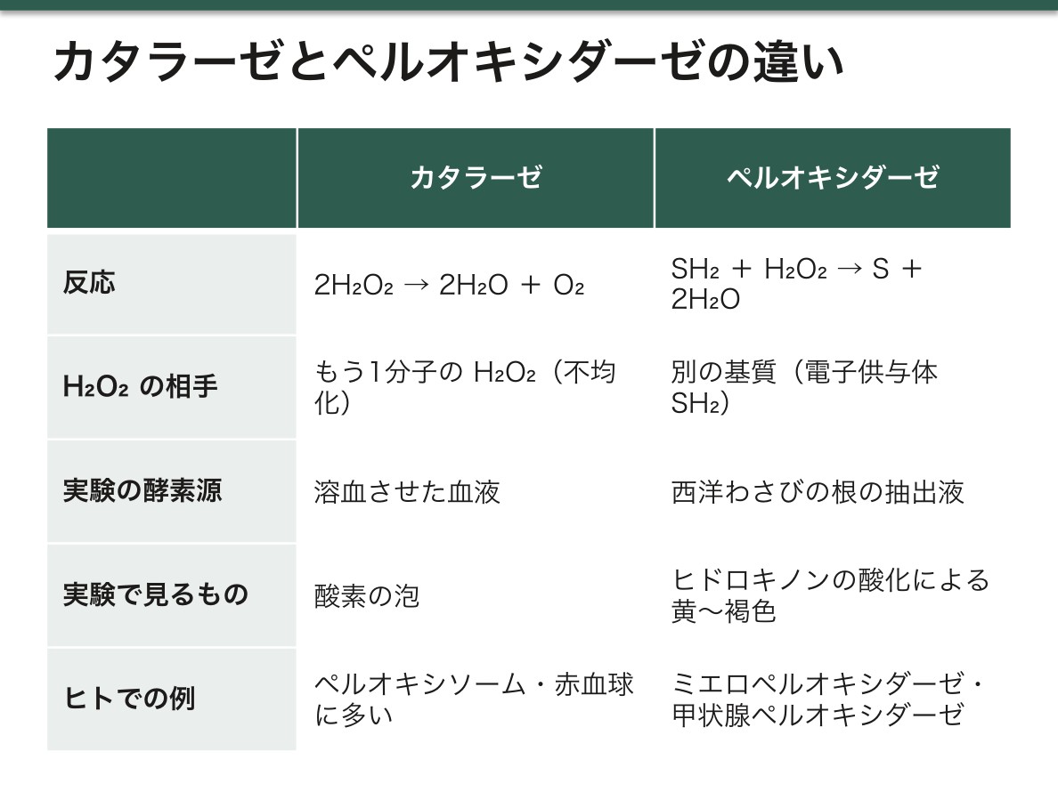
カタラーゼは、ヘム(鉄を含むポルフィリン)を活性中心にもつ酵素で、2H2O2 → 2H2O + O2 を触媒する。1分子の H2O2 が酸化剤、もう1分子が還元剤として働く(不均化)。1つの酵素分子が1秒間に数百万分子の H2O2 を処理する、最も速い酵素の1つである。体の中では肝臓や腎臓の細胞のペルオキシソームに多く、赤血球の細胞質にも大量に含まれている。
なぜ血液を溶血させるのか。カタラーゼは赤血球の中にある。細胞膜は H2O2 の出入りを遅らせ、酵素を細胞の中に閉じ込めている。水を加えて低張にすると赤血球は浸透圧で破れ(溶血)、中の酵素が液全体に出てくるので、加えた H2O2 と一度に出会える。反応が速く、はっきり見えるようになる。
泡が出たことを「酵素のせい」と言うには、次の2本を並べるとよい。煮沸した血液に H2O2 を加えると、酵素が変性しているのでほとんど泡は出ない。水に H2O2 を加えても、ほとんど泡は出ない。H2O2 は放っておいても分解するが、その速さは目に見えないほど遅い。酵素は反応を起こす力を与えているのではなく、もともと起こりうる反応を桁違いに速くしているだけである。
細胞が H2O2 を消す酵素を2種類もつ理由も押さえておくとよい。スーパーオキシドは SOD で H2O2 になり、H2O2 はカタラーゼかグルタチオンペルオキシダーゼで水になる。カタラーゼは H2O2 の濃度が高いときに大量に処理するのが得意で、グルタチオンペルオキシダーゼは細胞質の低い濃度の H2O2 や、膜の脂質にできた過酸化物の処理を受けもつ。
傷口にオキシドール(約 3% の H2O2)をつけると泡立つのは、血液と組織のカタラーゼのためである。細菌検査でも同じ反応を使う。コロニーを H2O2 に触れさせて泡が出ればカタラーゼ陽性で、グラム陽性球菌のうちブドウ球菌(陽性)とレンサ球菌(陰性)を分ける最初の一歩になる。カタラーゼが生まれつき欠けている無カタラーゼ血症は日本で最初に報告された病気で、口の中の細菌が出す H2O2 を処理できず、歯ぐきの潰瘍や壊疽を起こすことがある。
English version
Core
Over a sink, 1.5–2 mL of 2% hydrogen peroxide is quickly added to about 2 mL of haemolysed blood. The mixture foams violently at once, and the foam rises to the top of the tube. The gas is oxygen.
Catalase is an enzyme with haem (an iron-containing porphyrin) at its active centre. It catalyses 2H2O2 → 2H2O + O2: one H2O2 acts as the oxidising agent and the other as the reducing agent (disproportionation). One enzyme molecule handles millions of H2O2 molecules per second, making it one of the fastest enzymes. In the body it is abundant in the peroxisomes of liver and kidney cells, and there is a large amount in the cytoplasm of red blood cells.
Why haemolyse the blood? Catalase is inside the red cells. The cell membrane slows the movement of H2O2 and keeps the enzyme shut in. Adding water makes the solution hypotonic, the red cells burst by osmosis (haemolysis), and the enzyme spreads through the whole liquid, so it meets the added H2O2 all at once. The reaction becomes fast and easy to see.
Going further
To say the foam is “because of the enzyme”, add two more tubes. In boiled blood plus H2O2, almost no foam appears, because the enzyme is denatured. In water plus H2O2, almost no foam appears either. H2O2 does break down by itself, but far too slowly to see. The enzyme does not give the reaction its driving force; it only makes a reaction that could already happen many orders of magnitude faster.
It also helps to know why cells have two enzymes that remove H2O2. Superoxide is turned into H2O2 by SOD, and H2O2 is turned into water by catalase or glutathione peroxidase. Catalase is good at handling large amounts when H2O2 is high; glutathione peroxidase deals with low levels of H2O2 in the cytoplasm and with peroxides formed on membrane lipids.
In the clinic
Hydrogen peroxide (about 3%) foams on a wound because of catalase in blood and tissue. Microbiology uses the same reaction. If a colony touched with H2O2 gives bubbles, it is catalase-positive; this is the first step in separating staphylococci (positive) from streptococci (negative) among Gram-positive cocci. Acatalasaemia, an inborn lack of catalase, was first reported in Japan. Patients cannot remove the H2O2 made by mouth bacteria and may develop gum ulcers and gangrene.
ペルオキシダーゼ — 西洋わさびとヒドロキノン、4本の対照
ペルオキシダーゼは、H2O2 を水素(電子)の受け手にして、別の基質を酸化するヘム酵素である。一般式は SH2 + H2O2 → S + 2H2O。基質 SH2 は水素を差し出す電子供与体で、反応で酸化される。H2O2 は受け取って水に還元される。たとえば NADH を基質にする酵素(NADH ペルオキシダーゼ)なら NADH + H+ + H2O2 → NAD+ + 2H2O となる。カタラーゼとの違いは、H2O2 の相手がもう1分子の H2O2 ではなく別の基質であることだ。
実験では、ペルオキシダーゼを多く含む西洋わさびの根の抽出液を酵素源にし、基質にはヒドロキノン(ベンゼン環の向かい合う位置に OH が2つ、無色)を使う。ヒドロキノンは酸化されるとp-ベンゾキノン(黄色)になり、さらに反応が進むと褐色の物質ができるので、色がつけば酸化が起きたと分かる。
| 試験管 | 水 | 抽出液 | H2O2 | ヒドロキノン | 予想 | 意味 |
|---|---|---|---|---|---|---|
| 1 | − | 2 mL | 2滴 | − | 色はつかない | 抽出液と H2O2 だけでは色のもとが無い |
| 2 | − | 2 mL | 2滴 | 10滴 | すぐ黄〜褐色 | 全部そろった完全な系 |
| 3 | 2 mL | − | 2滴 | 10滴 | ゆっくり、薄く色づく | 酵素なしでも反応は起こりうるが遅い |
| 4 | − | 2 mL | − | 10滴 | 色はつかない(ほぼ) | H2O2 が無ければ酵素は働けない |
4本は、完全な系(2)から1つずつ成分を抜いた形になっている。1は「色のもとは抽出液ではなくヒドロキノンの酸化」、3は「速さの違いは酵素による」、4は「この酵素は H2O2 を必要とする」ことを示す。もし4がわずかに色づいたら、空気の酸素や抽出液に含まれる別の酸化酵素による酸化を疑う。
2と3の違いが、酵素の本質である。中身は酵素の有無だけで、反応そのものは3でも進む。酵素は活性化エネルギーを下げて反応を速めるだけで、反応が進むかどうかや最終的な行き先は変えない。抽出液は時間がたつと酵素が酸化や分解で弱るので、作りたてのほうが2と3の差がはっきりする。
ヒトのペルオキシダーゼの例として、セレンを含みグルタチオンを電子供与体にするグルタチオンペルオキシダーゼ(2GSH + H2O2 → GSSG + 2H2O)、好中球で H2O2 と Cl− から殺菌物質の次亜塩素酸を作るミエロペルオキシダーゼ、甲状腺でヨウ化物を酸化してサイログロブリンのチロシンにヨウ素を付ける甲状腺ペルオキシダーゼがある。
西洋わさびペルオキシダーゼ(HRP)は、抗体に付けてELISA・免疫組織化学・ウエスタンブロットの目印として広く使われる。抗体が結合した場所で HRP が H2O2 と色素の材料を反応させ、色(免疫組織化学なら DAB の茶色)を出す。ヒトのペルオキシダーゼは自己抗体の標的にもなり、抗 TPO 抗体は橋本病の、MPO-ANCA は顕微鏡的多発血管炎の代表的なマーカーである。ヘモグロビンもペルオキシダーゼに似た働きをもち、尿試験紙の潜血反応はこれで色を出す。だから筋肉が壊れてミオグロビンが出たときも陽性になり、顕微鏡で赤血球が見えないのに潜血陽性という食い違いが横紋筋融解症の手がかりになる。古い便潜血検査(グアヤック法)は食べた西洋わさびやカブのペルオキシダーゼでも陽性になったため、日本の大腸がん検診ではヒトのヘモグロビンだけに反応する免疫法が使われている。
English version
Core
Peroxidase is a haem enzyme that oxidises another substrate, using H2O2 as the acceptor of hydrogen (electrons). The general equation is SH2 + H2O2 → S + 2H2O. The substrate SH2 gives up hydrogen, so it is the electron donor, and it is oxidised. H2O2 accepts and is reduced to water. For an enzyme that uses NADH (NADH peroxidase), the reaction is NADH + H+ + H2O2 → NAD+ + 2H2O. The difference from catalase is that the partner of H2O2 is another substrate, not a second H2O2.
The enzyme source is horseradish root extract, which is rich in peroxidase, and the substrate is hydroquinone (a benzene ring with two OH groups opposite each other; colourless). When oxidised, hydroquinone becomes p-benzoquinone (yellow), and further reaction gives brown products, so colour means oxidation has happened.
| Tube | Water | Extract | H2O2 | Hydroquinone | Expected | Meaning |
|---|---|---|---|---|---|---|
| 1 | − | 2 mL | 2 drops | − | No colour | Extract and H2O2 alone have nothing to make colour |
| 2 | − | 2 mL | 2 drops | 10 drops | Quickly yellow to brown | The complete system |
| 3 | 2 mL | − | 2 drops | 10 drops | Slow, faint colour | The reaction can happen without enzyme, but slowly |
| 4 | − | 2 mL | − | 10 drops | No colour (almost) | Without H2O2 the enzyme cannot work |
Going further
The four tubes are the complete system (tube 2) with one component removed at a time. Tube 1 shows that the colour comes from oxidised hydroquinone, not from the extract. Tube 3 shows that the difference in speed is due to the enzyme. Tube 4 shows that this enzyme needs H2O2. If tube 4 turns slightly coloured, suspect oxidation by air oxygen or by another oxidising enzyme in the extract.
The difference between tubes 2 and 3 is the essence of an enzyme. They differ only in the enzyme, and the reaction still proceeds in tube 3. An enzyme lowers the activation energy and speeds up the reaction; it does not change whether the reaction can happen or where it ends. The enzyme in the extract weakens over time by oxidation and breakdown, so a freshly made extract shows the difference between tubes 2 and 3 more clearly.
Examples of human peroxidases are glutathione peroxidase, which contains selenium and uses glutathione as the electron donor (2GSH + H2O2 → GSSG + 2H2O); myeloperoxidase, which makes the bactericidal hypochlorous acid from H2O2 and Cl− in neutrophils; and thyroid peroxidase, which oxidises iodide in the thyroid and attaches iodine to the tyrosines of thyroglobulin.
In the clinic
Horseradish peroxidase (HRP) is widely attached to antibodies as a label in ELISA, immunohistochemistry and Western blotting. Where the antibody binds, HRP reacts H2O2 with a dye precursor and makes a colour (in immunohistochemistry, the brown of DAB). Human peroxidases are also targets of autoantibodies: anti-TPO antibodies are a typical marker of Hashimoto’s disease, and MPO-ANCA of microscopic polyangiitis. Haemoglobin also has peroxidase-like activity, which the blood reaction on urine dipsticks uses to make colour. So the test is also positive when damaged muscle releases myoglobin, and “blood-positive strip but no red cells under the microscope” is a clue to rhabdomyolysis. The old faecal occult blood test (guaiac method) also turned positive with peroxidase from eaten horseradish or turnip, so Japanese colorectal cancer screening uses an immunochemical test that reacts only to human haemoglobin.
まとめ
| 胆汁の pH と成分 — 酵素を含まない消化液 | 胆汁は肝細胞が休みなく作る黄緑〜褐色の液で、1日に 0.5〜1 L ほど分泌される。 |
|---|---|
| 胆汁はリパーゼを助ける — 牛乳の脂肪から出た脂肪酸を滴定で数える | 基質は牛乳の脂肪(トリアシルグリセロール)、リパーゼの源はパンクレアチン(膵臓から取った消化酵素の混合物)である。 |
| 表面張力 — 硫黄の粉が沈むのは、胆汁酸塩が水面に並ぶから | 2本の試験管に水 3 mL ずつを入れ、片方にだけ胆汁を3滴加える。 |
| コハク酸脱水素酵素とメチレンブルー — 人工の水素受容体で、脱水素を色に変える | コハク酸脱水素酵素(SDH)は、コハク酸から水素原子2個を外してフマル酸(トランス形の二重結合をもつ)に変える酸化還元酵素で、外した水素は酵素に結合したFAD が受け取って FAD… |
| 消える・戻る・また消える — 3相の観察と、足りない対照 | 恒温器から出した試験管は、時間とともに3つの相を見せる。 |
| カタラーゼ — 溶血させた血液に過酸化水素を注ぐと、泡があふれる | 流しの上で、溶血させた血液約 2 mL に2% 過酸化水素水 1.5〜2 mL を素早く加える。 |
| ペルオキシダーゼ — 西洋わさびとヒドロキノン、4本の対照 | ペルオキシダーゼは、H2O2 を水素(電子)の受け手にして、別の基質を酸化するヘム酵素である。 |
このノートは医学生が授業の内容を自分の言葉でまとめたもので、診療の判断に使うものではありません。誤りに気づいたらお知らせください。