核の見た目は、その細胞が今どうしているかを語る。
- 大きく明るく、核小体が目立つ——盛んに蛋白を作っている(活動している核)
- 小さく濃く、一様に黒い——休んでいるか、死につつある
- 核が濃縮して断片化する——死んだ後の像
だから病理の記述は、まず核から始まる——「核の大きさ・形・染まり方・核小体の3点を言えれば、所見の半分は書けている」。
そして悪性の判定も、核の所見が中心になる。核/細胞質比の増大・大小不同・クロマチンの増量・核小体の目立ち・核形の不整。
細胞の死に方は2つに分かれ、周りの反応がまったく違う——壊死は炎症を呼び、アポトーシスは呼ばない。
細胞小器官は細胞小器官の章、細胞死の病理は細胞傷害と細胞死の章、発癌は発癌の分子機構の章にある。
- 核・クロマチン・核小体
- 細胞周期・体細胞分裂・減数分裂
- Ki-67 — 細胞周期に入っている核だけが茶色になる
- アポトーシスと壊死 — 死に方が周りを変える
- 核の像から異型を読む — 良性と悪性を分ける手がかり
- 化生 — 上皮が、別の種類の上皮に置き換わる
- 制御された壊死 — 細胞の死に方は、2つでは足りない
- 封入体 — 核や細胞質の中の塊が、原因を名指しする
- 細胞質封入体(1)— 蓄えとしてのグリコーゲンと脂肪滴
- 細胞質封入体(2)— 色素:体の中で作られたものと、外から入ったもの
- 多核の巨細胞は、できかたで種類が分かれる
- 核・クロマチン・核小体Nucleus
- 細胞周期・体細胞分裂・減数分裂Cyclus cellularis
- Ki-67 — 細胞周期に入っている核だけが茶色になる
- アポトーシスと壊死 — 死に方が周りを変えるApoptosis
- 核の像から異型を読む — 良性と悪性を分ける手がかりAtypia
- 化生 — 上皮が、別の種類の上皮に置き換わるMetaplasia
- 制御された壊死 — 細胞の死に方は、2つでは足りない
- 封入体 — 核や細胞質の中の塊が、原因を名指しする
- 細胞質封入体(1)— 蓄えとしてのグリコーゲンと脂肪滴Inclusiones cytoplasmicae
- 細胞質封入体(2)— 色素:体の中で作られたものと、外から入ったものPigmentum
- 多核の巨細胞は、できかたで種類が分かれる
- まとめ
核・クロマチン・核小体Nucleus
核膜は二重膜で、外膜は粗面小胞体と連続する。核膜孔が物質の出入りを制御する。
クロマチン——DNAとヒストンの複合体。
- ヘテロクロマチン——凝縮していて転写されていない。濃く染まる。核膜の内側に張り付く
- ユークロマチン——ほどけていて転写されている。淡い
だから淡く明るい核=働いている核になる(小胞状核)。
核小体——リボソームRNAを作る場所。強い好塩基性。蛋白合成の盛んな細胞と悪性細胞で目立つ。
Barr小体——不活化したX染色体。女性の細胞で核膜に接する小塊。好中球では太鼓ばち状の付属体。
核の形が診断になる例——好中球の分葉・形質細胞の車軸状・Reed-Sternberg 細胞の鏡像核。
「淡い核は働いている」という原則が、標本を読む速度を大きく変える。リンパ球の核は濃く小さいが、活性化すると淡く大きくなる(リンパ節の章)——「反応性の胚中心が明るく見えるのは、核がほどけているからになる」。
だから胚中心の明るさは、免疫が動いている証拠になる。暗く小さい濾胞ばかりなら、反応していない——「明るさそのものが所見になる」。
核小体が目立つことは、悪性の手がかりの1つになる。ただし単独では決められない——再生している肝細胞でも目立つ(悪性の判定の章)——「1つの所見で悪性と言わない、が病理の基本になる」。
Barr 小体は、性染色体の数を推定する目安になる。Barr 小体の数 = X染色体の数 − 1(性染色体異常の章)——「Turner 症候群では0個、Klinefelter 症候群では1個になる」。
核膜孔を通る輸送は、薬の標的にもなっている。核外輸送を止める薬が多発性骨髄腫で使われる(分子標的薬の章)——「出入り口を塞ぐという発想が、治療になった」。
そして核の形態が、そのまま病名になる細胞がある。Reed-Sternberg 細胞のフクロウの眼、印環細胞の偏在核(Hodgkin リンパ腫の章)——「1個の細胞の顔で、診断が付くことがある」。
核小体が大きく目立つ=リボソームを盛んに作っている。増殖の速い細胞と悪性腫瘍の所見で、顕著な核小体はメラノーマや大細胞型リンパ腫を疑う手がかりになる。
クロマチンの状態が転写の状態を映す。ヘテロクロマチンが多く濃く染まる核=静かな細胞(小リンパ球)、ユークロマチンに富む明るい核=働いている細胞。
Barr 小体は不活化した X 染色体で、核膜の内側の濃い小塊として見える。好中球ではドラムスティックとして現れる。性染色体の数を推定するのに使われた。
核膜孔を通る輸送は薬の標的にもなる。抗核抗体(ANA)は膠原病の基本検査で、染色パターン(homogeneous・speckled・nucleolar・discrete)が疾患を絞る——核の構造がそのまま検査の読み方になっている。
English version
Core
The nuclear envelope is a double membrane whose outer leaflet is continuous with the rough ER. Nuclear pores control what enters and leaves.
Chromatin — DNA with histones.
- Heterochromatin — condensed, not transcribed, stains densely; lies against the inner nuclear membrane.
- Euchromatin — unwound, being transcribed, pale.
So a pale, open (vesicular) nucleus is a working nucleus.
Nucleolus — where ribosomal RNA is made; strongly basophilic, prominent in cells synthesising much protein and in malignant cells.
Barr body — the inactivated X chromosome; a small mass against the nuclear membrane in female cells, appearing as a drumstick in neutrophils.
Nuclear shape can itself be diagnostic — the lobes of a neutrophil, the cartwheel of a plasma cell, the mirror-image nuclei of the Reed–Sternberg cell.
Going further
The principle that a pale nucleus is a working nucleus greatly increases the speed of reading a slide. A lymphocyte nucleus is small and dense, but on activation becomes large and pale — a reactive germinal centre looks bright because the chromatin has unwound. So the brightness of a germinal centre is evidence that the immune system is active; follicles that are all small and dark are not reacting. Brightness itself is a finding.
A prominent nucleolus is one clue to malignancy, but it cannot decide the question alone — regenerating hepatocytes show it too. Never call something malignant on a single feature is the foundation of pathology.
The Barr body allows the number of sex chromosomes to be estimated: number of Barr bodies = number of X chromosomes − 1. None in Turner syndrome, one in Klinefelter syndrome.
Transport through the nuclear pore is also a drug target: an inhibitor of nuclear export is used in multiple myeloma — blocking the doorway became a treatment.
And some cells are diagnosed by nuclear form alone: the owl-eye of the Reed–Sternberg cell, the eccentric nucleus of the signet-ring cell. Sometimes the face of a single cell makes the diagnosis.
In the clinic
A large, prominent nucleolus means ribosomes are being made vigorously — a feature of rapidly proliferating cells and of malignancy. A strikingly prominent nucleolus raises the suspicion of melanoma or large B-cell lymphoma.
The state of the chromatin mirrors the state of transcription: a densely staining, heterochromatin-rich nucleus means a quiet cell (the small lymphocyte); a pale, euchromatin-rich nucleus means a working one.
The Barr body is the inactivated X chromosome, seen as a dense mass inside the nuclear membrane and as a drumstick in neutrophils; it was used to estimate the number of sex chromosomes.
Transport through the nuclear pore is a drug target. Antinuclear antibodies are the basic screening test in connective tissue disease, and the staining pattern — homogeneous, speckled, nucleolar, discrete — narrows the diagnosis. The structure of the nucleus is read directly as a laboratory result.
細胞周期・体細胞分裂・減数分裂Cyclus cellularis
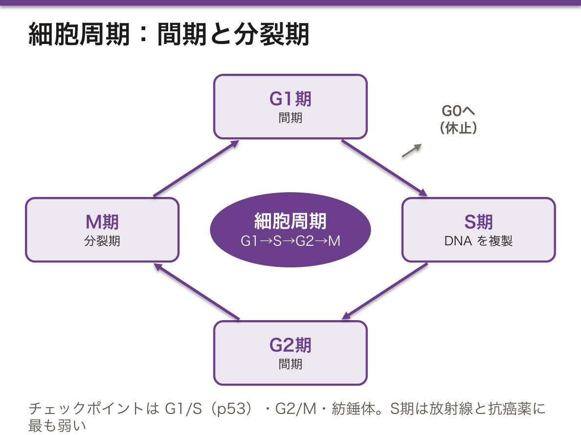
間期(G1・S・G2)と分裂期(M)。G0 は周期から外れた休止状態。
- S期——DNAを複製する。この時期の細胞だけが放射線と抗癌薬に最も弱い
- チェックポイント——G1/S(p53 が働く)・G2/M・紡錘体
分裂期の4相——前期・中期・後期・終期。中期に染色体が赤道面に並び、染色体分析はこの時期を止めて行う(コルヒチン)。
減数分裂——2回の分裂で染色体が半分になる。第一分裂で相同染色体が分かれ、第二分裂で姉妹染色分体が分かれる。
- 第一分裂前期で交叉(乗換え)が起き、多様性が生まれる
- 不分離が起きると異数体になる。母体年齢とともに増える
組織の分裂能——不安定細胞(表皮・腸上皮・骨髄)・安定細胞(肝・腎・線維芽細胞)・永久細胞(神経・心筋・骨格筋)。
3つの分裂能の区分が、修復の形を決めている。永久細胞は再生できないので、瘢痕で埋まる(修復と創傷治癒の章)——「心筋梗塞の後に線維化が残る理由が、この分類にある」。
逆に不安定細胞は、放射線と抗癌薬の副作用が最初に出る場所になる。骨髄抑制・粘膜炎・脱毛(抗癌薬の副作用の章)——「速く分裂する組織ほど、先に傷む」。
そして副作用の出る時期も、分裂の速さから予測できる。粘膜炎は1週前後、骨髄抑制は10〜14日——「時間の順番が、細胞の入れ替わりの速さの順番になっている」。
p53 は G1/S のチェックポイントを守っている。壊れると傷ついたDNAのまま複製が進む(癌抑制遺伝子の章)——「最も多くの癌で変異している遺伝子になる」。
減数分裂の不分離が、Down 症候群を含む異数性を生む。大半が母親の第一分裂で起きる(染色体異常の章)——「母体年齢との関係が、この段階の話になる」。
第一分裂は、胎生期に始まって排卵まで止まっている。つまり卵母細胞は何十年も途中で待機している(卵胞の章)——「待っている時間の長さが、そのまま誤りの起きやすさになる」。
抗癌薬と放射線は、細胞周期のどこを狙うかで分類される。
S 期 — 代謝拮抗薬(メトトレキサート・5-FU・シタラビン)。M 期 — ビンカアルカロイド・タキサン。周期非特異的 — アルキル化薬・白金製剤。
正常組織でも回転の速いところが同じように傷む——骨髄抑制・粘膜炎(口内炎・下痢)・脱毛という共通の副作用は、周期を止める薬である以上避けられない。
p53 は G1/S チェックポイントの門番で、ヒトの癌で最も高頻度に変異する遺伝子である。生殖細胞系列の変異が Li–Fraumeni 症候群。
減数分裂の不分離が異数性を生む。母体年齢が上がるほど増えるのは、卵母細胞が第一減数分裂の途中で何十年も止まっているためで、21トリソミーのリスク上昇の根拠になっている。
English version
Core
Interphase (G1, S, G2) and mitosis (M). G0 is a resting state outside the cycle.
- S phase — DNA is replicated. Cells in this phase are the most vulnerable to radiation and cytotoxic drugs.
- Checkpoints — G1/S (guarded by p53), G2/M, and the spindle checkpoint.
Mitosis has four stages — prophase, metaphase, anaphase, telophase. Chromosomes align at the equatorial plate in metaphase, and karyotyping is done by arresting cells there with colchicine.
Meiosis — two divisions halve the chromosome number: the first separates homologues, the second the sister chromatids. Crossing over in prophase I generates diversity; non-disjunction produces aneuploidy and increases with maternal age.
Proliferative capacity of tissues — labile (epidermis, intestinal epithelium, bone marrow), stable (liver, kidney, fibroblasts), permanent (neurons, cardiac and skeletal muscle).
Going further
These three categories determine the form that repair takes. Permanent cells cannot regenerate, so the defect is filled by scar — this classification is why fibrosis remains after myocardial infarction.
Conversely, labile cells are where the toxicity of radiation and chemotherapy appears first: marrow suppression, mucositis, alopecia. The faster a tissue turns over, the sooner it is damaged. The timing can be predicted from turnover as well — mucositis at about one week, marrow suppression at ten to fourteen days. The order in time is the order of the speed of renewal.
p53 guards the G1/S checkpoint; when it fails, replication proceeds with damaged DNA. It is the gene most frequently mutated across human cancers.
Non-disjunction in meiosis produces aneuploidy including Down syndrome, and most arises in the mother’s first division. That division begins in fetal life and is arrested until ovulation, so an oocyte waits, part-way through, for decades. The length of the wait is itself the liability to error — and the basis of the relationship with maternal age.
In the clinic
Cytotoxic drugs and radiation are classified by where in the cycle they act.
- S phase — antimetabolites (methotrexate, 5-FU, cytarabine).
- M phase — vinca alkaloids, taxanes.
- Cycle non-specific — alkylating agents, platinum compounds.
Normal tissues with rapid turnover are injured in the same way: marrow suppression, mucositis (stomatitis, diarrhoea) and alopecia are unavoidable for any drug that arrests the cycle.
p53 is the gatekeeper of the G1/S checkpoint and the most frequently mutated gene in human cancer; a germline mutation gives Li–Fraumeni syndrome.
Non-disjunction in meiosis produces aneuploidy. The rise with maternal age occurs because the oocyte remains arrested part-way through the first meiotic division for decades — the basis of the increasing risk of trisomy 21.
Ki-67 — 細胞周期に入っている核だけが茶色になる
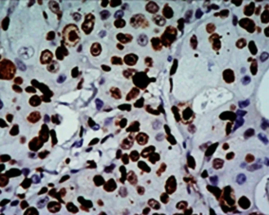
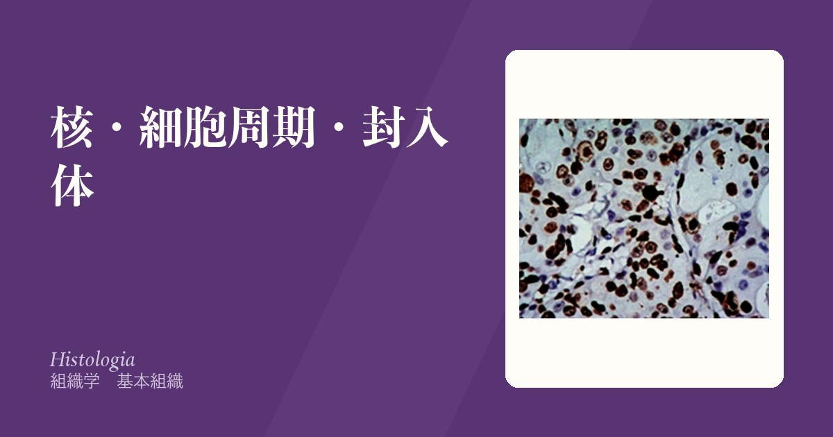
Ki-67 は核の蛋白で、細胞周期の G1・S・G2・M 期のすべてで発現し、休止している G0 期の細胞には無い。量は周期が進むほど増え、分裂期には染色体の表面を覆う。名前は、ドイツのキールで作られた抗体の67番目の株に由来する。
標本は免疫組織化学で作る。Ki-67 に結合する抗体に酵素を付けて、DAB という基質から茶色の沈殿を作らせ、背景の核をヘマトキシリンで青に染める。読み方は単純で、
- 茶色の核——周期に入っている細胞(陽性)
- 青い核——G0 で休んでいる細胞(陰性)
陽性の核が全体の何%かをKi-67 標識率(labeling index)として数える。
正常の腎尿細管では茶色の核はわずかで、ほとんどが青い。尿細管上皮はふだん G0 で休んでいるからである。
なぜ分裂像を数えるだけでは足りないのか。分裂期(M期)は周期の中で1時間ほどしかなく、標本に写る分裂像は少ない。Ki-67 は G1 から M まで全部を拾うので、増殖している細胞の割合(増殖分画)をそのまま見積もれる。
尿細管で陽性が少ないことには意味がある。尿細管上皮はふだんは休んでいるが、壊れれば周期に戻れる細胞である。急性尿細管壊死のあとでは Ki-67 陽性の核が一気に増え、残った細胞が分裂して尿細管を作り直していることが分かる。
Ki-67 は腫瘍の増える速さの指標として日常的に使われる。
- 乳癌——ホルモン受容体陽性の癌で、Ki-67 が低いもの(ルミナル A 型)と高いもの(B 型)を分け、化学療法を加えるかの判断材料にする
- 消化器の神経内分泌腫瘍——Ki-67 標識率で G1(3%未満)・G2(3〜20%)・G3(20%超)を決める
- リンパ腫——Burkitt リンパ腫ではほぼ100%になる
数える場所(最も陽性が多い領域)と数え方で値が変わるので、画像解析で数える施設も増えている。
English version
Core
Ki-67 is a nuclear protein expressed in all of G1, S, G2 and M, and absent in resting G0 cells. Its amount rises through the cycle, and in mitosis it coats the chromosome surface. The name comes from clone 67 of an antibody made in Kiel, Germany.
The slide is made by immunohistochemistry. An antibody against Ki-67 carries an enzyme that turns the substrate DAB into a brown precipitate, and the other nuclei are counterstained blue with haematoxylin. Reading it is simple:
- brown nucleus — a cell in the cycle (positive);
- blue nucleus — a cell resting in G0 (negative).
The percentage of positive nuclei is the Ki-67 labelling index.
In normal renal tubules few nuclei are brown and most are blue, because tubular cells usually rest in G0.
Going further
Why is counting mitoses not enough? Mitosis (M phase) lasts only about an hour of the cycle, so few mitotic figures are caught on a slide. Ki-67 picks up the whole span from G1 to M, so it estimates the fraction of proliferating cells (growth fraction) directly.
The few positive tubular cells mean something. Tubular epithelium usually rests but can return to the cycle when damaged. After acute tubular necrosis, Ki-67-positive nuclei rise sharply, showing that surviving cells divide to rebuild the tubule.
In the clinic
Ki-67 is used every day as a measure of how fast a tumour grows.
- Breast cancer — among hormone-receptor-positive cancers it separates low-Ki-67 (luminal A) from high-Ki-67 (luminal B) tumours and helps decide whether to add chemotherapy.
- Digestive neuroendocrine tumours — the labelling index sets the grade: G1 (<3%), G2 (3–20%), G3 (>20%).
- Lymphoma — almost 100% in Burkitt lymphoma.
The value changes with where (the most positive area) and how one counts, so more laboratories count with image analysis.
アポトーシスと壊死 — 死に方が周りを変えるApoptosis
アポトーシス——計画された死。細胞が縮み、核が凝縮・断片化し、小片(アポトーシス小体)になって食べられる。
- 膜は破れない。内容物が漏れない——だから炎症を起こさない
- 1個ずつ散発的に起きる
- 好酸性の丸い小体として見える(肝では Councilman 小体)
- 経路——内因性(ミトコンドリア・Bcl-2 家族)と外因性(Fas・TNF受容体)。どちらもカスパーゼに集まる
壊死——事故死。細胞が膨れ、膜が破れ、内容物が漏れる。
- 強い炎症を起こす。集団で起きる
- 核の変化——濃縮(pyknosis)→ 断片化(karyorrhexis)→ 融解(karyolysis)
壊死の型——凝固壊死(梗塞)・液状壊死(脳・膿瘍)・乾酪壊死(結核)・脂肪壊死(膵)・フィブリノイド壊死(血管炎)。
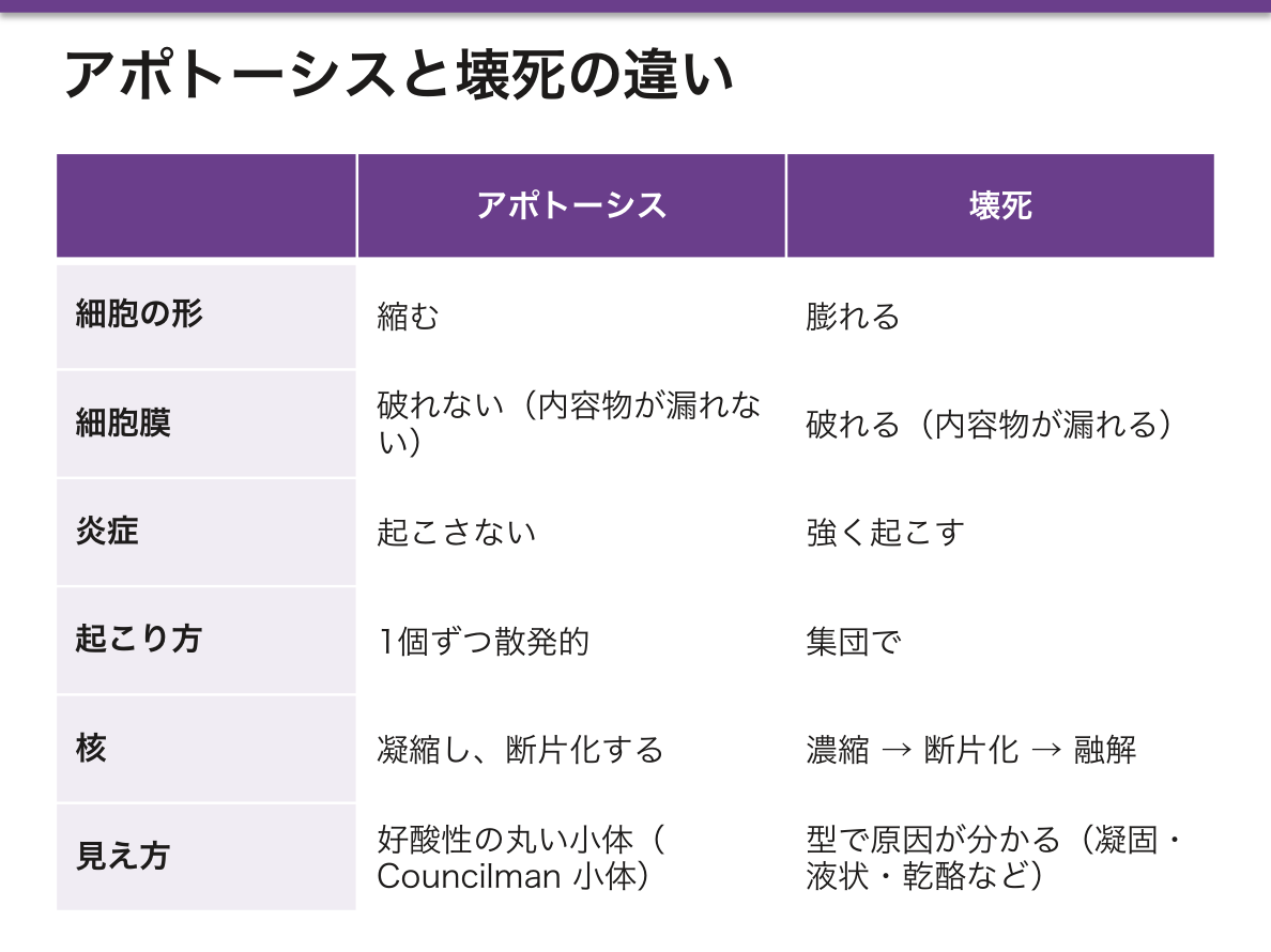
「炎症を起こすかどうか」が、この2つを分ける最も実用的な違いになる。だからアポトーシスが主体の肝炎では、肝細胞が死んでいても炎症が乏しいことがある(細胞死の章)——「死の量と、炎症の量は別に測る」。
脳だけが液状壊死になるのは、脂質が多く支持組織の膠原線維が乏しいからになる(脳梗塞の章)——「他の臓器では固まる梗塞が、脳では溶けて空洞になる」。
だから陳旧性の脳梗塞は、嚢胞として画像に写る(頭部CTの章)——「組織の死に方が、そのまま画像の所見になっている」。
乾酪壊死は、結核をほぼ言い当てる所見になる。類上皮細胞と Langhans 巨細胞に囲まれた、白いチーズ状の中心(結核の章)——「肉芽腫に乾酪があるかどうかが、鑑別を大きく絞る」。
アポトーシスの抑制が、癌の成立条件の1つになる。Bcl-2 の過剰発現が濾胞性リンパ腫を作る(発癌の章)——「増えるからではなく、死ななくなるから増える癌がある」。
そして治療の側も、この経路を使う。Bcl-2 阻害薬が慢性リンパ性白血病に効く(分子標的薬の章)——「死ねなくなった細胞に、死ぬ道を返す薬になる」。
死に方が違えば、後に残るものが違う。
アポトーシスは膜が破れないので炎症を起こさない。ウイルス感染細胞の排除・胚発生での指の形成・免疫寛容の成立はすべてこれによる。アポトーシスが足りなければ腫瘍と自己免疫、過剰なら変性疾患になる。
壊死は内容物が漏れて炎症を起こす。型で原因が推定できる。凝固壊死=虚血(心・腎・脾)、融解壊死=脳と化膿、乾酪壊死=結核、脂肪壊死=急性膵炎、フィブリノイド壊死=血管炎。壊死の顔つきを見れば、何が起きたかが分かる。
脳だけが融解壊死になるのは、脂質が多くコラーゲンの支持組織が乏しいためで、梗塞巣は最終的に嚢胞になる。
心筋逸脱酵素・トロポニンが測れるのは、壊死した細胞の中身が血中へ漏れ出すからである。
English version
Core
Apoptosis — programmed death. The cell shrinks, the nucleus condenses and fragments, and the remnants (apoptotic bodies) are engulfed. The membrane does not rupture, nothing leaks, and therefore no inflammation follows. It occurs in scattered single cells, appearing as round eosinophilic bodies (Councilman bodies in the liver). Two pathways — intrinsic (mitochondrial, the Bcl-2 family) and extrinsic (Fas, TNF receptor) — both converging on the caspases.
Necrosis — accidental death. The cell swells, the membrane ruptures, the contents leak, and intense inflammation follows. It occurs in groups of cells.
- Nuclear changes — pyknosis → karyorrhexis → karyolysis.
- Types — coagulative (infarct), liquefactive (brain, abscess), caseous (tuberculosis), fat (pancreas), fibrinoid (vasculitis).
Going further
Whether inflammation follows is the most practical difference between the two. In hepatitis dominated by apoptosis, hepatocytes may be dying with little inflammation — measure the amount of death and the amount of inflammation separately.
Only the brain undergoes liquefactive necrosis, because it is rich in lipid and poor in supporting collagen — an infarct that would solidify elsewhere dissolves into a cavity here. Hence an old cerebral infarct appears on imaging as a cyst: the way the tissue died becomes the radiological finding.
Caseous necrosis all but names tuberculosis: a white, cheesy centre surrounded by epithelioid cells and Langhans giant cells. Whether a granuloma is caseating narrows the differential sharply.
Suppression of apoptosis is one of the conditions for cancer. Overexpression of Bcl-2 produces follicular lymphoma — some cancers accumulate not because they divide but because they stop dying. Treatment uses the same pathway: a Bcl-2 inhibitor works in chronic lymphocytic leukaemia — a drug that gives back the route to death.
In the clinic
A different way of dying leaves something different behind.
Apoptosis does not rupture the membrane and so causes no inflammation. Elimination of virus-infected cells, formation of the digits in the embryo and the establishment of immune tolerance all depend on it. Too little gives tumours and autoimmunity; too much gives degenerative disease.
Necrosis spills the contents and provokes inflammation, and the type suggests the cause: coagulative = ischaemia (heart, kidney, spleen); liquefactive = brain and suppuration; caseous = tuberculosis; fat = acute pancreatitis; fibrinoid = vasculitis. Read the face of the necrosis and you know what happened.
Only the brain liquefies, because of its high lipid and low collagen content, and the infarct ends as a cyst.
Cardiac enzymes and troponin can be measured because the contents of necrotic cells leak into the blood.
核の像から異型を読む — 良性と悪性を分ける手がかりAtypia
「悪性らしさ」は、核の5つの所見に集約される。
- 核腫大・N/C比の上昇——細胞質に対して核が大きい
- 核の大小不同(多形性)——同じ集団の中で核の大きさが揃わない
- クロマチンの増量と粗大化——核が濃く染まる(濃染)
- 核小体の明瞭化・大型化——リボソーム産生が盛んなことの反映
- 核形不整・核膜の肥厚——切れ込み・溝
分裂像の多さと、異常な分裂像(三極分裂など)も重要になる。
細胞の配列も見る——極性の乱れ(核が並ぶ高さが揃わない)・重積。
ただし核の異型だけでは癌と言えない——浸潤(基底膜を越えているか)が最終的な根拠になる。
核の所見が「悪性らしさ」になるのは、それが増殖と転写の亢進の姿だからである。DNA 量が増えればクロマチンが増え、蛋白を大量に作れば核小体が目立つ(遺伝情報の流れの章)——「形態の異常は、代謝の亢進が見えているだけになる」。
だから再生や炎症でも同じ像が出る。修復中の上皮は核が大きく核小体も目立ち、悪性と紛らわしい(反応性異型)(修復と創傷治癒の章)——「勢いよく増えている細胞は、良性でも悪く見える」。
見分けの鍵は、変化に段階があるかどうかになる。反応性の異型は周囲へなだらかに移行し、腫瘍は境界で急に変わる——「連続していれば反応、途切れていれば腫瘍を疑う」。
そして核の所見が、そのまま診断名になっている腫瘍がある。甲状腺乳頭癌のすりガラス様核・核溝・核内細胞質封入体は、それ自体が診断根拠になる(甲状腺の章)——「浸潤を見なくても、核だけで決まる例外がある」。
核異型は「悪性らしさ」の指標だが、単独では診断にならない。浸潤の有無が最終的な決め手である。異型は強いが基底膜を越えていなければ上皮内癌、異型が乏しくても浸潤していれば癌(高分化腺癌)。
細胞診の判定はこの核所見で行う。子宮頸部細胞診(Pap)でN/C 比の上昇・クロマチンの増量・核形不整を見る。コイロサイト(核周囲の空洞化)はHPV 感染の印。
異型が炎症でも出ることに注意する。修復・再生(reactive atypia)では核が大きくなるが、核小体は目立っても核縁は滑らかで、細胞集団としての極性が保たれる。炎症の強い時期の生検は判定を誤りやすいので、治療してから採り直すことがある。
異型度(grade)は分化度、進行度(stage)は広がり——別の軸であり、混同しない。
English version
Core
"Looking malignant" reduces to five nuclear features.
- Nuclear enlargement and a raised N/C ratio — the nucleus is large relative to the cytoplasm.
- Anisonucleosis (pleomorphism) — nuclei within one population are not of uniform size.
- Increased and coarse chromatin — the nucleus stains darkly (hyperchromasia).
- Prominent, enlarged nucleoli — reflecting vigorous ribosome production.
- Irregular nuclear outline and thickened nuclear membrane — notches and grooves.
The number of mitoses and abnormal mitotic figures (tripolar, for example) also matter, as does the arrangement of the cells — loss of polarity and stratification.
Nuclear atypia alone does not make a carcinoma: invasion, whether the basement membrane has been crossed, is the final criterion.
Going further
These nuclear features signify malignancy because they are the appearance of increased proliferation and transcription. More DNA gives more chromatin; heavy protein synthesis gives prominent nucleoli. The morphological abnormality is simply raised metabolism made visible.
Hence the same picture appears in regeneration and inflammation. Repairing epithelium has large nuclei and prominent nucleoli and is easily mistaken for malignancy (reactive atypia) — a vigorously proliferating cell looks bad even when it is benign.
The key to distinguishing them is whether the change is graded. Reactive atypia merges gradually with the surrounding tissue; a tumour changes abruptly at its edge. Continuity suggests reaction, discontinuity suggests neoplasia.
And in some tumours the nuclear features are themselves the diagnosis: the ground-glass nuclei, nuclear grooves and intranuclear cytoplasmic inclusions of papillary thyroid carcinoma — an exception in which the nucleus decides without invasion having to be seen.
In the clinic
Nuclear atypia indicates malignancy but is not diagnostic on its own — invasion is the decisive criterion. Marked atypia without breach of the basement membrane is carcinoma in situ; slight atypia with invasion is still carcinoma (a well-differentiated adenocarcinoma).
Cytology is judged on these nuclear findings. Cervical cytology (Pap) looks for a raised N/C ratio, increased chromatin and irregular nuclear outline. The koilocyte, with perinuclear clearing, is the mark of HPV infection.
Beware that atypia also occurs in inflammation. In repair and regeneration the nuclei enlarge, but the nuclear outline stays smooth even when nucleoli are prominent, and the polarity of the cell group is preserved. A biopsy taken during intense inflammation is easily misread, so it is sometimes repeated after treatment.
Grade is differentiation and stage is extent — two separate axes; do not confuse them.
化生 — 上皮が、別の種類の上皮に置き換わるMetaplasia
分化した細胞が、別の分化した細胞に置き換わる可逆的な変化。すでに分化した細胞が変身するのではなく、幹細胞の分化の向きが変わる。
| 場所 | 元 | 化生後 | 刺激 |
|---|---|---|---|
| 気管支 | 多列線毛円柱上皮 | 重層扁平上皮 | 喫煙 |
| 食道下部 | 重層扁平上皮 | 円柱上皮(腸上皮化生) | 胃酸の逆流(Barrett 食道) |
| 胃 | 胃粘膜上皮 | 腸型の上皮(杯細胞を持つ) | Helicobacter pylori |
| 子宮頸部 | 円柱上皮 | 重層扁平上皮 | 腟の酸性環境(生理的) |
| 膀胱 | 尿路上皮 | 扁平上皮 | 慢性の結石・住血吸虫 |
刺激が去れば戻る。ただし化生した粘膜は、その場に本来ふさわしい機能を失っている。
異形成は化生と違い、細胞の異型を伴う——化生=種類が変わる/異形成=出来が悪くなる。
化生は「適応」であって、そこで話が終わらない。刺激に強い上皮に置き換わるが、代わりに元の仕事ができなくなる——「気管支が扁平上皮になれば煙には強いが、線毛が無いので痰を運べない」。
だから化生は、感染を呼ぶ。喫煙者の気道が感染を繰り返す理由の一部がここにある(呼吸器の章)——「守りを固めた場所が、掃除をやめている」。
そして化生した上皮からは、その化生した型の癌が出る。
| 化生 | そこから出る癌 |
|---|---|
| 気管支の扁平上皮化生 | 扁平上皮癌 |
| Barrett 食道 | 腺癌(本来の食道癌は扁平上皮癌) |
| 胃の腸上皮化生 | 腸型胃癌 |
(前がん病変の章)——「その場所に出るはずのない型の癌を見たら、化生を疑う」。
子宮頸部の扁平上皮化生だけは生理的だが、その移行帯こそが癌の好発部位になる。分化の向きが切り替わっている場所は、間違いも起きやすい(子宮頸癌の章)——「細胞診で狙って擦るのは、この帯になる」。
化生は適応であり、それ自体は可逆である。しかし異形成 → 上皮内癌 → 浸潤癌へ進む道の入口でもある。
Barrett 食道 — 食道下部の重層扁平上皮が円柱上皮(腸型)に置き換わる。胃食道逆流が原因で、食道腺癌のリスクになる。定期的な内視鏡監視の対象である。
気管支の扁平上皮化生 — 喫煙による。線毛を失うので粘液が運べなくなる。扁平上皮癌の前段階になる。
胃の腸上皮化生 — H. pylori による慢性萎縮性胃炎。胃癌(腸型)のリスク。除菌が予防になる。
子宮頸部の扁平上皮化生が起こる場所(移行帯)が、そのまま HPV の標的であり頸癌の発生部位である。細胞診で狙って擦る場所でもある——化生の場所を知っていることが、検診の手技そのものになっている。
English version
Core
A reversible change in which one differentiated cell type is replaced by another. An already differentiated cell does not transform; rather, the direction in which the stem cell differentiates changes.
| Site | Original | After metaplasia | Stimulus |
|---|---|---|---|
| Bronchus | Pseudostratified ciliated columnar | Stratified squamous | Smoking |
| Lower oesophagus | Stratified squamous | Columnar (intestinal) | Acid reflux (Barrett oesophagus) |
| Stomach | Gastric epithelium | Intestinal type, with goblet cells | Helicobacter pylori |
| Cervix | Columnar | Stratified squamous | The acid vaginal environment (physiological) |
| Bladder | Urothelium | Squamous | Chronic calculi, schistosomiasis |
It reverses if the stimulus is removed, but the metaplastic mucosa has lost the function proper to that site. Metaplasia = a change of type; dysplasia = a change in quality, with cellular atypia.
Going further
Metaplasia is an adaptation, but the story does not end there. The epithelium is replaced by one that resists the stimulus, but in exchange it can no longer do its original work — a squamous bronchus resists smoke but has no cilia and cannot move sputum.
So metaplasia invites infection: part of the reason a smoker’s airway is repeatedly infected. A place that has strengthened its defences has stopped cleaning itself.
And the cancer that arises from metaplastic epithelium is of the metaplastic type.
| Metaplasia | Cancer that arises |
|---|---|
| Bronchial squamous metaplasia | Squamous cell carcinoma |
| Barrett oesophagus | Adenocarcinoma (the native oesophageal cancer is squamous) |
| Gastric intestinal metaplasia | Intestinal-type gastric carcinoma |
If you see a type of cancer that should not occur at that site, suspect metaplasia.
Only cervical squamous metaplasia is physiological — and yet that transformation zone is precisely where carcinoma arises. Where the direction of differentiation is being switched, errors are also more likely. It is this zone that cytology deliberately samples.
In the clinic
Metaplasia is an adaptation and is in itself reversible, but it is also the entrance to the road dysplasia → carcinoma in situ → invasive carcinoma.
Barrett oesophagus — the stratified squamous epithelium of the lower oesophagus is replaced by columnar, intestinal-type epithelium. Caused by gastro-oesophageal reflux; a risk factor for oesophageal adenocarcinoma and an indication for endoscopic surveillance.
Bronchial squamous metaplasia — from smoking. Cilia are lost so mucus cannot be transported; it precedes squamous cell carcinoma.
Gastric intestinal metaplasia — from H. pylori chronic atrophic gastritis; a risk for intestinal-type gastric cancer, and eradication is preventive.
The site of cervical squamous metaplasia — the transformation zone — is the target of HPV and the site of cervical carcinoma, and it is where the smear is deliberately taken. Knowing where metaplasia occurs is the technique of screening itself.
制御された壊死 — 細胞の死に方は、2つでは足りない
長らく細胞死はアポトーシス(きれいに片づく死)と壊死(事故死)の2つとされてきた。しかし壊死に見える死のなかにも、遺伝子に書かれた手順どおりに進むものがあることが分かっている。
| ネクロプトーシス | RIPK1・RIPK3・MLKL が膜に穴を開ける | カスパーゼが働けないときの代替の死。ウイルスがカスパーゼを止めても細胞は死ねる |
| パイロトーシス | インフラマソーム → カスパーゼ1 → ガスダーミン D が孔を作る | 細胞が膨れて破裂し、IL-1β を放出する。強い炎症を伴う |
| フェロトーシス | 鉄依存性の脂質過酸化。GPX4 が守っている | 膜の脂質が酸化されて破綻する |
| ネトーシス | 好中球が自らのクロマチンを網として放出する | 菌を捕らえるが、血栓と自己免疫の材料にもなる |
組織像で「壊死」と読んだものが、実は制御された死である場合がある。この区別が意味を持つのは、止められるかどうかが変わるからである。事故なら防げないが、手順があるなら薬で介入できる。
炎症を起こすかどうかが、決定的な違いになる。
- アポトーシス——膜が保たれたままマクロファージに食べられる。中身が漏れないので炎症を起こさない
- 制御された壊死——膜が破れ、細胞内の分子(DAMPs:HMGB1・ATP・DNA)が漏れる。これが自然免疫の受容体を刺激して強い炎症を起こす
だから「無菌なのに激しい炎症が起きる」病態——虚血再灌流障害・急性膵炎・薬剤性肝障害・痛風——は、この経路で説明されるようになった。感染がなくても、細胞が破れれば免疫は動く。
疾患と結びつく例が増えている。
- フェロトーシス——虚血再灌流障害・神経変性。鉄と脂質過酸化に依存するので、鉄キレートや抗酸化で抑えられる可能性がある
- パイロトーシス——家族性地中海熱などの自己炎症性疾患。インフラマソームが暴走して IL-1β が出続けるので、IL-1 を標的とする薬が効く
- ネトーシス——敗血症の血栓症、抗好中球細胞質抗体関連血管炎、全身性エリテマトーデス。出した DNA が自己抗原になる
「感染していないのに CRP が高い」ときは、壊れた細胞そのものが炎症の原因である可能性を考える。抗菌薬を足す前に、壊死している臓器を探す。
English version
Core
Cell death was long divided into apoptosis (a tidy death) and necrosis (an accidental one). It is now clear that some deaths that look necrotic proceed by a genetically written programme.
| Form | Mechanism | Meaning |
|---|---|---|
| Necroptosis | RIPK1, RIPK3 and MLKL punch holes in the membrane | An alternative death when caspases cannot work — the cell can still die even if a virus blocks the caspases |
| Pyroptosis | Inflammasome → caspase-1 → gasdermin D forms pores | The cell swells and bursts, releasing IL-1β; intense inflammation |
| Ferroptosis | Iron-dependent lipid peroxidation, normally restrained by GPX4 | Membrane lipids are oxidised and the membrane fails |
| NETosis | A neutrophil casts out its own chromatin as a net | Traps organisms, but also becomes the material of thrombosis and autoimmunity |
Going further
What has been read on a slide as "necrosis" may in fact be a regulated death, and the distinction matters because it changes whether it can be stopped. An accident cannot be prevented; a programme can be interrupted with a drug.
Whether inflammation follows is the decisive difference.
- Apoptosis — the membrane stays intact and macrophages engulf the cell; nothing leaks, so no inflammation.
- Regulated necrosis — the membrane ruptures and intracellular molecules (DAMPs: HMGB1, ATP, DNA) escape, stimulating innate immune receptors and provoking intense inflammation.
So conditions in which violent inflammation occurs without infection — ischaemia–reperfusion injury, acute pancreatitis, drug-induced liver injury, gout — have come to be explained by this route. The immune system responds to a ruptured cell even in the absence of infection.
The disease associations are growing. Ferroptosis in ischaemia–reperfusion and neurodegeneration, potentially treatable by iron chelation or antioxidants. Pyroptosis in autoinflammatory disease such as familial Mediterranean fever, where a runaway inflammasome keeps releasing IL-1β — hence the effectiveness of IL-1 targeted drugs. NETosis in septic thrombosis, ANCA-associated vasculitis and SLE, where the expelled DNA becomes a self-antigen.
In the clinic
When the CRP is high without infection, consider that the broken cells themselves are the cause of the inflammation. Before adding another antibiotic, look for the organ that is dying.
封入体 — 核や細胞質の中の塊が、原因を名指しする
細胞の中に見える異常な塊を封入体と呼ぶ。形と場所で、原因がかなり絞れるので、HE 染色だけで診断に近づける数少ない所見になる。
| 核内・好酸性・周りが抜ける | Cowdry A 型 | 単純ヘルペス・水痘帯状疱疹 |
| 核内+細胞質にも/細胞が巨大 | フクロウの目 | サイトメガロウイルス |
| 細胞質・好酸性・神経細胞 | Negri 小体 | 狂犬病 |
| 細胞質・好酸性・肝細胞 | Mallory-Denk 体 | アルコール性肝炎 |
| 細胞質・すりガラス状の肝細胞 | HBs 抗原の蓄積 | B型慢性肝炎 |
封入体は「捨てられなかったもの」の姿である。由来で3つに分かれる。
- ウイルスの粒子そのもの——ヘルペス、CMV。組み立てられた粒子が核や細胞質に溜まっている
- 細胞が作った蛋白の凝集——Mallory-Denk 体は中間径フィラメント(ケラチン 8/18)の塊。神経変性疾患の Lewy 小体(α-シヌクレイン)、神経原線維変化(タウ)も同じ仲間で、折り畳みに失敗した蛋白を分解しきれずに集めたものである
- 取り込んだ異物——じん肺の粉塵、ヘモジデリン
2番目の見方が現代的で重要である。凝集した蛋白を1か所に集めること自体が、細胞の防御でもある——散らばった凝集体のほうが毒性が高い。封入体は病気の原因ではなく、細胞が対処しようとした跡として見るほうが実態に近い。
移植後や HIV で下痢・網膜炎・肝炎が出たら CMV を疑い、生検でフクロウの目を探す。免疫染色で確定させる。
English version
Core
An abnormal mass within a cell is an inclusion body. Because the form and the site narrow the cause considerably, it is one of the few findings that approaches a diagnosis on H&E alone.
| Appearance | Name | Cause |
|---|---|---|
| Intranuclear, eosinophilic, with a clear halo | Cowdry type A | Herpes simplex, varicella-zoster |
| Intranuclear and cytoplasmic; the cell is enlarged | Owl’s eye | Cytomegalovirus |
| Cytoplasmic, eosinophilic, in neurons | Negri body | Rabies |
| Cytoplasmic, eosinophilic, in hepatocytes | Mallory–Denk body | Alcoholic hepatitis |
| Cytoplasmic; ground-glass hepatocyte | Accumulated HBsAg | Chronic hepatitis B |
Going further
An inclusion body is the appearance of something that could not be disposed of, and there are three origins.
- Viral particles themselves — herpes, CMV: assembled particles accumulated in the nucleus or cytoplasm.
- Aggregates of protein made by the cell — the Mallory–Denk body is a mass of intermediate filaments (keratin 8/18). The Lewy body (α-synuclein) and the neurofibrillary tangle (tau) of neurodegenerative disease belong to the same family: misfolded protein gathered together because it could not be degraded.
- Ingested foreign material — the dust of pneumoconiosis, haemosiderin.
The second view is the modern and important one. Gathering aggregated protein into one place is itself a defence — dispersed aggregates are more toxic than collected ones. An inclusion body is closer to the trace of the cell’s attempt to cope than to the cause of the disease.
In the clinic
When diarrhoea, retinitis or hepatitis appears after transplantation or in HIV, suspect CMV and look for the owl’s eye on biopsy, then confirm by immunohistochemistry.
細胞質封入体(1)— 蓄えとしてのグリコーゲンと脂肪滴Inclusiones cytoplasmicae
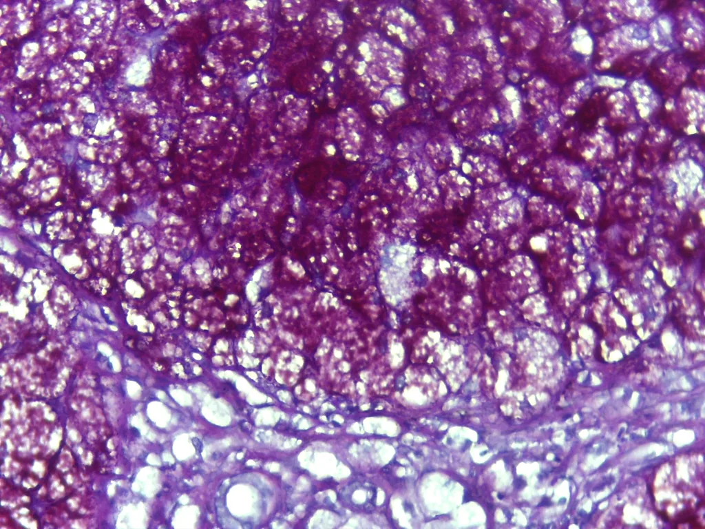
封入体は、細胞が作ったり取り込んだりした物質が一時的にたまったものである。小器官とちがって自分の仕事をする仕組みを持たず、細胞の状態によって現れたり消えたりする。正常の細胞でもふつうに見られる。ヨーロッパの教科書では次の4群に分ける。
| 群 | 例 |
|---|---|
| 栄養性(蓄え) | 肝細胞・筋のグリコーゲン、脂肪滴 |
| 分泌性 | 腺細胞の分泌顆粒(酵素原顆粒など) |
| 排泄性 | 細胞の外へ出す前の代謝産物 |
| 色素性 | メラニン、リポフスチン、ヘモジデリン、外から入った色素(次の項目) |
グリコーゲンは膜に包まれずに細胞質に散らばる粒で、肝細胞では花のように集まる(α粒子)。脂肪滴は中性脂肪とコレステロールエステルの塊で、膜はリン脂質の1層だけである。
どちらもふつうの HE 標本ではほとんど見えない。理由が違う。
グリコーゲンは水に溶けやすく、固定と染色のあいだに一部が流れ出て、細胞質が明るく抜けて見える。証明するにはPAS 反応を使う。過ヨウ素酸が糖の隣り合う水酸基を切ってアルデヒドを作り、それに Schiff 試薬が結合して赤紫に染まる。Best のカルミン法ではグリコーゲンが鮮やかな赤の粒になる。PAS は粘液や基底膜も染めるので、ジアスターゼ(アミラーゼ)で前処理すると色が消えることを対照に置いて、グリコーゲンだと確かめる。
脂肪滴はパラフィン標本を作るときのアルコールとキシレンに溶けて流れ出る。HE では丸い空胞が残るだけである。見るには凍結切片(またはオスミウム酸で固定)を使い、Sudan III・Oil red O(橙赤)やSudan Black B(黒)で染める。これらの色素は化学反応ではなく、溶媒より脂肪によく溶ける性質で脂肪に移っていく。
肝細胞のグリコーゲンは食後に増え、空腹で減る。封入体の量は細胞の今の状態を映す。
糖原病では、グリコーゲンを分解できずに肝細胞や筋にたまる。I型(von Gierke 病)は肝腫大と空腹時の低血糖、II型(Pompe 病)はリソソームの中にたまり心筋と骨格筋を侵す。
腎細胞癌の大半を占める淡明細胞型が「淡明」なのは、細胞質のグリコーゲンと脂肪が標本作製で抜けるからである。逆に α1-アンチトリプシン欠乏症の肝細胞には、ジアスターゼで消えない PAS 陽性の小球がたまる——ジアスターゼの対照がここで診断に使われる。
脂肪肝では脂肪滴の大きさが原因を示唆する。大きな1滴が核を押しのける大滴性はアルコールや肥満に、細かい滴が細胞質を埋める小滴性はミトコンドリアの脂肪酸酸化が止まる病態(妊娠性急性脂肪肝、Reye 症候群など)に多い。
English version
Core
An inclusion is a temporary store of material that a cell has made or taken in. Unlike an organelle it has no machinery of its own, and it appears and disappears with the cell’s state. Normal cells have them too. European textbooks divide them into four groups.
| Group | Examples |
|---|---|
| Trophic (stores) | glycogen in liver and muscle, lipid droplets |
| Secretory | secretory granules of gland cells (e.g., zymogen granules) |
| Excretory | metabolic products waiting to be removed |
| Pigment | melanin, lipofuscin, haemosiderin, pigments from outside (next item) |
Glycogen is scattered in the cytoplasm as particles with no membrane; in hepatocytes they gather into rosettes (α particles). Lipid droplets are masses of triglyceride and cholesterol ester, bounded only by a single layer of phospholipid.
Going further
Both are hard to see on routine H&E, for different reasons.
Glycogen dissolves easily in water, so part of it washes out during processing and the cytoplasm looks pale and empty. To prove it, use the PAS reaction: periodic acid cuts neighbouring hydroxyl groups of sugars into aldehydes, Schiff reagent binds them, and the result is magenta. With Best’s carmine, glycogen appears as bright red granules. PAS also stains mucins and basement membranes, so a control pre-treated with diastase (amylase), in which the colour disappears, confirms that it is glycogen.
Lipid droplets dissolve in the alcohol and xylene used for paraffin sections. H&E shows only round empty holes. To see them, use frozen sections (or osmium fixation) and stain with Sudan III or Oil red O (orange-red) or Sudan Black B (black). These dyes do not react chemically; they move into fat because they dissolve better in fat than in their solvent.
Hepatocyte glycogen rises after meals and falls in fasting. The amount of inclusions reflects the cell’s present state.
In the clinic
In glycogen storage diseases, glycogen cannot be broken down and collects in hepatocytes or muscle. Type I (von Gierke) causes an enlarged liver and fasting hypoglycaemia; type II (Pompe) stores it inside lysosomes and affects heart and skeletal muscle.
The most common kidney cancer, clear cell renal cell carcinoma, looks "clear" because its glycogen and lipid are washed out in processing. In contrast, hepatocytes in α1-antitrypsin deficiency hold PAS-positive globules that diastase does not remove — here the diastase control becomes a diagnostic test.
In fatty liver, droplet size hints at the cause. Macrovesicular fat (one large drop pushing the nucleus aside) suggests alcohol or obesity; microvesicular fat (many tiny drops) suggests failure of mitochondrial fatty-acid oxidation (acute fatty liver of pregnancy, Reye syndrome and others).
細胞質封入体(2)— 色素:体の中で作られたものと、外から入ったものPigmentum
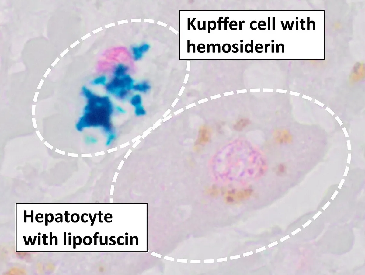
| 色素 | 色と見分け方 | どこに | |
|---|---|---|---|
| 内因性(ヘモグロビン由来) | ヘモジデリン | 黄金褐色の粒。Berlin 青(Perls)染色で青 | 出血のあとのマクロファージ、鉄過剰の肝・心 |
| ビリルビン | 黄〜緑 | 胆汁うっ滞の肝細胞・毛細胆管 | |
| 内因性(ヘモグロビン以外) | メラニン | 褐〜黒。銀で黒くなる(Fontana-Masson) | メラノサイト、角化細胞、網膜色素上皮 |
| リポフスチン | 黄褐色の細かい粒。蛍光を出す | 神経細胞・心筋・肝細胞。加齢で増える | |
| 外因性 | 炭粉 | 黒 | 肺と肺門リンパ節のマクロファージ |
| 入れ墨の色素 | 色はさまざま | 真皮のマクロファージ、所属リンパ節 | |
| トリパン青などの生体色素 | 青 | マクロファージだけが取り込む |
リポフスチンは、リソソームで分解しきれなかった脂質と蛋白の残り(残余小体)で、「消耗の色素」と呼ばれる。分裂しない長生きの細胞ほどたまる。
リンパ節の標本で、洞の中の大きな細胞が青い粒でいっぱいになり、周りのリンパ球は染まっていない像がある。これは生きた動物にトリパン青を注射し、色素を食べた細胞だけを浮かび上がらせた標本である(生体染色)。色素を取り込むのは、異物を食べてリソソームにためるマクロファージだけで、リンパ球は取り込まない。
この方法で、全身の肝・脾・リンパ節・骨髄にある「色素を食べる細胞」がひとつの系統としてまとめられた(かつての細網内皮系。今は単核食細胞系と呼ぶ)。
外から入った色素が一生残る理由も同じである。炭粉や入れ墨の色素は分解できないので、マクロファージのリソソームの中にとどまる。入れ墨が消えないのはこのためである。
ヘモジデリンの場所は原因を教える。慢性の左心不全や僧帽弁狭窄症で肺がうっ血すると、肺胞のマクロファージが漏れた赤血球を食べてヘモジデリンをためる(心不全細胞)。輸血を繰り返した鉄過剰ではまずマクロファージに、遺伝性ヘモクロマトーシスでは肝細胞そのものにたまり、肝硬変・糖尿病・皮膚の色素沈着の原因になる。
高齢者の心臓や肝臓が小さく茶色くなるのは、細胞が萎縮してリポフスチンが濃くなった状態である(褐色萎縮)。
都市生活者の肺門リンパ節は炭粉で黒い。入れ墨の色素で黒くなったリンパ節は、手術中に転移した黒色腫と見誤ることがあり、病理で確かめる。
English version
Core
| Pigment | Colour and test | Where | |
|---|---|---|---|
| Endogenous (from haemoglobin) | Haemosiderin | golden-brown granules; blue with Prussian blue (Perls) | macrophages after bleeding; liver and heart in iron overload |
| Bilirubin | yellow to green | hepatocytes and canaliculi in cholestasis | |
| Endogenous (not from haemoglobin) | Melanin | brown-black; black with silver (Fontana-Masson) | melanocytes, keratinocytes, retinal pigment epithelium |
| Lipofuscin | fine yellow-brown granules; fluorescent | neurons, heart muscle, hepatocytes; increases with age | |
| Exogenous | Carbon | black | macrophages of the lung and hilar nodes |
| Tattoo pigment | various | dermal macrophages, regional nodes | |
| Vital dyes such as trypan blue | blue | taken up only by macrophages |
Lipofuscin is the undigested remains of lipid and protein in lysosomes (residual bodies), called the "wear-and-tear pigment". Long-lived cells that do not divide collect the most.
Going further
In one lymph node slide, large cells in the sinuses are full of blue granules, while the lymphocytes around them are unstained. Trypan blue was injected into a living animal, and only the cells that ate the dye are shown (vital staining). Only macrophages, which eat foreign material and store it in lysosomes, take up the dye; lymphocytes do not.
With this method, the "dye-eating cells" of liver, spleen, lymph nodes and marrow were grouped as one system (formerly the reticuloendothelial system, now the mononuclear phagocyte system).
The same explains why pigments from outside stay for life. Carbon and tattoo ink cannot be digested, so they remain in macrophage lysosomes. That is why a tattoo does not fade away.
In the clinic
Where haemosiderin sits tells the cause. When chronic left heart failure or mitral stenosis congests the lung, alveolar macrophages eat leaked red cells and store haemosiderin (heart failure cells). In iron overload from repeated transfusion, it collects first in macrophages; in hereditary haemochromatosis it collects in hepatocytes themselves, causing cirrhosis, diabetes and skin pigmentation.
When an old person’s heart or liver becomes small and brown, the cells have shrunk and lipofuscin has become dense (brown atrophy).
Hilar lymph nodes of city dwellers are black with carbon. A node blackened by tattoo pigment can be mistaken during surgery for metastatic melanoma, so pathology must check it.
多核の巨細胞は、できかたで種類が分かれる
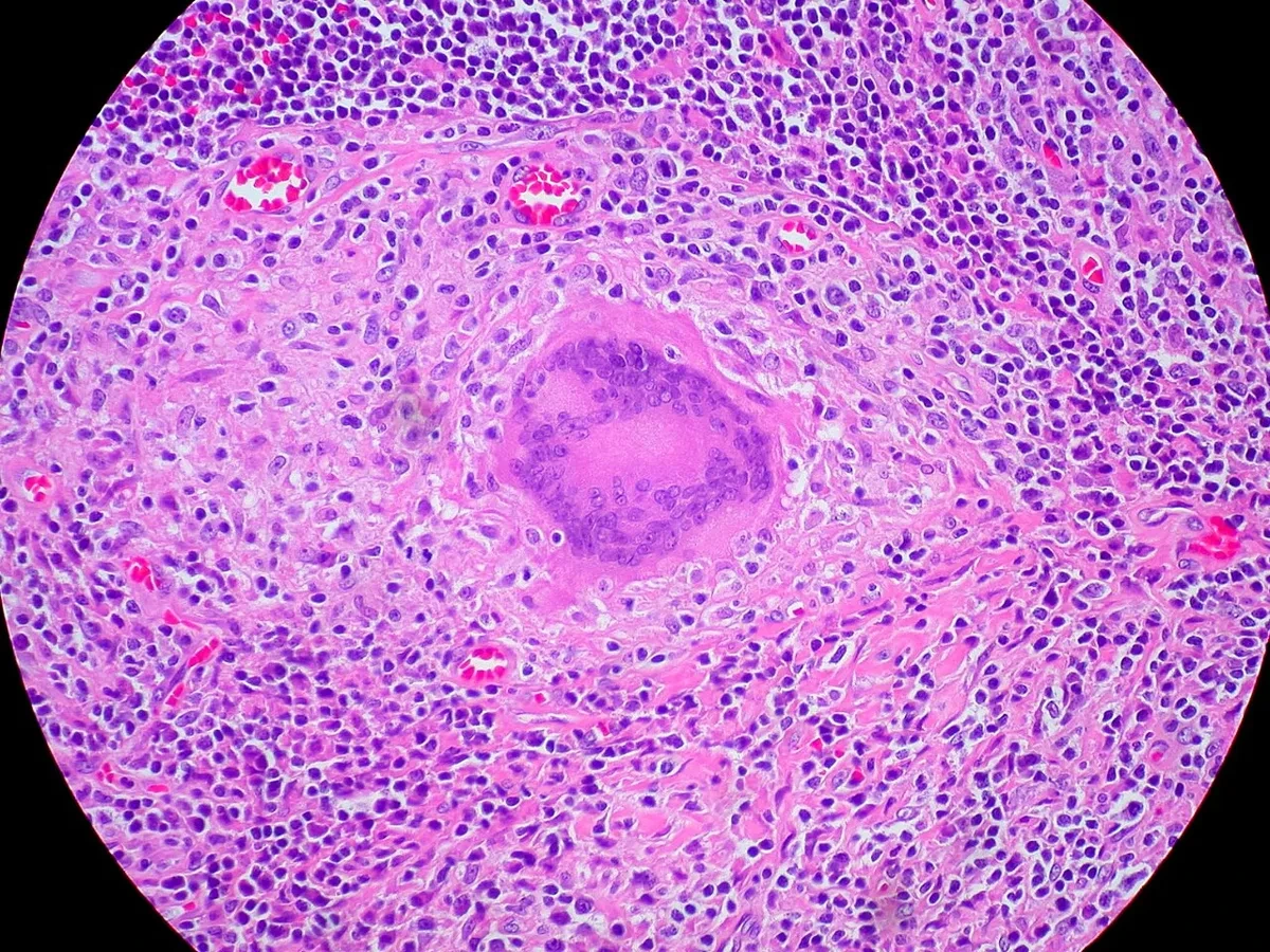
核が複数ある大きな細胞は、いくつかの由来がある。核の並び方と背景から、原因が絞れる。
| Langhans 型巨細胞 | 核が辺縁に馬蹄形/環状に並ぶ | 結核などの類上皮細胞肉芽腫。サルコイドーシスにも出る |
| 異物型巨細胞 | 核がばらばらに散在 | 縫合糸・人工物・尿酸結晶。偏光で異物が光ることがある |
| Touton 型巨細胞 | 核が環状に並び、その外側が泡沫状 | 黄色腫・脂肪織炎。脂質を貪食している |
| 破骨細胞型 | 核が非常に多い | 骨巨細胞腫・褐色腫(副甲状腺機能亢進症) |
| ウイルス性 | 合胞体を作る | 麻疹(Warthin-Finkeldey 細胞)・ヘルペス・RS ウイルス |
「Langhans 型を見たら結核」ではない。サルコイドーシス・真菌症・異物にも出るので、背景と染色で確かめる。
多核になる道筋は2つしかない。
- 細胞が融合する——マクロファージ由来の巨細胞、破骨細胞、骨格筋の筋線維、栄養膜の合胞体、ウイルス感染細胞
- 核だけ分裂して細胞質が分かれない——巨核球(正確には核内倍加)、肝細胞の一部、そして腫瘍細胞の異常分裂
後者は悪性を示唆することがある。Reed-Sternberg 細胞(Hodgkin リンパ腫)は2 つの大きな核と明瞭な核小体が向き合い、フクロウの目のように見える。「鏡像核」という言い方をする。
肉芽腫という語も、ここで整理しておく。マクロファージが上皮細胞のように変わったもの(類上皮細胞)が集まった塊で、「壊せないものを、壁で囲って封じ込める」反応である。中心が乾酪壊死になれば結核、ならなければサルコイドーシスや異物を考える——ただし例外が多いので、抗酸菌染色と培養を必ず添える。
⚠️ 肉芽腫と肉芽組織は別物である。肉芽組織は創傷治癒の毛細血管と線維芽細胞の集まりで、封じ込めの反応ではない。日本語が紛らわしいので、混同しない。
肉芽腫を見たら、抗酸菌染色・真菌染色・培養を出す。組織像だけで結核とサルコイドーシスは分けられない。
English version
Core
Large cells with several nuclei have several origins, and the arrangement of the nuclei together with the background narrows the cause.
| Type | Arrangement of nuclei | Setting |
|---|---|---|
| Langhans | Peripheral, in a horseshoe or ring | Epithelioid granuloma of tuberculosis; also sarcoidosis |
| Foreign body | Scattered irregularly | Suture, prosthetic material, urate crystals; the material may be birefringent |
| Touton | A ring of nuclei with foamy cytoplasm outside it | Xanthoma, panniculitis; the cell is phagocytosing lipid |
| Osteoclast-like | Very numerous nuclei | Giant cell tumour of bone, brown tumour of hyperparathyroidism |
| Viral syncytium | Fused cells | Measles (Warthin–Finkeldey cell), herpes, RSV |
"Langhans means tuberculosis" is wrong — it also occurs in sarcoidosis, fungal infection and foreign body reaction, so confirm with the background and with stains.
Going further
There are only two routes to becoming multinucleate.
- Cells fuse — macrophage-derived giant cells, osteoclasts, skeletal muscle fibres, the trophoblastic syncytium, virus-infected cells.
- The nucleus divides without the cytoplasm dividing — the megakaryocyte (strictly, endomitosis), some hepatocytes, and tumour cells after abnormal division.
The second can indicate malignancy. The Reed–Sternberg cell of Hodgkin lymphoma has two large nuclei with prominent nucleoli facing one another like owl’s eyes; they are called mirror-image nuclei.
The word granuloma is worth settling here: a mass of macrophages transformed to resemble epithelial cells (epithelioid cells), and the reaction means "wall off what cannot be destroyed". If the centre is caseous, think tuberculosis; if not, sarcoidosis or foreign body — but exceptions are many, so always add acid-fast stain and culture.
⚠️ A granuloma and granulation tissue are different things. Granulation tissue is the capillaries and fibroblasts of wound healing, not a walling-off reaction. The Japanese terms are confusingly similar; do not conflate them.
In the clinic
When you see a granuloma, send acid-fast and fungal stains and a culture. Tuberculosis and sarcoidosis cannot be separated on the histological picture alone.
まとめ
| 核・クロマチン・核小体 | 核膜は二重膜で、外膜は粗面小胞体と連続する。 |
|---|---|
| 細胞周期・体細胞分裂・減数分裂 | 間期(G1・S・G2)と分裂期(M)。G0 は周期から外れた休止状態。 |
| Ki-67 — 細胞周期に入っている核だけが茶色になる | Ki-67 は核の蛋白で、細胞周期の G1・S・G2・M 期のすべてで発現し、休止している G0 期の細胞には無い。 |
| アポトーシスと壊死 — 死に方が周りを変える | アポトーシス——計画された死。細胞が縮み、核が凝縮・断片化し、小片(アポトーシス小体)になって食べられる。 |
| 核の像から異型を読む — 良性と悪性を分ける手がかり | 「悪性らしさ」は、核の5つの所見に集約される。 |
| 化生 — 上皮が、別の種類の上皮に置き換わる | 分化した細胞が、別の分化した細胞に置き換わる可逆的な変化。 |
| 制御された壊死 — 細胞の死に方は、2つでは足りない | 長らく細胞死はアポトーシス(きれいに片づく死)と壊死(事故死)の2つとされてきた。 |
| 封入体 — 核や細胞質の中の塊が、原因を名指しする | 細胞の中に見える異常な塊を封入体と呼ぶ。 |
| 細胞質封入体(1)— 蓄えとしてのグリコーゲンと脂肪滴 | 封入体は、細胞が作ったり取り込んだりした物質が一時的にたまったものである。 |
| 細胞質封入体(2)— 色素:体の中で作られたものと、外から入ったもの | 色素色と見分け方どこに内因性(ヘモグロビン由来)ヘモジデリン黄金褐色の粒。 |
| 多核の巨細胞は、できかたで種類が分かれる | 核が複数ある大きな細胞は、いくつかの由来がある。 |
このノートは医学生が授業の内容を自分の言葉でまとめたもので、診療の判断に使うものではありません。誤りに気づいたらお知らせください。