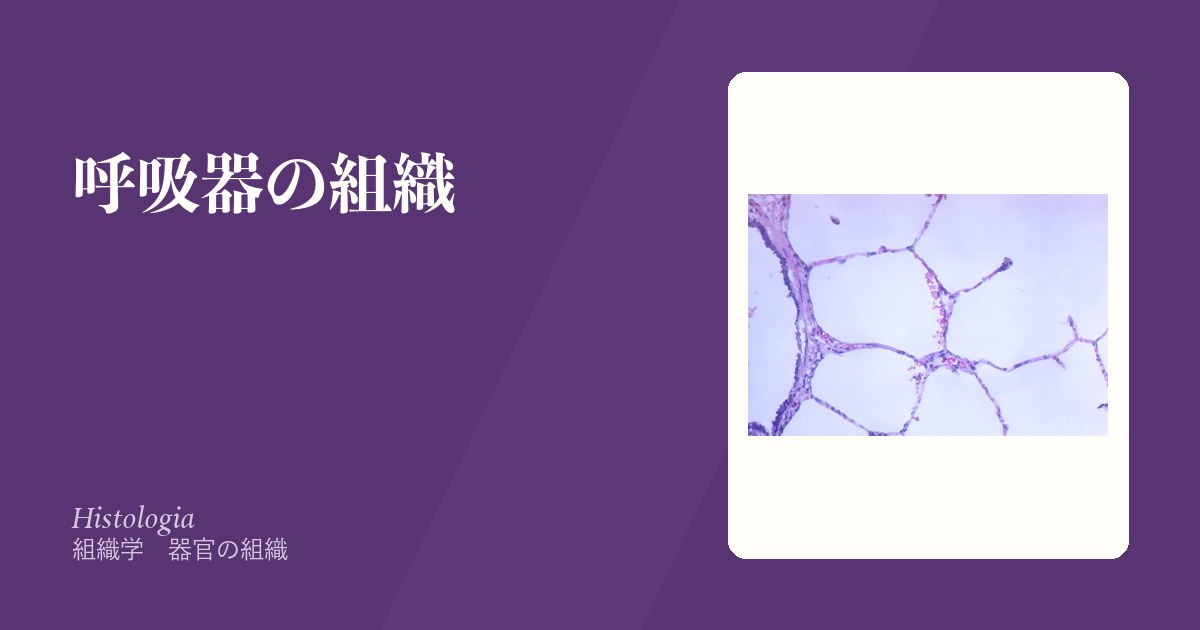
呼吸器は2つの部分に分かれる。導管部(鼻腔〜終末細気管支)は空気を運び、加温・加湿・除塵する。呼吸部(呼吸細気管支〜肺胞)でガス交換が起きる。
末梢へ進むにつれて壁の構成が規則正しく変わる——軟骨が消え、腺が消え、上皮が薄くなり、平滑筋の比重が上がる。この変化の順序を覚えると、どの高さの病変かが読める。
- 軟骨があるのは気管支まで。細気管支には無い
- だから細気管支は虚脱しやすく、呼気時に閉じる——喘息とCOPDが呼気性の障害である理由
交換の場である肺胞は厚さ0.2〜0.6μmしかない。この薄さが機能そのものであり、厚くなること(線維化・水腫)がそのまま低酸素になる。
- 導管部 — 末梢へ向かう壁の変化
- 肺胞
- 嗅上皮 — 中枢神経が体表に露出している唯一の場所
- 喉頭と声帯 — 上皮が切り替わる場所
- 胸膜と肺の二重の血管支配
- 鼻腔の粘膜 — 空気を作り変えてから、肺へ渡す
- 肺の間質 — 線維化は、どの隙間で起きているのか
- 肺胞マクロファージ — 吸い込んだものを、そのまま記録している
導管部 — 末梢へ向かう壁の変化
末梢へ向かうにつれて起きる変化を順に。
- 上皮——多列線毛円柱(気管・気管支)→ 単層線毛円柱(細気管支)→ 単層立方(終末細気管支)
- 杯細胞——細気管支より末梢では消える。代わりにクラブ細胞(分泌・解毒・幹細胞)が現れる
- 軟骨——気管はC字型、気管支は不規則な板状、細気管支では消失
- 腺——粘膜下腺は気管支まで。細気管支には無い
- 平滑筋——相対的に厚くなる
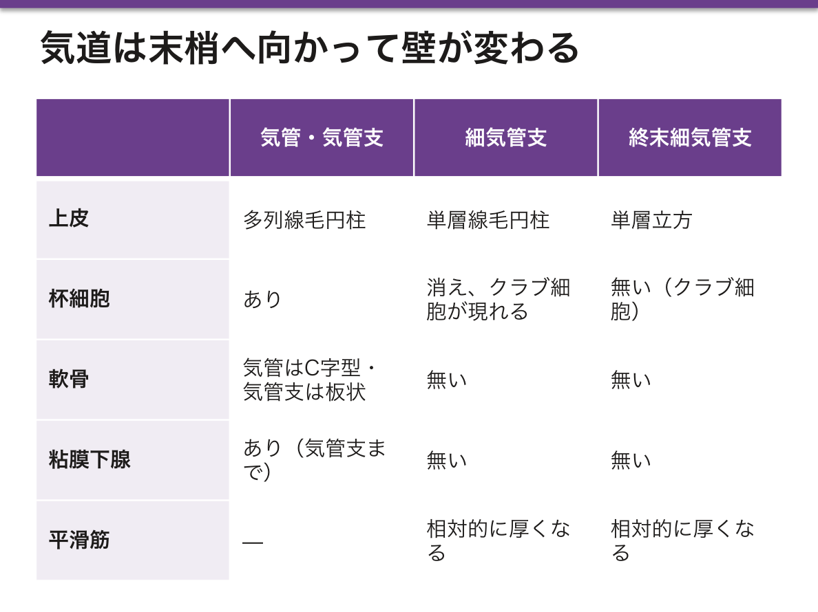
「細気管支=軟骨も腺も杯細胞も無く、平滑筋が相対的に厚い」——この一点から気道疾患が理解できる。
- 気管支喘息——平滑筋の収縮・粘膜浮腫・粘液栓。可逆的。組織では基底膜肥厚・好酸球浸潤・杯細胞過形成・平滑筋肥大(気道リモデリング)。喀痰にCurschmann 螺旋体・Charcot-Leyden 結晶
- COPD——喫煙による慢性炎症。慢性気管支炎では粘膜下腺が肥大(Reid 指数の上昇)、肺気腫では肺胞壁が破壊される
どちらも呼気で悪化するのは、吸気では胸腔内が陰圧で気道が引き広げられるのに対し、呼気では周囲の圧に押されて軟骨の無い細気管支が潰れるためである。1秒率の低下(閉塞性換気障害)という所見の実体がこれである。
気道の防御は粘液線毛クリアランスが担う。喫煙は線毛運動を止め、脱水は粘液を粘稠にする——どちらも同じ機能を別の側から壊す。
末梢へ行くほど軟骨が減り、平滑筋の比が上がる。これが喘息が「細気管支の病気」であることの理由になる。軟骨に支えられていないので、平滑筋が縮むと容易に閉じる。
呼気性の閉塞になるのは呼気では胸腔内圧が上がって外から気道を押しつぶすからである。喘息と COPD で呼気が延長するのはこの力学による。
クラブ細胞(Clara 細胞)は細気管支の分泌細胞で、界面活性物質と解毒酵素を出し、幹細胞にもなる。杯細胞は細気管支より末梢には無い——末梢に粘液が出ていれば異常である。
喫煙で杯細胞が末梢まで増える(杯細胞化生)と粘液が溜まり、線毛は失われて運べない。慢性気管支炎の本体になる。
English version
Core
The changes on the way to the periphery, in order.
- Epithelium — pseudostratified ciliated columnar (trachea, bronchi) → simple ciliated columnar (bronchioles) → simple cuboidal (terminal bronchioles).
- Goblet cells — disappear beyond the bronchioles; club cells (secretion, detoxification, stem cells) appear instead.
- Cartilage — C-shaped in the trachea, irregular plates in the bronchi, absent in the bronchioles.
- Glands — submucosal glands as far as the bronchi; none in the bronchioles.
- Smooth muscle — relatively thicker.
Going further
From the single point that a bronchiole has no cartilage, no glands and no goblet cells, with relatively thick smooth muscle, the airway diseases can be understood.
- Asthma — contraction of the smooth muscle, mucosal oedema and mucus plugging. Reversible. Histologically: thickened basement membrane, eosinophilic infiltration, goblet cell hyperplasia and smooth muscle hypertrophy (airway remodelling). Curschmann’s spirals and Charcot–Leyden crystals in the sputum.
- COPD — chronic inflammation from smoking. In chronic bronchitis the submucosal glands hypertrophy (a raised Reid index); in emphysema the alveolar walls are destroyed.
Both worsen on expiration because on inspiration the negative intrathoracic pressure pulls the airways open, whereas on expiration the surrounding pressure crushes the bronchioles, which have no cartilage. This is the physical substance of the finding of a reduced FEV1 ratio (obstructive ventilatory defect).
The defence of the airway is mucociliary clearance. Smoking stops the cilia and dehydration thickens the mucus — each destroys the same function from a different side.
In the clinic
The further peripheral, the less cartilage and the higher the proportion of smooth muscle. This is why asthma is a disease of the bronchioles: unsupported by cartilage, they close easily when the muscle contracts.
The obstruction is expiratory because on expiration the intrathoracic pressure rises and crushes the airway from outside. The prolonged expiration of asthma and COPD is this mechanics.
Club (Clara) cells are the secretory cells of the bronchiole, releasing surfactant and detoxifying enzymes, and serving as stem cells.
Goblet cells are absent beyond the bronchiole — mucus in the periphery is abnormal. When smoking increases goblet cells into the periphery (goblet cell metaplasia), mucus accumulates and the cilia, being lost, cannot move it. This is the substance of chronic bronchitis.
肺胞Alveolus pulmonis
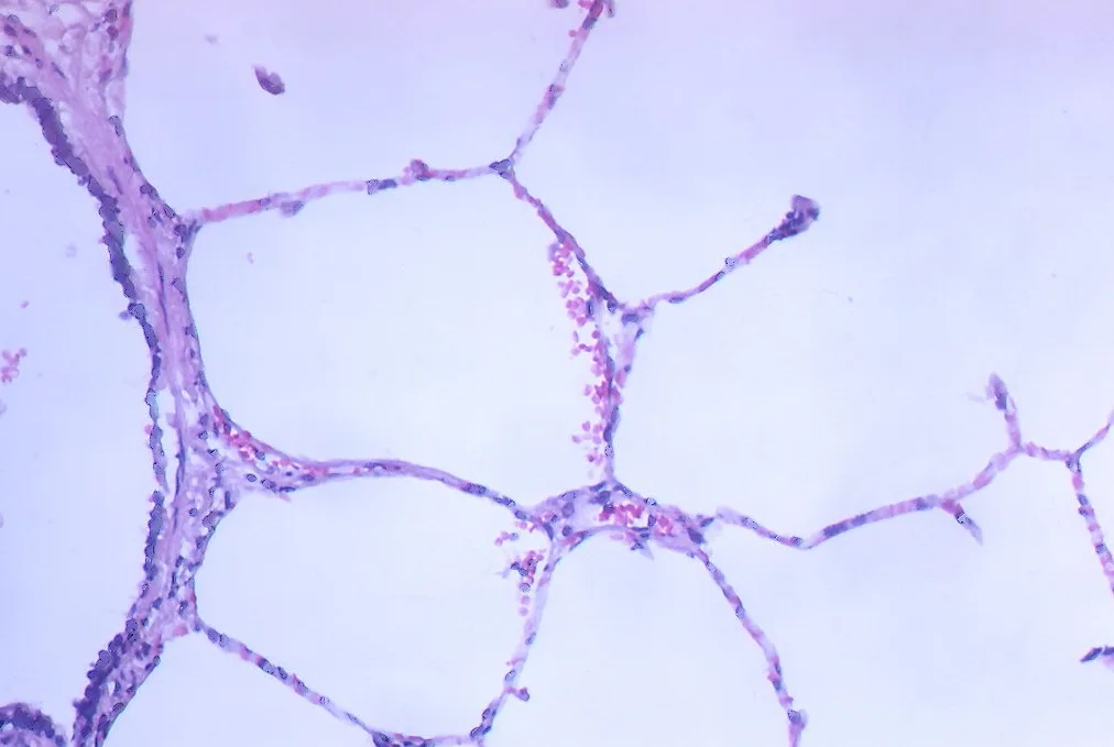
ガス交換の場。壁を作るのは2種の細胞。
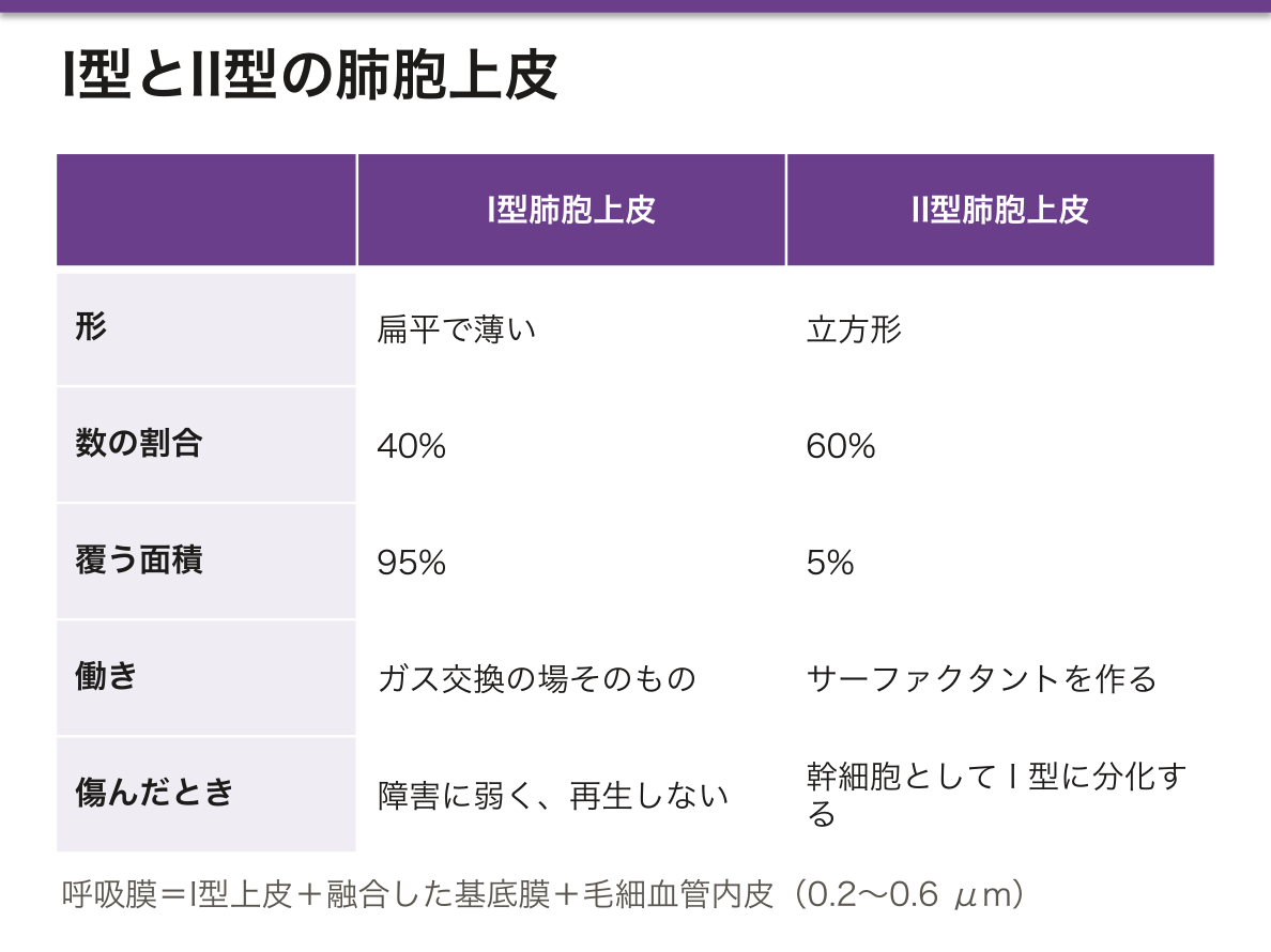
- I型肺胞上皮——扁平で薄い。面積の95%を覆うが数は40%。ガス交換の場そのもの。障害に弱く再生しない
- II型肺胞上皮——立方形。数は60%だが面積は5%。サーファクタントを作る。幹細胞としてI型に分化する
ほかに肺胞大食細胞が肺胞腔を巡回する。
呼吸膜=I型上皮+融合した基底膜+毛細血管内皮、0.2〜0.6μm。
サーファクタント(主成分ジパルミトイルホスファチジルコリン)は表面張力を下げ、Laplace の法則(P = 2T/r)による小さい肺胞の虚脱を防ぐ。半径が小さいほど内圧が高くなるので、サーファクタントが無ければ小さい肺胞が大きい肺胞に吸い込まれて潰れる。
産生は在胎34〜35週で十分になる。だから早産児は新生児呼吸窮迫症候群を起こす。羊水のL/S比(レシチン/スフィンゴミエリン比)2以上で成熟と判定し、切迫早産では母体にステロイドを投与して産生を促す。
肺胞大食細胞は所見として現れる。うっ血性心不全では肺胞へ漏れた赤血球を貪食し、ヘモジデリンを抱えた心不全細胞となる。喫煙者では褐色の色素を持つ。肺胞腔を掃除した記録が細胞に残る。
急性呼吸窮迫症候群(ARDS)の組織像がびまん性肺胞傷害である。I型上皮と内皮が傷害され、透過性が亢進して蛋白に富む滲出液が肺胞腔を満たし、硝子膜を形成する。
この結果、コンプライアンスが低下(硬い肺)し、換気血流比の不均等とシャントにより酸素投与に反応しにくい低酸素血症になる。回復期にはII型上皮が増殖して修復するが、線維化が残れば拘束性障害が残る。「なぜ酸素を上げても上がらないのか」の答えは、肺胞が水で埋まっている=換気されていないからである。
English version
Core
The site of gas exchange. Two cell types make the wall.
- Type I pneumocyte — flat and thin. Covers 95 per cent of the area but is 40 per cent of the number. The site of gas exchange itself. Vulnerable to injury and does not regenerate.
- Type II pneumocyte — cuboidal. 60 per cent of the number but 5 per cent of the area. Makes surfactant. As a stem cell it differentiates into type I.
Alveolar macrophages patrol the alveolar space in addition.
The respiratory membrane = type I epithelium + fused basal laminae + capillary endothelium, 0.2–0.6 µm.
Going further
Surfactant (chiefly dipalmitoylphosphatidylcholine) lowers the surface tension and prevents the collapse of small alveoli predicted by Laplace’s law (P = 2T/r). The smaller the radius the higher the internal pressure, so without surfactant the small alveoli would be sucked into the large ones and collapse.
Production becomes adequate at 34–35 weeks’ gestation, so the preterm infant develops neonatal respiratory distress syndrome. Maturity is judged by an amniotic L/S ratio (lecithin/sphingomyelin) of 2 or more, and in threatened preterm labour the mother is given steroids to promote production.
The alveolar macrophage appears as a finding. In congestive heart failure it phagocytoses red cells that have leaked into the alveoli and becomes the heart failure cell laden with haemosiderin. In smokers it carries brown pigment. A record of what has been cleaned from the alveolar space remains in the cell.
In the clinic
The histology of acute respiratory distress syndrome (ARDS) is diffuse alveolar damage. Type I epithelium and endothelium are injured, permeability increases, protein-rich exudate fills the alveolar spaces and forms hyaline membranes.
The result is reduced compliance (a stiff lung) and hypoxaemia poorly responsive to oxygen, from ventilation–perfusion mismatch and shunt. In recovery, type II cells proliferate and repair the surface, but if fibrosis remains a restrictive defect persists.
The answer to "why does raising the oxygen not raise the saturation?" is that the alveoli are filled with water — they are not being ventilated.
嗅上皮 — 中枢神経が体表に露出している唯一の場所Regio olfactoria
鼻腔の上壁と上鼻甲介・鼻中隔の上部にある、切手ほどの面積の特殊な上皮。多列円柱上皮で、3種類の細胞からなる。
| 細胞 | 役割 |
|---|---|
| 嗅細胞 | 双極性の神経細胞そのもの。樹状突起の先に嗅小胞と嗅線毛(運動性がなく、におい分子の受容面)。軸索は束になって篩板を貫き、嗅球へ入る |
| 支持細胞 | 細胞を支え、リポフスチンを含んで黄褐色に見える |
| 基底細胞 | 幹細胞——嗅細胞を一生涯にわたり作り替える |
粘膜固有層に Bowman 腺(漿液腺)があり、におい分子を溶かす漿液を出す。
この上皮が特異なのは「受容器がそのまま神経細胞であること」。他の感覚では、受容細胞(味蕾の味細胞、内耳の有毛細胞、網膜の視細胞)が別の神経に信号を渡すが、嗅覚だけは受容細胞の軸索が直接中枢へ届く。
もう一つの特異性が「成体でも神経細胞が再生する」こと。基底細胞という幹細胞が嗅細胞を供給し続ける——中枢神経系で神経新生が確実に起こる数少ない場所のひとつで、再生医学の題材になっている。
嗅覚だけが視床を経ないで大脳皮質(嗅内皮質・扁桃体・海馬)へ届くのも例外。においが情動と記憶を強く呼び起こすのは、この配線に由来する。
嗅覚障害の3つの型——①気導性(鼻閉でにおい分子が届かない:慢性副鼻腔炎・鼻茸・アレルギー性鼻炎) ②嗅神経性(上皮そのものの障害:感冒後・薬剤・有毒ガス) ③中枢性(頭部外傷で篩板を貫く嗅神経が剪断される、腫瘍、神経変性疾患)。
嗅覚低下は Parkinson病と Alzheimer病の早期症状として現れ、運動症状より数年先行することがある。Kallmann症候群では嗅球の形成不全と GnRH ニューロンの遊走障害が同時に起こり、嗅覚脱失+低ゴナドトロピン性性腺機能低下症となる——発生学的に同じ場所から出発する細胞だからこそ揃って欠ける。
English version
Core
A specialised epithelium the area of a postage stamp, on the roof of the nasal cavity, the superior concha and the upper septum. Pseudostratified columnar, of three cell types.
| Cell | Role |
|---|---|
| Olfactory cell | A bipolar neurone itself. An olfactory vesicle with olfactory cilia (non-motile, the receptive surface for odour molecules) at the end of the dendrite; the axons bundle together, pierce the cribriform plate and enter the olfactory bulb |
| Supporting cell | Supports the cells; contains lipofuscin and looks yellow-brown |
| Basal cell | Stem cell — renews the olfactory cells throughout life |
Bowman’s glands (serous) in the lamina propria secrete the serous fluid in which odour molecules dissolve.
Going further
What is peculiar about this epithelium is that the receptor is itself the neurone. In every other sense the receptor cell (the taste cell of the taste bud, the hair cell of the inner ear, the photoreceptor of the retina) hands its signal to a separate neurone; in olfaction alone the axon of the receptor cell reaches the centre directly.
The other peculiarity is that the neurones regenerate even in the adult. The basal cells, as stem cells, go on supplying olfactory cells — one of the few places in the nervous system where neurogenesis certainly occurs, and a subject of regenerative medicine.
Olfaction alone also reaches the cortex (entorhinal cortex, amygdala, hippocampus) without passing through the thalamus. That a smell so strongly evokes emotion and memory derives from this wiring.
In the clinic
Three types of olfactory disturbance — ① conductive (nasal obstruction prevents odour molecules arriving: chronic sinusitis, polyps, allergic rhinitis) ② sensorineural (injury to the epithelium itself: post-viral, drugs, toxic gases) ③ central (head injury shearing the olfactory nerves as they pierce the cribriform plate, tumour, neurodegeneration).
Reduced smell appears as an early symptom of Parkinson’s and Alzheimer’s disease and may precede the motor symptoms by years.
In Kallmann syndrome hypoplasia of the olfactory bulb and failure of migration of the GnRH neurones occur together, giving anosmia with hypogonadotrophic hypogonadism — they are lost together precisely because the cells set out from the same place in development.
喉頭と声帯 — 上皮が切り替わる場所Plica vocalis
喉頭は気道の入口であり発声器でもある。組織学の要点は上皮の切り替わりにある。
| 部位 | 上皮 |
|---|---|
| 喉頭蓋の舌面・喉頭面上部 | 重層扁平上皮(食物が当たるため) |
| 声帯(声帯ヒダ) | 重層扁平上皮——振動と摩擦に耐える |
| それ以外(仮声帯・喉頭室・声門下) | 多列線毛円柱上皮 |
声帯の粘膜固有層は3層(浅層=Reinke腔・中間層・深層)に分かれ、その下に声帯筋がある。浅層は疎で、上皮が自由に滑る——これが振動を生む仕組み(粘膜波動)。
Reinke 浮腫は、喫煙と音声酷使によりこの浅層に浮腫が起こるもの。声が低くなるのが特徴で、中年女性の嗄声で「男性のような低い声」なら真っ先に疑う。禁煙が治療の第一。
声帯には毛細リンパ管がほとんど無い——これが臨床的に決定的な意味を持つ。声門癌(声帯にできた癌)はリンパ節転移が少なく、早期に嗄声が出るため見つかりやすい。だから頭頸部癌の中で最も予後が良い。逆に声門上部・下咽頭はリンパ管が豊富で、症状が出にくく転移も早いため予後が悪い。同じ喉頭癌でも、数センチの違いで運命が変わる。
喫煙は線毛円柱上皮を扁平上皮化生させる——防御としては合理的(摩擦に強くなる)だが、粘液線毛クリアランスを失い、扁平上皮癌の母地になる。
声帯は重層扁平上皮——気道でありながら、こすれるので扁平上皮である。その上下は多列線毛円柱上皮なので、上皮が2度切り替わる。
切り替わる場所に癌ができる。喉頭癌の大半は扁平上皮癌で、喫煙と飲酒が危険因子。声門癌は早期から嗄声が出るので発見が早く、リンパ管が乏しいので転移しにくく、予後がよい。声門上癌は症状が遅れ、リンパ節転移が早い——組織の違いがそのまま予後の違いになる。
声帯ポリープ・結節はReinke 腔(上皮直下の粗な層)の浮腫で、音声酷使による。沈黙療法と音声治療が基本になる。
喉頭蓋炎は急速に気道を閉じる。咽頭を診ようとして刺激すると閉塞を誘発するので気道確保の準備をしてから診る。
English version
Core
The larynx is both the entrance to the airway and the organ of voice. The histological point is where the epithelium changes over.
| Site | Epithelium |
|---|---|
| Lingual surface of the epiglottis and upper laryngeal surface | Stratified squamous (food strikes it) |
| Vocal fold | Stratified squamous — to withstand vibration and friction |
| Elsewhere (vestibular fold, ventricle, subglottis) | Pseudostratified ciliated columnar |
The lamina propria of the vocal fold is in three layers (superficial = Reinke’s space, intermediate, deep), with the vocalis muscle beneath. The superficial layer is loose and lets the epithelium slide freely — the mechanism that produces the mucosal wave.
Going further
Reinke’s oedema is oedema of that superficial layer from smoking and vocal overuse. Characteristically the voice becomes lower, so in a middle-aged woman with hoarseness and a "man’s low voice" it is the first thing to suspect. Stopping smoking is the first treatment.
The vocal fold has almost no lymphatic capillaries — and this is decisive clinically. Glottic carcinoma (arising on the fold) seldom metastasises to nodes and causes hoarseness early, so it is found early. It therefore has the best prognosis of the head and neck cancers.
Conversely the supraglottis and hypopharynx are rich in lymphatics, produce symptoms late and metastasise early, and have a poor prognosis. In the same larynx, a few centimetres change the fate.
Smoking causes squamous metaplasia of the ciliated columnar epithelium — reasonable as a defence (it resists friction) but it loses mucociliary clearance and becomes the seedbed of squamous carcinoma.
In the clinic
The vocal fold is stratified squamous — airway though it is, it is squamous because it is rubbed. Above and below it the epithelium is pseudostratified ciliated columnar, so the epithelium changes over twice.
Cancer arises where it changes over. Most laryngeal cancer is squamous carcinoma, with smoking and alcohol as risk factors. Glottic carcinoma causes hoarseness early so it is found early, and with few lymphatics it seldom metastasises: the prognosis is good. Supraglottic carcinoma presents late and metastasises early — a difference in tissue becoming directly a difference in prognosis.
Vocal polyps and nodules are oedema of Reinke’s space (the loose layer just beneath the epithelium) from vocal overuse. Voice rest and speech therapy are the basis of treatment.
Epiglottitis closes the airway rapidly. Stimulating the pharynx in an attempt to look at it can precipitate obstruction, so examine only after preparing to secure the airway.
胸膜と肺の二重の血管支配Pleura
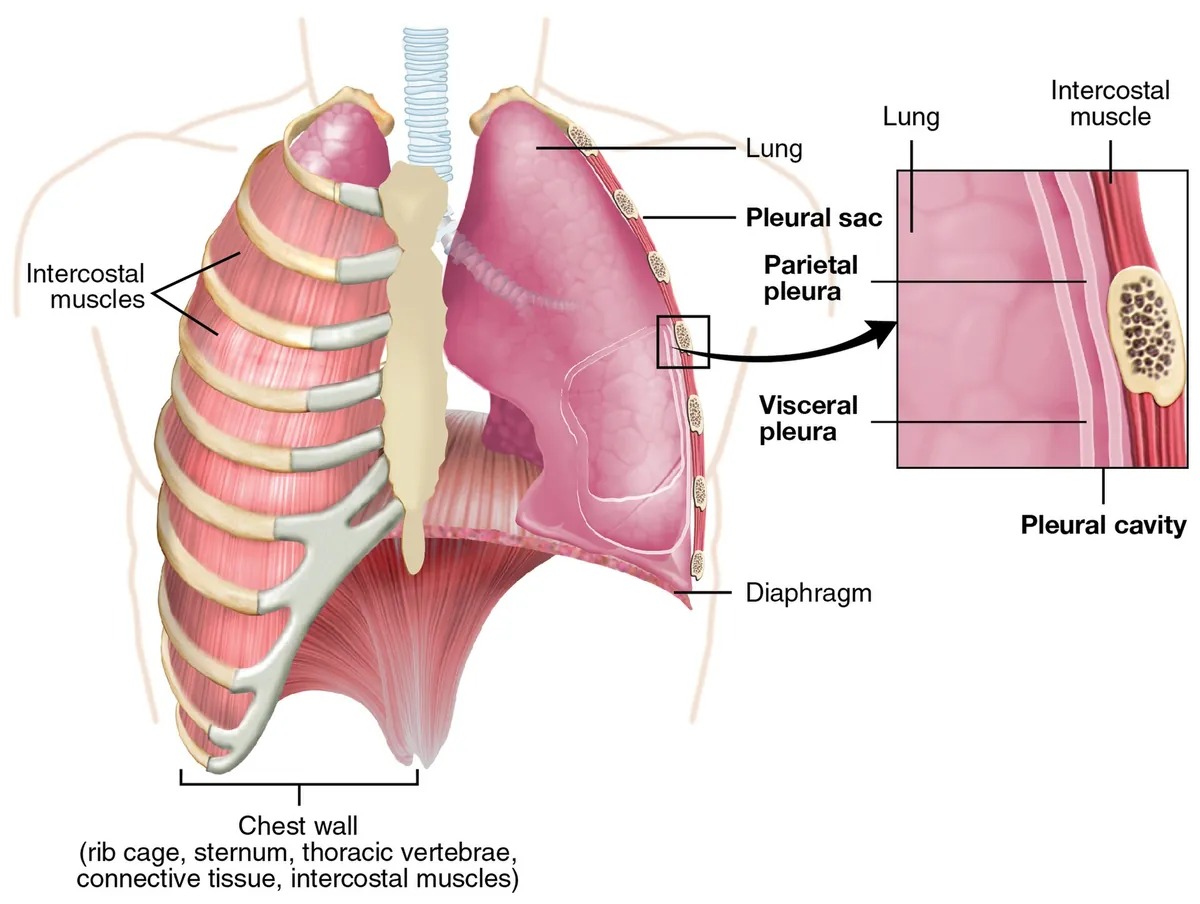
胸膜は単層扁平の中皮とその下の疎性結合組織からなる。臓側胸膜は肺表面を覆って肺門で折り返し、壁側胸膜となって胸壁・横隔膜・縦隔を裏打ちする。間が胸膜腔で、少量の漿液が入る。
肺は2系統の血管を持つ。
| 肺循環(機能血管) | 気管支循環(栄養血管) | |
|---|---|---|
| 由来 | 肺動脈(右室から) | 気管支動脈(胸大動脈から) |
| 運ぶ血 | 静脈血——ガス交換される側 | 動脈血——気道と肺の組織を養う |
| 圧 | 低圧(収縮期25 mmHg前後) | 体循環と同じ高圧 |
神経支配の違いが症状を決める。 臓側胸膜には痛覚が無く、壁側胸膜には肋間神経が分布する——だから胸膜痛は壁側胸膜に炎症が及んで初めて生じる。肺の中に大きな腫瘍があっても痛くないのに、小さな炎症が胸壁に接すると激しく痛むのはこのため。
横隔膜の胸膜は中央部を横隔神経(C3-5)、辺縁部を肋間神経が支配する。中央部の刺激は肩へ放散する(C4皮膚分節)——横隔膜下膿瘍・脾破裂の肩の痛み〈Kehr徴候〉の解剖学的な根拠になる。
二重支配の帰結——肺塞栓では必ずしも肺梗塞が起こらない。肺動脈が詰まっても気管支動脈から血流があるため。梗塞になるのは、もともと心不全などで気管支循環も不十分な場合に限られる。逆に気管支拡張症では気管支動脈が拡張・増生し、大量喀血の源になる——止血には気管支動脈塞栓術を行う。
中皮腫は胸膜の中皮から発生し、アスベスト曝露から30〜40年の潜伏期を経る。びまん性に胸膜を包み込むように進展し、胸水を伴う。喫煙とは相乗しない(肺癌はアスベスト×喫煙で相乗する)点が対照的。
胸水の Light 基準——①胸水/血清の蛋白比 > 0.5 ②胸水/血清の LD 比 > 0.6 ③胸水 LD > 血清 LD 正常上限の2/3。1つでも満たせば滲出性で、胸膜そのものの病変(感染・悪性・膠原病)を探す。
English version
Core
The pleura is a simple squamous mesothelium over loose connective tissue. The visceral pleura covers the lung surface and reflects at the hilum to become the parietal pleura lining chest wall, diaphragm and mediastinum. The pleural cavity between them holds a small amount of serous fluid.
The lung has two vascular systems.
| Pulmonary circulation (functional) | Bronchial circulation (nutritive) | |
|---|---|---|
| Origin | Pulmonary artery (from the right ventricle) | Bronchial arteries (from the thoracic aorta) |
| Blood carried | Venous — the side that is exchanged | Arterial — nourishes the airways and lung tissue |
| Pressure | Low (systolic around 25 mmHg) | High, as in the systemic circulation |
Going further
The difference in innervation decides the symptoms. The visceral pleura has no pain sensation; the parietal pleura is supplied by the intercostal nerves — so pleuritic pain arises only once inflammation reaches the parietal pleura. This is why a large tumour within the lung is painless while a small inflammation touching the chest wall hurts severely.
The diaphragmatic pleura is supplied centrally by the phrenic nerve (C3–5) and peripherally by the intercostal nerves. Central irritation radiates to the shoulder (the C4 dermatome) — the anatomical basis of the shoulder pain of a subphrenic abscess or a ruptured spleen (Kehr’s sign).
A consequence of the double supply — pulmonary embolism does not necessarily cause pulmonary infarction, because blood still arrives from the bronchial arteries. Infarction occurs only when the bronchial circulation is also inadequate, as in heart failure. Conversely, in bronchiectasis the bronchial arteries dilate and proliferate and become the source of massive haemoptysis — treated by bronchial artery embolisation.
In the clinic
Mesothelioma arises from the pleural mesothelium after a latency of 30–40 years from asbestos exposure. It spreads diffusely to envelop the pleura and is accompanied by an effusion. It is not synergistic with smoking (whereas lung cancer is synergistic for asbestos × smoking) — a notable contrast.
Light’s criteria for a pleural effusion — ① pleural/serum protein ratio > 0.5 ② pleural/serum LD ratio > 0.6 ③ pleural LD > two thirds of the upper limit of normal serum LD. Meeting any one makes it an exudate, and a lesion of the pleura itself (infection, malignancy, connective tissue disease) is sought.
鼻腔の粘膜 — 空気を作り変えてから、肺へ渡すTunica mucosa nasi
上皮は多列線毛円柱上皮(呼吸上皮)+杯細胞。鼻前庭だけが重層扁平上皮で、鼻毛がある。
粘膜固有層の特徴は、静脈の多さになる——下鼻甲介の粘膜には海綿静脈叢(膨大な静脈洞)があり、自律神経で充血・収縮する。
3つの仕事——加温(約 32〜34 ℃へ)・加湿(ほぼ 100% へ)・濾過。ここを 1 秒足らずで済ませる。
Kiesselbach 部位——鼻中隔前下部。4本の動脈が吻合し、粘膜が薄い。鼻出血の約 90%。
副鼻腔の粘膜は鼻腔より薄く、線毛は自然口へ向かって一定方向に打つ。
「静脈が多い」という一点で、鼻の症状の大半が説明できる。鼻づまりの実体は、粘膜の腫れではなく静脈洞の充血になる(鼻の構造の章)——「だから血管収縮薬の点鼻が、数分で効く」。
そして同じ理由で、使い続けると効かなくなる。反跳性の充血が起こり、薬剤性鼻炎になる(呼吸器の薬の章)——「市販の点鼻薬を手放せなくなる患者は、この機序で作られている」。
左右の鼻が交互に詰まるのも、この静脈洞の周期的な変化になる(ネーザルサイクル)——「片方ずつ休ませて、粘膜を乾かさない仕組みになっている」。
線毛が自然口へ向かって打つことは、副鼻腔炎の治療方針を決めている。排出は重力ではなく線毛が担うので、自然口が塞がると内容が出ていかない(副鼻腔の章)——「穴を開け直すのではなく、自然口の通りを回復させる手術になる」。
口呼吸が体に悪いと言われる理由も、この章から出てくる。加温・加湿・濾過を経ずに冷たく乾いた空気が下気道へ届く(喘息の章)——「運動誘発性の喘息が、寒い日に強く出る」。
下鼻甲介の海綿静脈叢が、鼻づまりを作っている。粘膜の腫れではなく、静脈叢の充血である。だから血管収縮薬(点鼻)がよく効く。
ただし使い続けると反跳性に悪化する(薬剤性鼻炎)。市販の点鼻薬の連用で起こる、実務でよく出会う問題である。
鼻閉が左右で交代する(ネーザルサイクル)のは自律神経が左右の静脈叢を交互に開閉している正常な現象である。
Kiesselbach 部位は鼻中隔前下部の動脈の吻合で、鼻出血の約90%がここから起こる。小鼻を圧迫し、坐位で前傾させる。
嗅上皮は鼻腔の天井にあるので、鼻閉があると匂いが届かない(呼吸性嗅覚障害)。ウイルス感染後や外傷では嗅神経そのものが傷む(嗅神経性)。篩板の骨折では髄液鼻漏になる。
English version
Core
The epithelium is pseudostratified ciliated columnar (respiratory epithelium) with goblet cells. Only the vestibule is stratified squamous, with the vibrissae.
The feature of the lamina propria is the abundance of veins — the mucosa of the inferior concha contains cavernous venous plexuses (large venous sinuses) that engorge and contract under autonomic control.
Three jobs — warming (to about 32–34 °C), humidifying (to nearly 100 per cent) and filtering, all completed in under a second.
Kiesselbach’s area — the anteroinferior septum, where four arteries anastomose and the mucosa is thin. About 90 per cent of nosebleeds.
The mucosa of the paranasal sinuses is thinner than that of the nose, and the cilia beat consistently towards the ostium.
Going further
The single point that there are many veins explains most of the symptoms of the nose. The substance of a blocked nose is not swelling of the mucosa but engorgement of the venous sinuses — which is why a vasoconstrictor spray works within minutes.
And for the same reason it stops working with continued use. Rebound congestion occurs and rhinitis medicamentosa follows — the patient who cannot give up an over-the-counter spray is made by this mechanism.
That the nostrils block alternately is also a cyclical change in these sinuses (the nasal cycle) — an arrangement that rests one side at a time and keeps the mucosa from drying.
That the cilia beat towards the ostium decides the strategy in sinusitis. Drainage is by cilia, not by gravity, so if the ostium is blocked the contents cannot leave — the operation restores the patency of the natural ostium rather than making a new hole.
Why mouth breathing is said to be bad also comes out of this chapter: cold dry air reaches the lower airway without being warmed, humidified or filtered — exercise-induced asthma is worse on a cold day.
In the clinic
The cavernous plexus of the inferior concha creates nasal obstruction — engorgement of the plexus, not swelling of the mucosa. So a vasoconstrictor spray works well, but with continued use it rebounds and worsens (rhinitis medicamentosa). A problem met often in practice from continuous use of over-the-counter sprays.
Alternating obstruction (the nasal cycle) is a normal phenomenon in which the autonomic nerves open and close the two plexuses in turn.
Kiesselbach’s area is the arterial anastomosis of the anteroinferior septum, the source of about 90 per cent of nosebleeds. Compress the ala and sit the patient leaning forward.
The olfactory epithelium lies in the roof, so nasal obstruction prevents odours reaching it (conductive olfactory loss). After viral infection or trauma the olfactory nerves themselves are damaged (sensorineural). A fracture of the cribriform plate gives CSF rhinorrhoea.
肺の間質 — 線維化は、どの隙間で起きているのかInterstitium pulmonis
間質とは、肺胞上皮と毛細血管内皮のあいだ、および小葉間・血管周囲・胸膜下の結合組織を指す。
肺胞壁には厚い側と薄い側がある。
| 薄い側 | 厚い側 | |
|---|---|---|
| 構造 | I型肺胞上皮・融合した基底膜・内皮のみ | あいだに膠原線維・弾性線維・線維芽細胞が入る |
| 役割 | ガス交換 | 水の移動・支持 |
間質にいる細胞——線維芽細胞・筋線維芽細胞・肺胞マクロファージ(腔内)・リンパ球・肥満細胞。
肺の弾性収縮力は、弾性線維とサーファクタントによる表面張力の両方から生まれる。
「厚い側と薄い側がある」ことが、間質性肺炎の症状の順番を決めている。線維化はまず厚い側で進むので、ガス交換より先に肺が硬くなる——「息が吸いにくいという訴えが、低酸素より早く来る」。
だから拘束性換気障害になる。%肺活量が下がり、1秒率は保たれる。拡散能(DLCO)が早期から落ちる(呼吸の数字の章)——「スパイロメトリーの型が、間質のどこが壊れたかを映している」。
肺水腫の初期に間質へ水が溜まるのも、同じ構造による。厚い側に余裕があるので、まずそこが膨れる(間質性肺水腫)。次に肺胞腔へ溢れる(肺胞性肺水腫)(心不全の章)——「胸部写真の Kerley B 線が、肺胞に水が来る前の段階を示している」。
間質のリンパ管が、この水を汲み出している。だから間質の浮腫はある程度まで代償される(リンパ管の章)——「症状が出た時点で、排水の能力を超えている」。
線維化した肺が元に戻らないのは、筋線維芽細胞が消えずに残るためになる(修復と創傷治癒の章)——「治癒の仕組みが、止まらなくなった状態として理解する」。
薄い側で交換し、厚い側で水を扱う。この非対称が肺水腫の起こり方を決める。
心原性肺水腫 — 左房圧の上昇により、まず厚い側の間質に水が溜まる(間質性肺水腫)。Kerley B 線・血管影の増強。進むと肺胞へあふれる(肺胞性肺水腫)——蝶形陰影・泡沫状の血痰。間質から肺胞へという順序が画像所見の順序そのものである。
急性呼吸窮迫症候群(ARDS) — 透過性の亢進による非心原性肺水腫。びまん性肺胞傷害・硝子膜形成。心原性と違い、心不全では説明できないことが定義に入る。
間質性肺炎は間質の線維化で、拘束性障害・拡散障害・労作時低酸素・fine crackles・ばち指。蜂巣肺は不可逆である。閉塞性(喘息・COPD)とは呼吸機能検査の型が正反対になる。
English version
Core
The interstitium means the space between alveolar epithelium and capillary endothelium, and the connective tissue of the interlobular septa, around the vessels and beneath the pleura.
The alveolar wall has a thick side and a thin side.
| Thin side | Thick side | |
|---|---|---|
| Structure | Type I epithelium, fused basal laminae and endothelium only | Collagen, elastic fibres and fibroblasts lie between |
| Role | Gas exchange | Movement of water, and support |
Cells of the interstitium — fibroblasts, myofibroblasts, alveolar macrophages (in the space), lymphocytes, mast cells.
The elastic recoil of the lung arises from both the elastic fibres and the surface tension modified by surfactant.
Going further
That there is a thick side and a thin side decides the order of symptoms in interstitial pneumonia. Fibrosis advances first on the thick side, so the lung stiffens before gas exchange fails — the complaint of difficulty in breathing in comes earlier than the hypoxia.
So the defect is restrictive: the vital capacity falls while the FEV1 ratio is preserved, and the diffusing capacity (DLCO) falls early — the pattern on spirometry mirrors which part of the interstitium has been destroyed.
That water collects in the interstitium early in pulmonary oedema follows from the same structure. The thick side has room, so it swells first (interstitial oedema) and the fluid then overflows into the alveoli (alveolar oedema) — Kerley B lines on the chest film mark the stage before water reaches the alveoli.
The lymphatics of the interstitium pump that water out, so interstitial oedema is compensated up to a point — by the time symptoms appear the drainage capacity has been exceeded.
A fibrosed lung does not return to normal because the myofibroblasts do not disappear — understand it as the mechanism of healing that has stopped being able to stop.
In the clinic
Exchange on the thin side, water handled on the thick side. This asymmetry decides how pulmonary oedema develops.
Cardiogenic pulmonary oedema — a rising left atrial pressure first fills the interstitium of the thick side (interstitial oedema): Kerley B lines, prominent vascular markings. Progressing, it overflows into the alveoli (alveolar oedema) — a butterfly shadow and frothy blood-stained sputum. The sequence interstitium → alveolus is precisely the sequence of the radiological findings.
ARDS — non-cardiogenic pulmonary oedema from increased permeability. Diffuse alveolar damage with hyaline membranes. Unlike the cardiogenic form, its definition includes that it cannot be explained by heart failure.
Interstitial pneumonia is fibrosis of the interstitium: a restrictive defect, impaired diffusion, hypoxia on exertion, fine crackles and clubbing. Honeycombing is irreversible. The pattern on lung function testing is the exact opposite of the obstructive diseases (asthma, COPD).
肺胞マクロファージ — 吸い込んだものを、そのまま記録しているMacrophagus alveolaris
肺胞の内腔には、常にマクロファージがいる。気道が外界と繋がっている以上、届いた粒子を片づける細胞が要るからである。そして食べたものが、そのまま細胞の見た目になる。
| ヘモジデリンを含む | Berlin blue で青く染まる | 心不全細胞。左心不全・肺うっ血・肺出血。喀痰にも出る |
| 黒い粒子 | 炭粉 | 喫煙・大気汚染。リンパ節まで運ばれて黒くなる(炭粉沈着) |
| 細胞質が泡沫状 | 脂質を含む | 脂肪誤嚥・閉塞性肺炎・アミオダロン |
| 茶褐色で喫煙者に多い | 喫煙関連の色素 | 剥離性間質性肺炎・呼吸細気管支炎関連間質性肺疾患 |
喀痰の細胞診で肺胞マクロファージが見えるかどうかは、その検体が本当に下気道から出たものかの判定にも使われる。
気管支肺胞洗浄(BAL)は、この細胞たちを回収して調べる検査である。生理食塩水を注入して吸い出し、細胞の種類と比率を見る。
| リンパ球が増加 | サルコイドーシス・過敏性肺炎・薬剤性 | CD4/CD8 比が上がればサルコイドーシス、下がれば過敏性肺炎を示唆する |
| 好酸球が増加 | 好酸球性肺炎・薬剤性・寄生虫 | |
| 好中球が増加 | 感染・急性呼吸窮迫症候群・特発性肺線維症 | |
| 回収液が回を追って血性になる | びまん性肺胞出血 | ヘモジデリン貪食マクロファージも増える |
| 乳白色で PAS 陽性の物質 | 肺胞蛋白症 | サーファクタントが片づけられずに溜まる |
肺胞蛋白症は、この細胞の機能不全そのものが病気になった例である。マクロファージが GM-CSF の信号を受け取れず(自己抗体による)、サーファクタントを分解できなくなる。治療が全肺洗浄(生理食塩水で肺を洗い流す)というのも、掃除する細胞が働かないので、外から洗うという理屈である。
肺胞マクロファージには弱点がある。結核菌は貪食されても殺されず、その中で生き延びる——細胞性免疫(T 細胞からの活性化)が無いと処理できない。だから免疫抑制で結核が再燃する。
びまん性の陰影で診断がつかないときは、BAL を検討する。細胞の比率だけでも鑑別がかなり絞れる。
English version
Core
Macrophages are always present in the alveolar space, because so long as the airway connects with the outside world, a cell is needed to clear away what arrives. And what it has eaten becomes the appearance of the cell.
| Appearance | Content | Meaning |
|---|---|---|
| Contains haemosiderin | Stains blue with Berlin blue | Heart failure cell. Left heart failure, pulmonary congestion, pulmonary haemorrhage. Also appears in the sputum |
| Black particles | Carbon | Smoking, air pollution. Carried as far as the lymph nodes, which blacken (anthracosis) |
| Foamy cytoplasm | Lipid | Lipid aspiration, obstructive pneumonia, amiodarone |
| Brown, common in smokers | Smoking-related pigment | Desquamative interstitial pneumonia, respiratory bronchiolitis-associated interstitial lung disease |
Whether alveolar macrophages are seen on sputum cytology is also used to judge whether the specimen really came from the lower airway.
Going further
Bronchoalveolar lavage (BAL) is an investigation that recovers these cells and examines them: saline is instilled and aspirated, and the types and proportions of cells assessed.
| Finding | Suggests |
|---|---|
| Lymphocytosis | Sarcoidosis, hypersensitivity pneumonitis, drug-induced. A raised CD4/CD8 ratio suggests sarcoidosis, a low one hypersensitivity pneumonitis |
| Eosinophilia | Eosinophilic pneumonia, drug-induced, parasitic |
| Neutrophilia | Infection, ARDS, idiopathic pulmonary fibrosis |
| Successive aliquots increasingly bloody | Diffuse alveolar haemorrhage. Haemosiderin-laden macrophages also increase |
| Milky, PAS-positive material | Alveolar proteinosis — surfactant accumulating because it is not cleared |
Alveolar proteinosis is an example of dysfunction of this very cell becoming the disease. The macrophage cannot receive the GM-CSF signal (because of an autoantibody) and cannot degrade surfactant. That the treatment is whole-lung lavage — washing the lung out with saline — follows the same logic: the cell that cleans does not work, so it is washed from outside.
The alveolar macrophage has a weakness. Mycobacterium tuberculosis is phagocytosed but not killed, and survives inside it — it cannot be dealt with without cell-mediated immunity (activation by T cells). This is why tuberculosis reactivates under immunosuppression.
In the clinic
When a diffuse shadow cannot be diagnosed, consider BAL. The proportions of the cells alone narrow the differential considerably.
まとめ
| 導管部 — 末梢へ向かう壁の変化 | 末梢へ向かうにつれて起きる変化を順に。上皮——多列線毛円柱(気管・気管支)→ 単層線毛円柱(細気管支)→ 単層立方(終末細気管支)杯細胞——細気管支より末梢では消える。 |
|---|---|
| 肺胞 | ガス交換の場。壁を作るのは2種の細胞。 |
| 嗅上皮 — 中枢神経が体表に露出している唯一の場所 | 鼻腔の上壁と上鼻甲介・鼻中隔の上部にある、切手ほどの面積の特殊な上皮。 |
| 喉頭と声帯 — 上皮が切り替わる場所 | 喉頭は気道の入口であり発声器でもある。組織学の要点は上皮の切り替わりにある。 |
| 胸膜と肺の二重の血管支配 | 胸膜は単層扁平の中皮とその下の疎性結合組織からなる。 |
| 鼻腔の粘膜 — 空気を作り変えてから、肺へ渡す | 上皮は多列線毛円柱上皮(呼吸上皮)+杯細胞。 |
| 肺の間質 — 線維化は、どの隙間で起きているのか | 間質とは、肺胞上皮と毛細血管内皮のあいだ、および小葉間・血管周囲・胸膜下の結合組織を指す。 |
| 肺胞マクロファージ — 吸い込んだものを、そのまま記録している | 肺胞の内腔には、常にマクロファージがいる。 |
このノートは医学生が授業の内容を自分の言葉でまとめたもので、診療の判断に使うものではありません。誤りに気づいたらお知らせください。