卵巣は使い切る器官である。精巣が作り続けるのと対照的に、卵母細胞は胎生期に数が決まり、以後は減る一方になる。子宮内膜はそのサイクルに合わせて毎月作り直される粘膜で、どの時期の像かを言えるようにしておく。
- 卵胞 — 二細胞説と黄体
- 子宮内膜 — 毎月作り直される粘膜
- 卵子の減数分裂は、2度止まる
- 卵管・子宮頸部・腟 — 接合部が問題になる
- 腟の上皮 — グリコーゲンが、自浄作用を作っている
卵胞 — 二細胞説と黄体Folliculus ovaricus
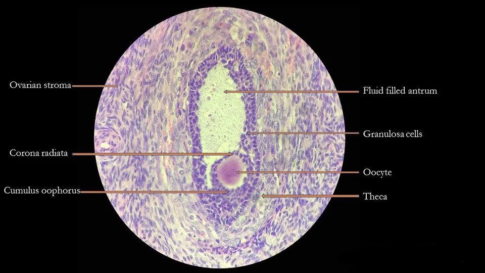
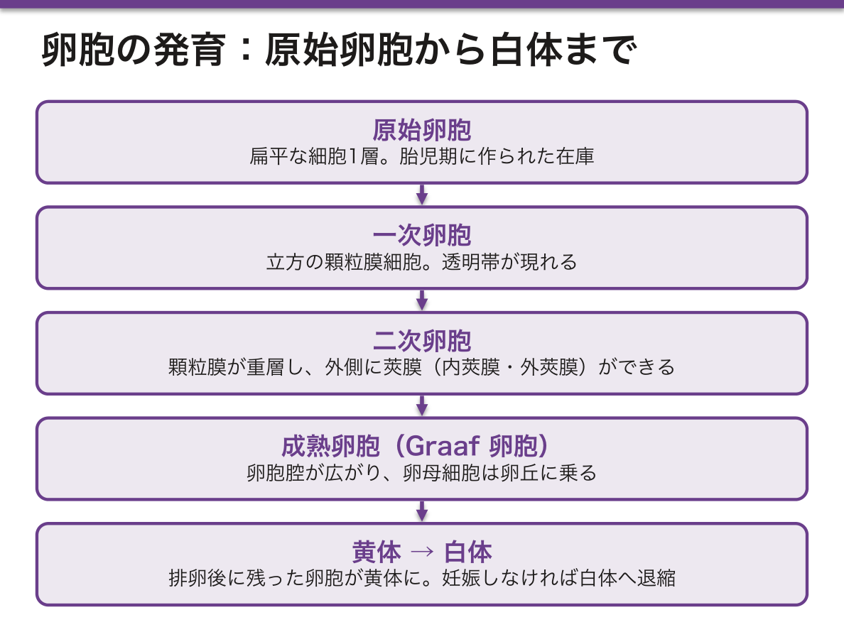
卵巣皮質に埋まる、卵母細胞とそれを包む細胞の集まり。発育の順に。
- 原始卵胞——扁平な細胞1層。胎児期に作られた在庫
- 一次卵胞——立方の顆粒膜細胞。透明帯が現れる
- 二次卵胞——顆粒膜が重層し、外側に莢膜(内莢膜・外莢膜)ができる
- 胞状卵胞 → 成熟卵胞(Graaf 卵胞)——卵胞腔が広がり、卵母細胞は卵丘に乗る
排卵後、残った卵胞が黄体になり、妊娠しなければ白体へ退縮する。
エストロゲンは1種類の細胞では作れない——二細胞説がこの章の核心である。
- 莢膜細胞——黄体形成ホルモンを受け、コレステロールからアンドロゲンまで作る。アロマターゼを持たないのでそこで止まる
- 顆粒膜細胞——卵胞刺激ホルモンを受け、アロマターゼでアンドロゲンをエストロゲンに変える。血管が無いのでコレステロールを取り込めず、最初から作ることはできない
材料を作る細胞と、仕上げる細胞が分かれている。だからアロマターゼ阻害薬は顆粒膜側だけを止め、閉経後乳癌の治療になる。逆に多嚢胞性卵巣症候群では黄体形成ホルモン優位で莢膜が働きすぎ、アンドロゲン過剰(多毛・痤瘡)と卵胞が育ちきらず並ぶ(ネックレスサイン)という像になる。
減数分裂が2回止まるのも卵巣に特有である。第一分裂は胎児期に始まって前期で停止し、排卵直前に再開する。第二分裂は中期で停止し、受精で初めて完了する。
つまり40歳の卵子は40年間分裂の途中で待っていたことになる。染色体を引く紡錘糸の装置がそれだけ長く維持されるため、加齢とともに不分離が増え、21トリソミーなどの染色体異常が増える。母体年齢が効く理由が、この「止まったまま待つ」という仕組みそのものである。
二細胞説がそのまま多嚢胞性卵巣症候群の説明になる。莢膜細胞が LH でアンドロゲンを作り、顆粒膜細胞が FSH とアロマターゼでエストロゲンに変える。
多嚢胞性卵巣症候群 — LH/FSH 比の上昇とインスリン抵抗性によりアンドロゲンが過剰になる。月経異常(無排卵)・多毛やにきび・多嚢胞卵巣。無排卵で黄体ができず、プロゲステロンが出ないためエストロゲン単独の刺激が続き、子宮内膜癌のリスクが上がる。ホルモンの経路を追えば、合併症まで導ける。
黄体はプロゲステロンを出して内膜を維持する。妊娠すれば hCG が黄体を保つ。黄体機能不全は不育症の一因になる。
顆粒膜細胞腫はエストロゲン産生腫瘍で、閉経後出血や思春期早発症で発症し、Call–Exner 小体が組織学的特徴である。
English version
Core
An oocyte with its investing cells, embedded in the ovarian cortex. In order of development:
- Primordial follicle — a single layer of flattened cells. The stock laid down in fetal life.
- Primary follicle — cuboidal granulosa cells; the zona pellucida appears.
- Secondary follicle — the granulosa becomes multilayered and a theca (interna and externa) forms outside it.
- Antral → mature (Graafian) follicle — the antrum enlarges and the oocyte rides on the cumulus.
After ovulation the remaining follicle becomes the corpus luteum, and if there is no pregnancy it regresses to the corpus albicans.
Going further
Oestrogen cannot be made by one cell type — the two-cell theory is the core of this chapter.
- Theca cell — responds to LH and makes androgen from cholesterol. Having no aromatase, it stops there.
- Granulosa cell — responds to FSH and converts androgen to oestrogen with aromatase. Being avascular it cannot take up cholesterol and cannot start from the beginning.
The cell that makes the material and the cell that finishes it are separate. So an aromatase inhibitor blocks only the granulosa side and becomes a treatment for postmenopausal breast cancer. Conversely in polycystic ovary syndrome LH predominance overworks the theca, giving androgen excess (hirsutism, acne) and follicles that never finish growing, lined up as the "string of pearls".
That meiosis arrests twice is also peculiar to the ovary. The first division begins in fetal life and stops in prophase, resuming just before ovulation; the second stops in metaphase and is completed only at fertilisation. An egg in a woman of 40 has waited 40 years in mid-division. The spindle apparatus that pulls the chromosomes must be maintained for that long, so non-disjunction increases with age and chromosomal abnormalities such as trisomy 21 become commoner. The reason maternal age matters is this very arrangement of waiting while arrested.
In the clinic
The two-cell theory is itself the explanation of polycystic ovary syndrome. The theca cell makes androgen under LH and the granulosa cell converts it to oestrogen with FSH and aromatase.
Polycystic ovary syndrome — a raised LH/FSH ratio and insulin resistance make androgen excessive. Menstrual disturbance (anovulation), hirsutism and acne, polycystic ovaries. With no ovulation there is no corpus luteum and no progesterone, so unopposed oestrogen stimulation continues and the risk of endometrial carcinoma rises. Follow the hormonal pathway and even the complication can be derived.
The corpus luteum secretes progesterone and maintains the endometrium; in pregnancy hCG sustains it. Luteal insufficiency is one cause of recurrent pregnancy loss.
Granulosa cell tumour is oestrogen-producing, presenting as postmenopausal bleeding or precocious puberty, with Call–Exner bodies as its histological feature.
子宮内膜 — 毎月作り直される粘膜Endometrium
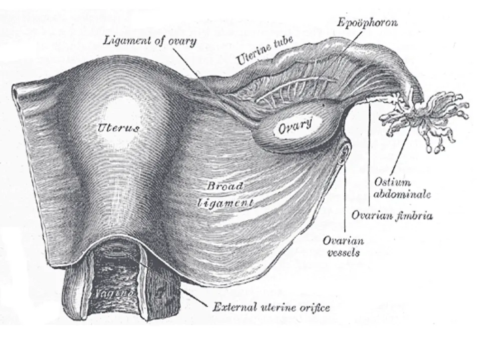
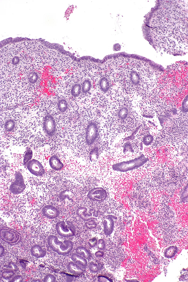
子宮体部の粘膜。2層に分かれる。
- 機能層——内腔側。月経で剥がれ落ちる。らせん動脈が栄養する
- 基底層——筋層側。残って再生の元になる。直動脈が栄養する
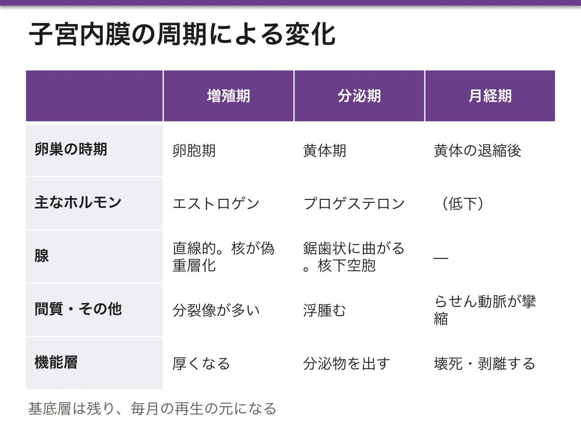
周期による変化。
- 増殖期(卵胞期)——エストロゲンで厚くなる。腺は直線的、核が偽重層化して分裂像が多い
- 分泌期(黄体期)——プロゲステロンで腺が鋸歯状に曲がり、核下空胞のあと分泌物が出る。間質が浮腫む
- 月経期——黄体退縮でらせん動脈が攣縮し、機能層が壊死・剥離する
血管を2系統に分けてあることが、毎月出血しても止まる仕組みである。らせん動脈はプロゲステロンの低下に反応して攣縮し、機能層だけを虚血に落とす。基底層を養う直動脈は反応しないので再生の元は必ず残る。捨てる範囲と残す範囲が、血管の走り方であらかじめ決めてある。
この二重構造から、臨床の判断が導ける。
- 子宮内膜が「増殖期のまま」続くのは、プロゲステロンが出ていない=排卵していないということ。無排卵周期・多嚢胞性卵巣症候群でこうなる
- エストロゲン単独の刺激が続くと内膜が過剰に増える——子宮内膜増殖症、そして類内膜癌へ進む。だから子宮を持つ人へのホルモン補充では、必ずプロゲスチンを併用する
- 組織を見れば周期のどこかが分かる——腺の形と核下空胞は日付の目安になる
タモキシフェンが乳腺では抗エストロゲン、子宮内膜ではエストロゲン様に働くという組織による違いも、内膜癌のリスクとして現実の問題になる。
子宮内膜症は、内膜組織が子宮の外(卵巣・ダグラス窩・腹膜)に存在する疾患である。異所性の内膜も同じホルモンに反応して周期的に出血するが、逃げ場が無いため、その場に血液が溜まり炎症と癒着を起こす。
卵巣にできればチョコレート嚢胞、症状は月経困難症・性交痛・排便痛・不妊。「組織の場所だけが違い、性質は正常」なのに病気になる——出口の有無が病態を決めている。
治療が排卵と月経を止める方向(低用量エストロゲン・プロゲスチン配合薬、ジエノゲスト、GnRH アナログ)に向かうのは、周期的な反応そのものが病態だからである。
English version
Core
The mucosa of the body of the uterus, in two layers.
- Functional layer — towards the lumen. Shed at menstruation. Supplied by the spiral arteries.
- Basal layer — towards the myometrium. Remains, and is the source of regeneration. Supplied by the straight arteries.
Changes through the cycle.
- Proliferative (follicular) phase — thickens under oestrogen. The glands are straight, the nuclei pseudostratified, mitoses numerous.
- Secretory (luteal) phase — under progesterone the glands become saw-toothed; subnuclear vacuoles appear and then secretion. The stroma becomes oedematous.
- Menstrual phase — as the corpus luteum regresses the spiral arteries constrict and the functional layer necroses and is shed.
Going further
Dividing the blood supply into two systems is the mechanism by which monthly bleeding stops. The spiral arteries constrict in response to the fall in progesterone and render only the functional layer ischaemic; the straight arteries feeding the basal layer do not respond, so the source of regeneration always survives. What is discarded and what is kept has been decided in advance by the course of the vessels.
From this double structure the clinical judgements follow.
- An endometrium that stays in the proliferative phase means no progesterone — that is, no ovulation. It occurs in anovulatory cycles and in polycystic ovary syndrome.
- Continued unopposed oestrogen makes the endometrium grow excessively — endometrial hyperplasia, and on to endometrioid carcinoma. So in hormone replacement for a woman with a uterus, a progestogen is always added.
- Looking at the tissue tells you where in the cycle it is — the shape of the glands and the subnuclear vacuoles date it.
That tamoxifen is anti-oestrogenic in the breast but oestrogen-like in the endometrium — a difference made by the tissue — is a real problem as a risk of endometrial carcinoma.
In the clinic
Endometriosis is the presence of endometrial tissue outside the uterus (ovary, pouch of Douglas, peritoneum). The ectopic endometrium responds to the same hormones and bleeds cyclically, but with nowhere to escape the blood collects where it is and causes inflammation and adhesions. In the ovary it forms a chocolate cyst; the symptoms are dysmenorrhoea, dyspareunia, dyschezia and infertility.
Only the location of the tissue differs and its nature is normal, yet it is a disease — the presence or absence of an exit decides the pathology.
Treatment is directed at stopping ovulation and menstruation (combined low-dose oestrogen–progestogen, dienogest, GnRH analogues) because the cyclical response itself is the disease.
卵子の減数分裂は、2度止まるOocytus
卵母細胞の減数分裂は、途中で長く停止する。
| 時期 | 状態 | 止まっている期間 |
|---|---|---|
| 胎生 5 か月頃 | 第一減数分裂の前期(複糸期)で停止 | 排卵まで(最長 50 年) |
| 排卵の直前 | 第一減数分裂を完了し、第一極体を放出 | — |
| 排卵時 | 第二減数分裂の中期で再び停止 | 受精まで |
| 受精 | 第二減数分裂を完了し、第二極体を放出 | — |
卵母細胞の数——胎生 5 か月に約 700 万で最多、出生時 約 200 万、思春期 約 30 万、排卵するのは生涯で約 400 個。
残りは閉鎖する。作り足されることは無い。
「40 年以上、分裂の途中で止まったままである」ことが、加齢による染色体異常を説明する。紡錘糸と接着蛋白(コヒーシン)が長い年月で劣化し、染色体の分離が誤りやすくなる(染色体異常の章)——「母体年齢とともに 21 トリソミーが増える理由になる」。
だから不分離は、圧倒的に母由来で起こる——「精子は 74 日で作り直されるので、時間の影響を受け方が違う」。
卵の在庫が有限であることは、卵巣機能の評価に直結する(不妊の章)——「抗ミュラー管ホルモン(AMH)が、残っている前胞状卵胞の量を反映する」。
抗腫瘍薬と放射線は、この在庫を減らす(放射線生物学の章)——「治療前の妊孕性温存を、治療を始める前に切り出す必要がある」。
第二減数分裂の中期で止まっているので、受精しなければ完了しない(受精の章)——「未受精卵は、分裂の途中で老化していく」。
減数分裂が長く止まっていることが、母体年齢と染色体異常の関係を作る。
第一減数分裂の前期で、胎生期から排卵まで最長 50 年止まっている。紡錘体と接着蛋白(コヒーシン)が経年的に劣化するため、不分離が増える。21トリソミー・18トリソミー・13トリソミーの母体年齢依存性はここから来る。
だから出生前診断の説明では「年齢とともにリスクが上がる」ことを機序とともに伝えられる。NIPT・羊水検査・絨毛検査の選択肢を示す。
卵子は在庫を使い切る——胎児期に作られた原始卵胞が減り続け、尽きると閉経になる。AMH が卵巣予備能の指標になる。抗癌薬・放射線で在庫が失われると早発閉経になるので、治療前の卵子・胚・卵巣組織の凍結保存を検討する。
第二減数分裂は受精で初めて完了する。
English version
Core
Meiosis in the oocyte stops for long periods along the way.
| Time | State | Duration of the arrest |
|---|---|---|
| About 5 months’ gestation | Arrested in prophase of the first division (diplotene) | Until ovulation — up to 50 years |
| Just before ovulation | Completes the first division, extrudes the first polar body | — |
| At ovulation | Arrested again in metaphase of the second division | Until fertilisation |
| At fertilisation | Completes the second division, extrudes the second polar body | — |
Numbers of oocytes — a maximum of about 7 million at 5 months’ gestation, about 2 million at birth, about 300,000 at puberty, and about 400 ovulated in a lifetime. The rest become atretic. None are ever added.
Going further
That it stays arrested in mid-division for over 40 years explains the chromosomal abnormalities of maternal age. The spindle and the cohesion proteins (cohesins) degrade over the years and the chromosomes separate less reliably — the reason trisomy 21 increases with maternal age.
So non-disjunction is overwhelmingly of maternal origin — sperm are rebuilt every 74 days and are affected by time differently.
That the stock of eggs is finite bears directly on assessment of ovarian function — anti-Müllerian hormone (AMH) reflects the number of pre-antral follicles remaining.
Cytotoxic drugs and radiation reduce that stock — fertility preservation must be raised before the treatment begins.
And being arrested in metaphase of the second division, it is not completed unless fertilisation occurs — an unfertilised egg ages in mid-division.
In the clinic
The long arrest of meiosis creates the relation between maternal age and chromosomal abnormality. The arrest is in prophase of the first division, from fetal life until ovulation — up to 50 years. The spindle and the cohesins degrade with time, so non-disjunction increases. The maternal-age dependence of trisomies 21, 18 and 13 comes from here.
So in counselling about prenatal testing, the rise in risk with age can be conveyed together with its mechanism, and the options of NIPT, amniocentesis and chorionic villus sampling set out.
The egg supply is used up — the primordial follicles laid down in fetal life fall continuously, and when they are exhausted, menopause. AMH is the index of ovarian reserve. Chemotherapy and radiation destroy the stock and cause premature ovarian insufficiency, so freezing of oocytes, embryos or ovarian tissue is considered before treatment.
The second meiotic division is completed only at fertilisation.
卵管・子宮頸部・腟 — 接合部が問題になるTuba uterina
卵管——単層円柱上皮。線毛細胞と分泌細胞(釘状細胞)が混じる。粘膜のひだが複雑で、膨大部で最も入り組む。
- 筋層は内輪・外縦。受精は膨大部で起きる
子宮頸部——頸管は単層円柱上皮+粘液腺、腟部は重層扁平上皮。
- 扁平円柱上皮接合部(SCJ)——2つが接する線。思春期以降、円柱上皮が外へ出て扁平上皮化生を起こす(移行帯)
- ここが HPV の標的になり、子宮頸癌の発生部位になる
- 閉塞した腺が嚢胞になったものがNaboth 嚢胞
腟——重層扁平上皮(非角化)・腺は無い・平滑筋・外膜。上皮にグリコーゲンが蓄えられる。
移行帯が癌の発生部位であることが、細胞診の採取法を決めている。ブラシで頸管内と外子宮口の両方を擦る(子宮頸部細胞診の章)——「病変のある場所を取らなければ、検査は陰性になる」。
そして移行帯の位置は年齢で動く。閉経後は頸管内へ引っ込むので、採取が難しくなる——「同じ手技が、年齢によって当たらなくなる」。
化生そのものは正常な変化になる。異形成と区別する(前癌病変の章)——「置き換わることと、異型を帯びることは別になる」。
腟のグリコーゲンが、乳酸菌により乳酸に変えられて pH を 4 前後に保つ(グリコーゲンの章)——「上皮が蓄えた糖が、細菌叢を通じて防御になっている」。
だからエストロゲンが下がる時期(小児・閉経後)は、pH が上がって感染しやすくなる(腟炎の章)——「年齢によって、起きやすい腟炎の種類が変わる」。
卵管の線毛が損なわれると、受精卵の輸送が滞る。クラミジア感染後の卵管炎が、異所性妊娠と不妊の主要な原因になる(異所性妊娠の章)——「無症状の感染が、何年も後に妊娠の形で表れる」。
接合部(扁平円柱上皮境界・SCJ)が病気の場所である。移行帯で扁平上皮化生が起き、そこが HPV の標的になる。子宮頸癌はここから発生するので、細胞診はこの場所を狙って擦る。
SCJ は年齢で移動する。若年では外側(外子宮口の外)、閉経後は頸管内へ引っ込む。だから閉経後は細胞診で SCJ を採りにくく、偽陰性が増える——解剖の加齢変化が、検診の限界を作っている。
卵管の線毛が失われると異所性妊娠が増える。骨盤内炎症性疾患(クラミジア・淋菌)が最大の原因で、卵管性不妊にもつながる。
腟は非角化重層扁平上皮で腺を持たない。分泌は頸管腺と漏出による。
English version
Core
- Uterine tube — simple columnar epithelium, a mixture of ciliated cells and secretory (peg) cells. The mucosal folds are complex and most intricate in the ampulla. The muscle is inner circular, outer longitudinal. Fertilisation occurs in the ampulla.
- Cervix — the canal is simple columnar with mucous glands; the vaginal part is stratified squamous.
- Squamocolumnar junction (SCJ) — the line where the two meet. After puberty the columnar epithelium everts and undergoes squamous metaplasia (the transformation zone).
- This is the target of HPV and the site where cervical carcinoma arises.
- An obstructed gland becomes a Nabothian cyst.
- Vagina — stratified squamous (non-keratinised), no glands, smooth muscle and adventitia. The epithelium stores glycogen.
Going further
That the transformation zone is where cancer arises decides how the smear is taken: brush both the endocervical canal and the ectocervix — if the site of the lesion is not sampled, the test comes back negative.
And the position of the transformation zone moves with age. After the menopause it retreats into the canal and sampling becomes difficult — the same technique stops reaching its target as the patient ages.
The metaplasia itself is a normal change, to be distinguished from dysplasia — being replaced and becoming atypical are two different things.
The glycogen of the vagina is converted to lactic acid by lactobacilli and holds the pH near 4 — a sugar stored by the epithelium becoming, by way of the flora, a defence. So at times of low oestrogen (childhood, after the menopause) the pH rises and infection is easier — the kind of vaginitis that occurs changes with age.
If the cilia of the tube are damaged, transport of the fertilised egg stalls. Salpingitis after chlamydial infection is a major cause of ectopic pregnancy and infertility — an asymptomatic infection presenting years later in the form of a pregnancy.
In the clinic
The junction (squamocolumnar junction, SCJ) is where the disease is. Squamous metaplasia occurs in the transformation zone and that is the target of HPV. Cervical carcinoma arises there, so the smear is taken by brushing that place deliberately.
The SCJ moves with age — laterally in the young (outside the external os), retreating into the canal after the menopause. So after the menopause the SCJ is harder to sample and false negatives increase — an age-related change in anatomy creating the limits of a screening programme.
Loss of the tubal cilia increases ectopic pregnancy. Pelvic inflammatory disease (chlamydia, gonococcus) is the commonest cause, and leads to tubal infertility as well.
The vagina is non-keratinised stratified squamous and has no glands; its secretion comes from the cervical glands and by transudation.
腟の上皮 — グリコーゲンが、自浄作用を作っているVagina
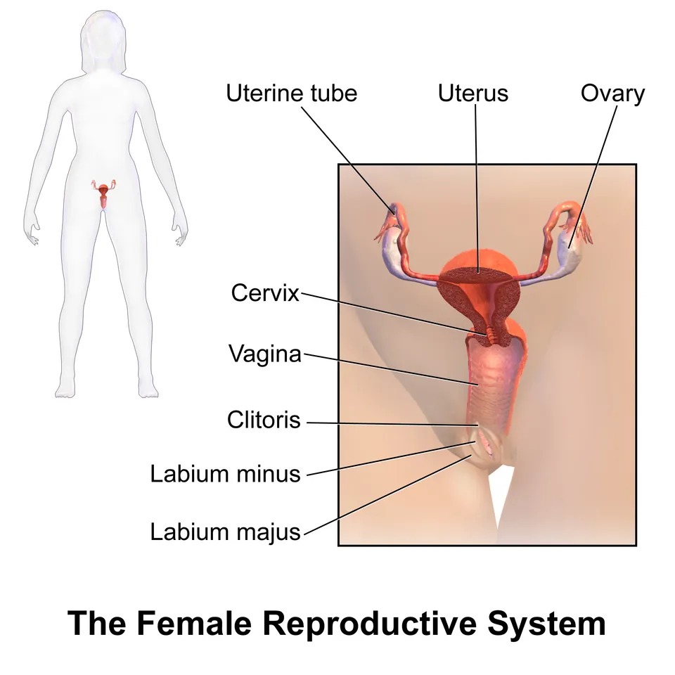
非角化重層扁平上皮。腺を持たない——分泌液は子宮頸管腺と血管からの漏出でまかなわれる。
壁は3層——粘膜(ひだが多い)・筋層(平滑筋)・外膜。
エストロゲンが上皮にグリコーゲンを蓄えさせる。剥がれた上皮のグリコーゲンを Lactobacillus(Döderlein 桿菌)が乳酸に変え、腟内は pH 3.8〜4.5 に保たれる。
だから腟の環境は、年齢とともに変わる。
| 時期 | エストロゲン | 上皮 | pH |
|---|---|---|---|
| 小児期 | 低い | 薄い・グリコーゲン少 | 中性に近い |
| 性成熟期 | 高い | 厚い・グリコーゲン多 | 酸性 |
| 閉経後 | 低い | 薄く萎縮 | 中性に近い |
「上皮の厚さと pH がエストロゲンで決まる」という一本の筋で、小児期と閉経後の疾患が同じ形になる。どちらも上皮が薄く、乳酸桿菌が育たず、感染しやすい(更年期の章)——「幼女腟炎と萎縮性腟炎は、原因が同じである」。
だから閉経後の萎縮性腟炎には、局所エストロゲンが理にかなう。上皮を厚くすれば、菌叢も pH も戻る——「症状ではなく、環境を戻している」。
腟に腺が無いことは、分泌物を診るときの前提になる。おりものの由来は頸管腺なので、性状の変化は頸部の状態を反映する(月経周期の章)——「排卵期に透明で糸を引くのは、頸管粘液の変化になる」。
細菌性腟症は、乳酸桿菌が減って嫌気性菌が増えた状態になる。pH が上がり、灰白色でにおいの強い帯下が出る。KOH でアミン臭が立つ(whiff テスト)、clue cell が見える(嫌気性菌の章)——「炎症ではなく、菌叢の入れ替わりなので『炎』と呼ばない」。
そして酸性であること自体が、精子の通過を制限している。精液がアルカリ性なのは、この酸を中和するためになる(精嚢の章)——「2つの器官の pH が、互いを前提に設計されている」。
エストロゲンがなければ、自浄作用は働かない。
萎縮性腟炎(閉経後) — 上皮が薄くなりグリコーゲンが減り、乳酸桿菌が住めず pH が上がる。乾燥・性交痛・易感染。局所エストロゲンが有効である。小児期も同じ理由で感染しやすい。
細菌性腟症 — 乳酸桿菌が減り、嫌気性菌が増える。pH 4.5 以上・魚臭・clue cell。早産のリスクになる。
腟カンジダ症はpH が正常のままで、白いカッテージチーズ様の帯下と強い掻痒。抗菌薬使用後・妊娠・糖尿病で増える。pH を測るだけで鑑別が進む。
細胞診の成熟指数はエストロゲンの状態を上皮の成熟度から読む方法である。
English version
Core
Non-keratinised stratified squamous epithelium. It has no glands — the fluid comes from the cervical glands and from vascular transudate. The wall is three layers — mucosa (much folded), muscle (smooth) and adventitia.
Oestrogen makes the epithelium store glycogen. Lactobacillus (Döderlein’s bacillus) converts the glycogen of the shed cells to lactic acid, and the vagina is held at pH 3.8–4.5.
So the vaginal environment changes with age.
| Period | Oestrogen | Epithelium | pH |
|---|---|---|---|
| Childhood | Low | Thin, little glycogen | Near neutral |
| Reproductive years | High | Thick, much glycogen | Acid |
| After the menopause | Low | Thin and atrophic | Near neutral |
Going further
The single thread that the thickness of the epithelium and the pH are set by oestrogen makes the diseases of childhood and of the postmenopause take the same form. In both the epithelium is thin, lactobacilli do not grow, and infection is easy — vulvovaginitis of the child and atrophic vaginitis have the same cause.
So topical oestrogen makes sense for postmenopausal atrophic vaginitis: thicken the epithelium and both flora and pH return — restoring the environment rather than the symptom.
That the vagina has no glands is the premise for examining a discharge. The discharge comes from the cervical glands, so a change in its character reflects the state of the cervix — that it becomes clear and stringy at ovulation is a change in cervical mucus.
Bacterial vaginosis is a state in which lactobacilli have fallen and anaerobes increased. The pH rises and a grey, strong-smelling discharge appears; KOH releases an amine odour (whiff test) and clue cells are seen — it is not an inflammation but an exchange of flora, which is why it is not called a "-itis".
And the acidity itself limits the passage of sperm. Semen is alkaline in order to neutralise that acid — the pH of two organs designed on the assumption of each other.
In the clinic
Without oestrogen there is no self-cleansing.
Atrophic vaginitis (postmenopausal) — the epithelium thins, glycogen falls, lactobacilli cannot live there and the pH rises. Dryness, dyspareunia, susceptibility to infection. Topical oestrogen is effective. Childhood is susceptible for the same reason.
Bacterial vaginosis — lactobacilli fall and anaerobes increase. pH above 4.5, a fishy odour, clue cells. A risk factor for preterm birth.
Vaginal candidiasis leaves the pH normal, with a white cottage-cheese discharge and severe itching. It increases after antibiotics, in pregnancy and in diabetes. Measuring the pH alone advances the differential.
The maturation index on cytology reads the oestrogen state from the maturity of the epithelium.
まとめ
| 卵胞 — 二細胞説と黄体 | 卵巣皮質に埋まる、卵母細胞とそれを包む細胞の集まり。 |
|---|---|
| 子宮内膜 — 毎月作り直される粘膜 | 子宮体部の粘膜。2層に分かれる。 |
| 卵子の減数分裂は、2度止まる | 卵母細胞の減数分裂は、途中で長く停止する。 |
| 卵管・子宮頸部・腟 — 接合部が問題になる | 卵管——単層円柱上皮。線毛細胞と分泌細胞(釘状細胞)が混じる。 |
| 腟の上皮 — グリコーゲンが、自浄作用を作っている | 非角化重層扁平上皮。腺を持たない——分泌液は子宮頸管腺と血管からの漏出でまかなわれる。 |
このノートは医学生が授業の内容を自分の言葉でまとめたもので、診療の判断に使うものではありません。誤りに気づいたらお知らせください。