ホルモンを出す細胞は、内分泌腺の中だけにあるのではない。消化管・気道・泌尿路の上皮に散らばって存在する細胞群があり、これをびまん性神経内分泌系(DNES)と呼ぶ。腺という「場所」を持たないので、染色しなければ見えないのが特徴である。
- 傍濾胞細胞とびまん性神経内分泌系 — 腺の外にもホルモンを出す細胞がいる
- 頸動脈小体 — 化学を測る、もう一つの受容器
傍濾胞細胞とびまん性神経内分泌系 — 腺の外にもホルモンを出す細胞がいるCellula parafollicularis
甲状腺の傍濾胞細胞(C細胞)——濾胞のあいだ、または濾胞上皮の基底側にある。明るい細胞質。
- カルシトニンを出す。血中カルシウムを下げる(破骨細胞を抑える)
- 第4咽頭嚢(鰓後体)由来で、濾胞上皮とは起源が違う
びまん性神経内分泌系——消化管・気道・膵などに散在する内分泌細胞の総称。
| 細胞 | 出すもの | 場所 |
|---|---|---|
| G細胞 | ガストリン | 胃前庭部 |
| S細胞 | セクレチン | 十二指腸 |
| I細胞 | コレシストキニン | 十二指腸・空腸 |
| 腸クロム親和性細胞 | セロトニン | 消化管全体 |
| L細胞 | GLP-1 | 回腸・大腸 |
共通の目印——クロモグラニンA・シナプトフィジン・NSE で染まる。
「起源が違う細胞が、同じ器官に同居している」ことが、腫瘍の型を分ける。甲状腺髄様癌は C細胞由来なので、カルシトニンと CEA を出し、放射性ヨウ素が効かない(甲状腺の章)——「同じ臓器の癌でも、由来が違えば治療が変わる」。
だから多発性内分泌腫瘍症2型に含まれる(遺伝性腫瘍の章)——「RET 変異・褐色細胞腫・副甲状腺機能亢進症と組で覚える」。
びまん性神経内分泌系は、「散らばった内分泌腺」として扱うと理解が早い。1つの腺に集まっていないだけで、性質はどれも内分泌細胞になる(内分泌腺の型の項)——「だから同じ免疫染色で、まとめて識別できる」。
神経内分泌腫瘍は、どの細胞から出たかで症状が決まる(膵の嚢胞と腫瘍の章)——「ガストリノーマは難治性の潰瘍、インスリノーマは低血糖、カルチノイドは紅潮と下痢になる」。
カルチノイド症候群が肝転移で初めて出るのも、この解剖から導ける(セロトニンの章)——「門脈から入ったセロトニンは肝で分解されるので、肝を迂回して初めて全身症状になる」。
びまん性神経内分泌系の細胞は、全身に散っているから腫瘍も全身に出る。
神経内分泌腫瘍 — クロモグラニン A・シナプトフィジンが共通のマーカー。Ki-67 と核分裂数で grade を決める。消化管・膵・肺に多い。
カルチノイド症候群 — セロトニンなどが全身循環に出て、皮膚潮紅・下痢・喘鳴・右心系の弁膜症。肝転移があって初めて症状が出るのは、肝で初回通過的に分解されなくなるからである——門脈系の解剖がそのまま症候の条件になっている。尿中 5-HIAA で診断する。
C 細胞(傍濾胞細胞)は神経堤由来で、甲状腺髄様癌になる。カルシトニンと CEA が腫瘍マーカー、MEN2(RET 変異)の一部として褐色細胞腫を必ず先に除外してから手術する。
English version
Core
Parafollicular cells (C cells) of the thyroid — lie between the follicles, or on the basal side of the follicular epithelium. Pale cytoplasm.
- Secrete calcitonin, which lowers blood calcium (by inhibiting osteoclasts).
- Derived from the fourth pharyngeal pouch (ultimobranchial body) — a different origin from the follicular epithelium.
The diffuse neuroendocrine system — the collective name for the endocrine cells scattered through gut, airway and pancreas.
| Cell | Secretes | Site |
|---|---|---|
| G cell | Gastrin | Gastric antrum |
| S cell | Secretin | Duodenum |
| I cell | Cholecystokinin | Duodenum, jejunum |
| Enterochromaffin cell | Serotonin | Throughout the gut |
| L cell | GLP-1 | Ileum, colon |
Common markers — chromogranin A, synaptophysin, NSE.
Going further
That cells of different origin live together in one organ separates the types of tumour. Medullary thyroid carcinoma arises from the C cell, so it secretes calcitonin and CEA and does not respond to radioactive iodine — even in the same organ, a different origin changes the treatment. Hence its inclusion in multiple endocrine neoplasia type 2 — remember it as a set with the RET mutation, phaeochromocytoma and hyperparathyroidism.
The diffuse neuroendocrine system is quickest understood as a scattered endocrine gland. It simply is not gathered into one gland; in nature every one of them is an endocrine cell — which is why a single set of immunostains identifies them all.
In a neuroendocrine tumour the symptoms are decided by which cell it came from — a gastrinoma gives intractable ulcers, an insulinoma hypoglycaemia, a carcinoid flushing and diarrhoea.
That carcinoid syndrome appears only with liver metastases follows from the same anatomy — serotonin entering by the portal vein is degraded in the liver, so systemic symptoms require the liver to be bypassed.
In the clinic
The cells of the diffuse neuroendocrine system are scattered throughout the body, so their tumours appear throughout the body too.
Neuroendocrine tumour — chromogranin A and synaptophysin are the shared markers. The grade is set by Ki-67 and the mitotic count. Commonest in the gut, pancreas and lung.
Carcinoid syndrome — serotonin and others reach the systemic circulation, giving flushing, diarrhoea, wheeze and right-sided valvular disease. Symptoms appear only once there are liver metastases, because first-pass degradation in the liver is lost — the anatomy of the portal system becomes directly the condition for the syndrome. Diagnosed by urinary 5-HIAA.
The C (parafollicular) cell is neural crest derived and gives medullary thyroid carcinoma. Calcitonin and CEA are the tumour markers, and as part of MEN2 (RET mutation) phaeochromocytoma must always be excluded before operating.
頸動脈小体 — 化学を測る、もう一つの受容器Glomus caroticum
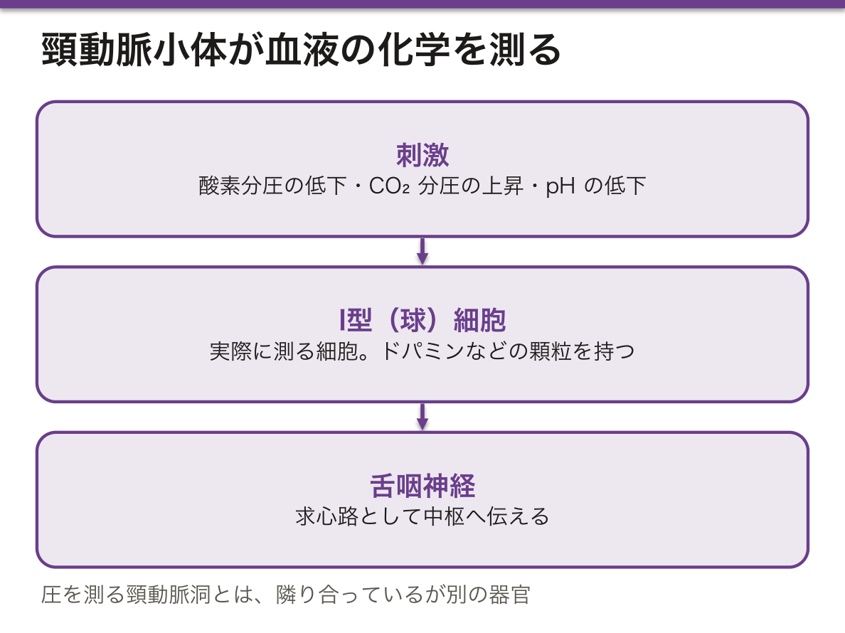
総頸動脈の分岐部にある、数ミリの化学受容器。単位重量あたりの血流量が体で最も多い。
- I型(球)細胞——神経堤由来。ドパミンなどを含む顆粒を持つ。実際に測る細胞
- II型(鞘)細胞——グリアに似た支持細胞
測っているもの——動脈血の酸素分圧の低下・二酸化炭素分圧の上昇・pH の低下。
求心路——頸動脈小体・頸動脈洞は舌咽神経、大動脈小体・大動脈弓の圧受容器は迷走神経。
圧を測る頸動脈洞(分岐部の膨らみ)と、隣り合っているが別の器官になる。
「酸素分圧を測っている」という点が、貧血との違いを説明する。末梢化学受容器が見ているのは溶存酸素の分圧であって、酸素の含量ではない(呼吸の章)——「重症の貧血や一酸化炭素中毒でも、呼吸が速くならない理由がここにある」。
そして中枢化学受容器との役割分担がはっきりしている。ふだんの換気は延髄の中枢化学受容器が二酸化炭素で決めており、末梢は低酸素のときにだけ主役になる——「低酸素の刺激だけが、末梢にしか無い」。
だから慢性の二酸化炭素貯留がある患者で、酸素を大量に投与すると換気が落ちうる(呼吸器の章)——「残っていた低酸素という刺激を、取り上げてしまうことになる」。
神経堤由来であることは、腫瘍の名前に現れる。頸動脈小体腫瘍(傍神経節腫)は、頸部の拍動性腫瘤として現れ、SDH 遺伝子の変異と関連する(頸部の腫瘤の章)——「副腎の褐色細胞腫と、同じ系列の腫瘍になる」。
隣の頸動脈洞は圧受容器なので、押すと徐脈になる(不整脈の章)——「同じ場所にある2つの受容器が、まったく別の量を測っている」。
頸動脈小体は酸素分圧を測っている。血流量あたりの代謝が体で最も高いため、動脈血の酸素分圧の低下に鋭敏に反応できる。
低酸素換気応答の主役である。COPD で慢性的に CO2 が高い患者では、中枢の CO2 感受性が鈍り、低酸素刺激が呼吸の主な駆動になる。高濃度酸素を漫然と投与すると換気が落ちて CO2 ナルコーシスになりうる——目標 SpO2 を 88〜92% に設定する根拠である。
頸動脈小体腫瘍(傍神経節腫) — 頸部分岐部の拍動性腫瘤。血管造影で lyre sign(内外頸動脈が開く)。神経堤由来で、SDH 遺伝子変異と関連し褐色細胞腫を合併しうる。
頸動脈洞(圧受容器)は別のものである——小体は化学、洞は圧と分けて覚える。
English version
Core
A chemoreceptor a few millimetres across at the bifurcation of the common carotid artery. It has the highest blood flow per unit weight in the body.
- Type I (glomus) cells — neural crest derived, with granules containing dopamine among others. These are the cells that actually measure.
- Type II (sustentacular) cells — supporting cells resembling glia.
What it measures — a fall in arterial oxygen tension, a rise in carbon dioxide tension, a fall in pH.
Afferents — the carotid body and carotid sinus by the glossopharyngeal nerve; the aortic bodies and aortic arch baroreceptors by the vagus.
It lies next to the carotid sinus (the dilatation at the bifurcation), which measures pressure — adjacent, but a different organ.
Going further
That it measures oxygen tension explains the difference from anaemia. What the peripheral chemoreceptor sees is the partial pressure of dissolved oxygen, not the oxygen content — here is why breathing does not quicken even in severe anaemia or carbon monoxide poisoning.
And the division of labour with the central chemoreceptor is clear. Ordinary ventilation is set by the medullary central chemoreceptors responding to carbon dioxide; the peripheral receptors take the lead only in hypoxia — the hypoxic stimulus exists only peripherally.
So in a patient with chronic carbon dioxide retention, giving a large amount of oxygen can reduce ventilation — it takes away the one stimulus that was left.
The neural crest origin shows in the name of the tumour. A carotid body tumour (paraganglioma) presents as a pulsatile neck mass and is associated with mutations of the SDH genes — a tumour of the same lineage as the adrenal phaeochromocytoma.
The neighbouring carotid sinus is a baroreceptor, so pressing on it causes bradycardia — two receptors in the same place measuring entirely different quantities.
In the clinic
The carotid body measures oxygen tension. Having the highest metabolism per unit blood flow in the body, it can respond sensitively to a fall in arterial oxygen tension. It is the principal agent of the hypoxic ventilatory response.
In COPD with chronic hypercapnia, central CO₂ sensitivity is blunted and the hypoxic stimulus becomes the main drive to breathe. Indiscriminate high-concentration oxygen can reduce ventilation and cause CO₂ narcosis — the basis for a target SpO₂ of 88–92 per cent.
Carotid body tumour (paraganglioma) — a pulsatile mass at the bifurcation, with the lyre sign (splaying of the internal and external carotid arteries) on angiography. Neural crest derived, associated with SDH mutations, and may accompany phaeochromocytoma.
The carotid sinus is a different thing — remember the body as chemical and the sinus as pressure.
まとめ
| 傍濾胞細胞とびまん性神経内分泌系 — 腺の外にもホルモンを出す細胞がいる | 甲状腺の傍濾胞細胞(C細胞)——濾胞のあいだ、または濾胞上皮の基底側にある。 |
|---|---|
| 頸動脈小体 — 化学を測る、もう一つの受容器 | 総頸動脈の分岐部にある、数ミリの化学受容器。 |
このノートは医学生が授業の内容を自分の言葉でまとめたもので、診療の判断に使うものではありません。誤りに気づいたらお知らせください。