20種のアミノ酸を全部覚える必要は無い。試験と臨床で効くのは側鎖の性質による分類だけになる。
- 荷電——酸性(Asp・Glu)/塩基性(Lys・Arg・His)。電気泳動での動きを決める
- 疎水性——蛋白の内側に埋まる。膜貫通部位を作る
- 特殊な役割——Cys(S-S結合)・Pro(折れ曲がる)・Gly(最小・コラーゲン)
そして「必須/非必須」と「糖原性/ケト原性」の2つの分類が、栄養と代謝で使われる。
蛋白質を分ける技術は、そのまま臨床検査になる——血清蛋白分画・免疫固定電気泳動・Western blot はすべて、荷電と大きさで分けている。
- 側鎖で分けるアミノ酸 — 覚えるべきは例外だけ
- 蛋白質を分ける — 電気泳動が臨床検査になるとき
- アミノ酸の分類 — 側鎖ひとつで、性質がすべて決まる
- 等電点と緩衝 — アミノ酸が、pH を支えている
- 両性イオンの反応式 — アミノ酸は酸とも塩基とも反応する
- ペプチド結合の作り方と、N 末端・C 末端の見分け方
- 血漿アミノ酸分析 — 数字の並びから、どの経路が詰まったかを読む
側鎖で分けるアミノ酸 — 覚えるべきは例外だけ
必須アミノ酸9種——バリン・ロイシン・イソロイシン・トレオニン・メチオニン・リジン・フェニルアラニン・トリプトファン・ヒスチジン。
- 分岐鎖アミノ酸(BCAA)——バリン・ロイシン・イソロイシン。骨格筋で代謝される(肝ではない)
特殊な側鎖。
- システイン——S-S 結合を作る。細胞外の蛋白の安定性を担う
- プロリン——環状。αヘリックスを折る。コラーゲンでは水酸化される
- グリシン——側鎖が H のみ。コラーゲンの3残基ごとに必要
- ヒスチジン——pKa が生理的 pH に近く、緩衝と酵素の触媒に使われる
糖原性/ケト原性——ケト原性のみは ロイシンとリジンの2つだけ。
等電点(pI)——正味の電荷が0になる pH。pH が pI より高ければ蛋白は陰性になる。
「ケト原性のみはロイシンとリジンの2つ」が、飢餓の理解に効く。他のアミノ酸は糖新生の材料になれる(糖新生の章)——「筋を壊して血糖を保てる理由が、この分類にある」。
BCAA が骨格筋で代謝されることが、肝不全での使い方につながる。肝硬変では BCAA が減り、芳香族アミノ酸が増える(Fischer比の低下)(肝性脳症の章)——「アミノ酸の比が、脳症の指標と治療の両方になる」。
フェニルアラニンの代謝が止まるのがフェニルケトン尿症になる。チロシンが必須になり、メラニンも作れず色白になる(アミノ酸代謝異常症の章)——「1つの酵素が止まると、必須/非必須の区分が入れ替わる」。
だから新生児マススクリーニングの対象になっている(新生児マススクリーニングの章)——「食事だけで防げる知的障害があるので、全員に測る」。
プロリンの水酸化にビタミンCが要る。だから壊血病では、コラーゲンが正しく作れず出血する(ビタミンCの章)——「1アミノ酸の修飾の失敗が、全身の結合組織を弱くする」。
等電点の考え方が、血清蛋白分画の読み方を決めている。アルカリ性の緩衝液中で、アルブミンが最も陰性に帯電して速く動く(蛋白分画の章)——「泳動の順番は、等電点の順番になっている」。
English version
Core
Nine essential amino acids — valine, leucine, isoleucine, threonine, methionine, lysine, phenylalanine, tryptophan, histidine.
Branched-chain amino acids (BCAA) — valine, leucine, isoleucine. Metabolised in skeletal muscle, not in the liver.
Special side chains
- Cysteine — forms S–S bonds; provides the stability of extracellular proteins.
- Proline — cyclic; breaks the α helix. In collagen it is hydroxylated.
- Glycine — side chain only H. Required at every third residue of collagen.
- Histidine — its pKa is near physiological pH, so it serves in buffering and in enzyme catalysis.
Glucogenic / ketogenic — only two are purely ketogenic: leucine and lysine.
Isoelectric point (pI) — the pH at which the net charge is zero. Above the pI the protein is negatively charged.
Going further
That leucine and lysine are the only purely ketogenic amino acids tells in understanding starvation. All the others can be substrate for gluconeogenesis — the reason the blood glucose can be maintained by breaking down muscle lies in this classification.
That the BCAA are metabolised in skeletal muscle bears on their use in liver failure. In cirrhosis the BCAA fall and the aromatic amino acids rise (a fall in Fischer’s ratio) — the ratio of amino acids serving as both the index of encephalopathy and the treatment.
A block in the metabolism of phenylalanine is phenylketonuria: tyrosine becomes essential, melanin cannot be made and the skin is pale — one enzyme stopping and the division into essential and non-essential changing over. Hence its place in newborn screening — an intellectual disability preventable by diet alone, so everyone is tested.
The hydroxylation of proline requires vitamin C, so in scurvy collagen cannot be made correctly and bleeding follows — failure to modify one amino acid weakening the connective tissue of the whole body.
The idea of the isoelectric point decides how a serum protein electrophoresis is read. In an alkaline buffer, albumin is the most negatively charged and moves fastest — the order of migration is the order of the isoelectric points.
蛋白質を分ける — 電気泳動が臨床検査になるとき
血清蛋白分画(セルロースアセテート膜電気泳動)——アルブミン・α₁・α₂・β・γ の5分画。
| パターン | 読み |
|---|---|
| アルブミン低下+γ上昇(多クローン性) | 肝硬変・慢性炎症 |
| γ分画に鋭い山(M蛋白) | 多発性骨髄腫・MGUS |
| α₂上昇+アルブミン低下 | ネフローゼ症候群 |
| α₁が消える | α₁-アンチトリプシン欠損症 |
| 全体に低下 | 低栄養・蛋白漏出 |
SDS-PAGE——SDS で荷電を揃え、大きさだけで分ける。
免疫固定電気泳動——M蛋白の重鎖と軽鎖の型を決める。
Western blot——電気泳動 → 膜に転写 → 抗体で検出。蛋白を見る。
ELISA——抗原抗体反応を酵素で発色させ、定量する。
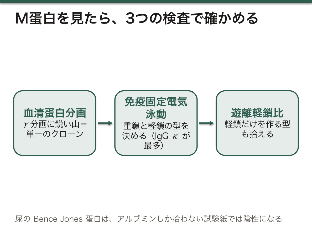
「鋭い山(M蛋白)」が見えたら、単一のクローンが増えていることになる(多発性骨髄腫の章)——「なだらかな山は反応、鋭い山は腫瘍と読む」。
だから次は、免疫固定電気泳動で型を決める。IgG κ が最多になる——「あることの確認と、何であるかの確認が別の検査になる」。
そして遊離軽鎖比を測る。軽鎖のみを作る型では、血清の総蛋白が上がらない(骨髄腫腎の章)——「蛋白分画が正常でも、骨髄腫を否定できない」。
尿では、この軽鎖が Bence Jones 蛋白になる。試験紙法ではアルブミンしか拾えないので、陰性になる(尿検査の章)——「試験紙が見ているものを知らないと、見落とす」。
α₁分画の消失が、α₁-アンチトリプシン欠損症を示す。若年の肺気腫と肝硬変が組み合わさる(肺気腫の章)——「1本の帯の欠如が、2つの臓器の病気を説明する」。
ELISA が定量、Western blot が確認という役割分担が、感染症の検査で使われてきた(検査の組み立ての章)——「感度の高い検査でふるい、特異度の高い検査で確かめる」。
English version
Core
Serum protein electrophoresis (on cellulose acetate) — five fractions: albumin, α₁, α₂, β, γ.
| Pattern | Read as |
|---|---|
| Low albumin with raised γ (polyclonal) | Cirrhosis, chronic inflammation |
| A sharp peak in γ (M protein) | Myeloma, MGUS |
| Raised α₂ with low albumin | Nephrotic syndrome |
| α₁ absent | α₁-antitrypsin deficiency |
| All fractions low | Malnutrition, protein loss |
- SDS-PAGE — SDS equalises the charge so that separation is by size alone.
- Immunofixation — determines the heavy and light chain type of an M protein.
- Western blot — electrophoresis → transfer to a membrane → detection with antibody. Looks at protein.
- ELISA — an antigen–antibody reaction developed with an enzyme, giving a quantity.
Going further
A sharp peak (an M protein) means a single clone is expanding — read a broad hump as a reaction and a sharp peak as a tumour.
So the next step is immunofixation to determine the type, IgG κ being the commonest — confirming that it is there and confirming what it is are two different tests.
Then the free light chain ratio is measured. In the light-chain-only type the total serum protein does not rise — a normal electrophoresis does not exclude myeloma.
In the urine those light chains are Bence Jones protein. The dipstick detects only albumin and reads negative — miss it unless you know what the strip is looking at.
Loss of the α₁ fraction indicates α₁-antitrypsin deficiency, combining emphysema in a young person with cirrhosis — the absence of one band explaining disease in two organs.
The division of labour — ELISA to quantify, Western blot to confirm — has been used in infectious disease serology: screen with the sensitive test and confirm with the specific one.
アミノ酸の分類 — 側鎖ひとつで、性質がすべて決まる
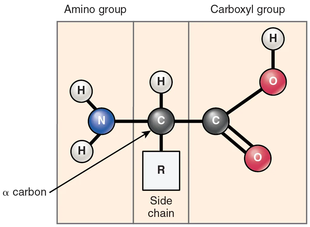
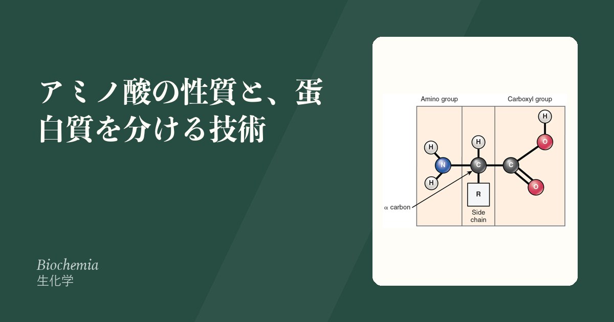
20種のアミノ酸は骨格が同じ(アミノ基・カルボキシ基・水素・側鎖がα炭素に付く)。違うのは側鎖 R だけで、そこから性質・立体構造・機能のすべてが導かれる。
| 分類 | アミノ酸 | 性質 |
|---|---|---|
| 非極性・脂肪族 | Gly, Ala, Val, Leu, Ile, Pro, Met | 蛋白の内側に埋まる(疎水性相互作用) |
| 芳香族 | Phe, Tyr, Trp | 280 nm の吸光——蛋白の定量に使う |
| 極性・非荷電 | Ser, Thr, Cys, Asn, Gln | Ser/Thr/Tyr はリン酸化を受ける |
| 酸性(負電荷) | Asp, Glu | — |
| 塩基性(正電荷) | Lys, Arg, His | His の pKa は約6——生理的pH付近で解離が変わり緩衝と触媒に働く |
必須アミノ酸は9種——Phe, Val, Thr, Trp, Ile, Met, His, Leu, Lys(「風呂場椅子独占」)。ヒトが炭素骨格を作れないもの。
「側鎖が構造を決め、構造が機能を決める」——生化学の中心的な考え方が、この分類にすでに現れている。
| アミノ酸 | 特別な役割 |
|---|---|
| グリシン | 側鎖が水素だけ=最も小さい。コラーゲンの3重らせんの内側に来られる唯一のアミノ酸。3残基ごとに必ず Gly で、他に置換すると骨形成不全症になる |
| プロリン | 側鎖が主鎖に環を作る=αヘリックスを折る(helix breaker)。コラーゲンでは水酸化されてヒドロキシプロリンとなり、鎖間の水素結合を作る——この水酸化にビタミンCが要る(壊血病) |
| システイン | SH基が2つでジスルフィド結合——分泌蛋白の三次構造を固定する(細胞内は還元的なのでできにくい) |
| ヒスチジン | pKa≒6 でプロトンを出し入れできる——酵素の触媒残基、ヘモグロビンの Bohr 効果 |
| チロシン | カテコラミン・甲状腺ホルモン・メラニンの原料。リン酸化で増殖シグナル |
| トリプトファン | セロトニン・メラトニン・ナイアシンの原料 |
アミノ酸は両性電解質で、正味の電荷が0になるpH=等電点〈pI〉を持つ。pI では溶解度が最小になり沈殿する——これを利用したのが等電点電気泳動で、血清蛋白分画やHb の異常(HbS は Glu→Val で負電荷を1つ失い、泳動が遅れる)の検出につながる。
ヒトの蛋白はすべて L型(グリシンを除き不斉炭素を持つ)。D型アミノ酸は細菌の細胞壁(D-Ala, D-Glu)にあり、ヒトに無いからこそ抗菌薬の標的になる——バンコマイシンは D-Ala-D-Ala に結合し、耐性菌はこれを D-Ala-D-Lac に変えて逃れる。
English version
Core
All twenty share the same skeleton (amino group, carboxyl group, hydrogen and side chain on the α carbon). Only the side chain R differs, and from it follow all the properties, the conformation and the function.
| Class | Amino acids | Property |
|---|---|---|
| Non-polar, aliphatic | Gly, Ala, Val, Leu, Ile, Pro, Met | Buried inside the protein (hydrophobic interaction) |
| Aromatic | Phe, Tyr, Trp | Absorbance at 280 nm — used to quantify protein |
| Polar, uncharged | Ser, Thr, Cys, Asn, Gln | Ser, Thr and Tyr are phosphorylated |
| Acidic (negative) | Asp, Glu | — |
| Basic (positive) | Lys, Arg, His | The pKa of His is about 6 — its ionisation changes near physiological pH, serving in buffering and catalysis |
Nine are essential — Phe, Val, Thr, Trp, Ile, Met, His, Leu, Lys: those whose carbon skeleton humans cannot make.
Going further
"The side chain decides the structure and the structure decides the function" — the central idea of biochemistry already appears in this classification.
| Amino acid | Its special role |
|---|---|
| Glycine | The side chain is a hydrogen alone = the smallest. The only amino acid that can lie on the inside of the collagen triple helix. Every third residue must be Gly, and substitution gives osteogenesis imperfecta |
| Proline | Its side chain rings back onto the main chain = a helix breaker. In collagen it is hydroxylated to hydroxyproline, forming hydrogen bonds between the chains — and that hydroxylation requires vitamin C (scurvy) |
| Cysteine | Two SH groups make a disulphide bond, fixing the tertiary structure of secreted proteins (the cytosol is reducing, so it forms poorly there) |
| Histidine | With a pKa near 6 it can take up and release protons — the catalytic residue of enzymes, and the Bohr effect of haemoglobin |
| Tyrosine | The raw material of catecholamines, thyroid hormone and melanin. Phosphorylated in growth signalling |
| Tryptophan | The raw material of serotonin, melatonin and niacin |
Amino acids are amphoteric and have an isoelectric point (pI) at which the net charge is zero. At the pI the solubility is least and they precipitate — the basis of isoelectric focusing, and so of serum protein electrophoresis and of the detection of abnormal haemoglobins (HbS, Glu→Val, loses one negative charge and migrates more slowly).
All human proteins are L (every amino acid but glycine has an asymmetric carbon). D-amino acids occur in bacterial cell walls (D-Ala, D-Glu), and because they are absent from us they are a target for antibiotics — vancomycin binds D-Ala-D-Ala, and resistant organisms escape by changing it to D-Ala-D-Lac.
等電点と緩衝 — アミノ酸が、pH を支えている
アミノ酸は、同じ分子の中に酸(-COOH)と塩基(-NH₃⁺)を持つ(両性電解質)。
等電点(pI)——正味の電荷がゼロになる pH。この pH で溶解度が最も低く、電気泳動で動かない。
| 側鎖 | pI | 生理的 pH(7.4)での電荷 |
|---|---|---|
| 中性 | 約 6 | ほぼ中性 |
| 酸性(Asp・Glu) | 低い(約 3) | 負 |
| 塩基性(Lys・Arg) | 高い(Lys 約 9.7・Arg 約 10.8) | 正 |
| ヒスチジン | 約 7.6 | ほとんどが中性(側鎖の pKa は約 6.0。正に荷電しているのは数%だが、生理的 pH に最も近い pKa を持つ側鎖) |
緩衝作用が最も強いのは、pKa が pH と一致するときになる——だから生理的 pH で緩衝できるアミノ酸は、実質的にヒスチジンだけになる。
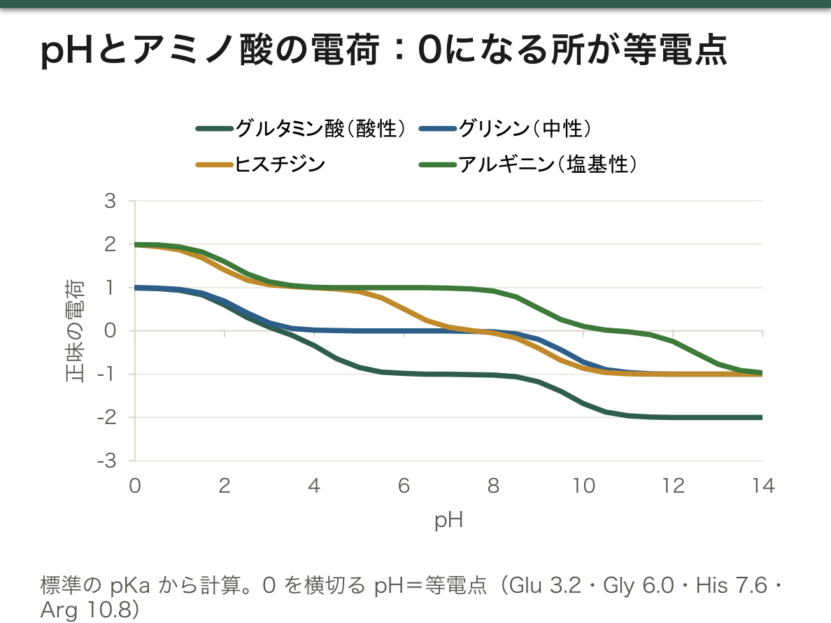
「ヒスチジンだけが、生理的 pH で緩衝できる」という一点が、血液の緩衝を説明する。ヘモグロビンが優れた緩衝剤なのは、ヒスチジン残基を多く持つからになる(緩衝系の章)——「重炭酸系に次ぐ第2の緩衝が、赤血球の中にある」。
だから脱酸素化したヘモグロビンほど、水素イオンをよく受け取る(Haldane 効果)(酸素解離曲線の項)——「組織で酸素を放した瞬間に、二酸化炭素と酸を引き受けられる」。
等電点が溶解度の最低点であることは、結石と沈殿を説明する(結石の化学の章)——「シスチン結石が酸性尿で析出するのは、この性質による」。
電気泳動でアルブミンが最も陽極へ動くのも、pI が低い(約 4.7)からになる(この章の蛋白質を分ける項)——「泳動像の並び順は、電荷の順そのものになる」。
薬の吸収も、同じ理屈で pH に左右される(薬物動態の章)——「イオン型は膜を通れない。弱酸性の薬は胃で、弱塩基性の薬は小腸で吸収されやすい」。
English version
Core
An amino acid carries within one molecule both an acid (–COOH) and a base (–NH₃⁺): it is amphoteric.
Isoelectric point (pI) — the pH at which the net charge is zero. At that pH the solubility is least and it does not move on electrophoresis.
| Side chain | pI | Charge at pH 7.4 |
|---|---|---|
| Neutral | About 6 | Nearly neutral |
| Acidic (Asp, Glu) | Low (about 3) | Negative |
| Basic (Lys, Arg) | High (Lys about 9.7, Arg about 10.8) | Positive |
| Histidine | About 7.6 | Mostly neutral (side-chain pKa about 6.0; only a few per cent are positively charged, but no other side chain has a pKa closer to physiological pH) |
Buffering is strongest when the pKa equals the pH — so the only amino acid that can effectively buffer at physiological pH is histidine.
Going further
The single point that histidine alone can buffer at physiological pH explains the buffering of the blood. Haemoglobin is an excellent buffer because it is rich in histidine residues — the second buffer after the bicarbonate system lies inside the red cell.
So the more deoxygenated the haemoglobin, the better it accepts protons (the Haldane effect) — the moment it releases oxygen in the tissues, it can take on carbon dioxide and acid.
That the isoelectric point is the point of least solubility explains stones and precipitates — cystine stones crystallise in acid urine because of this property.
That albumin migrates furthest towards the anode is likewise because its pI is low (about 4.7) — the order on the electrophoretogram is the order of charge itself.
Drug absorption depends on pH by the same reasoning — the ionised form cannot cross membranes, so weak acids are absorbed in the stomach and weak bases in the small intestine.
両性イオンの反応式 — アミノ酸は酸とも塩基とも反応する
アミノ酸は、水の中では –COOH が H⁺ を放し、–NH₂ が H⁺ を受け取った両性イオン(H₃N⁺–CHR–COO⁻)として存在する。だから結晶は塩のように融点が高く、水によく溶ける。
アラニン(側鎖 –CH₃、pKa は –COOH が約2.3、–NH₃⁺ が約9.7、pI 約6.0)を例に、pH を変えたときの形を並べる。
| pH | 主な形 | 正味の電荷 | 電気泳動で動く向き |
|---|---|---|---|
| 1(強い酸性) | H₃N⁺–CH(CH₃)–COOH | +1 | 陰極 |
| 6(pI 付近) | H₃N⁺–CH(CH₃)–COO⁻ | 0 | 動かない |
| 12(強い塩基性) | H₂N–CH(CH₃)–COO⁻ | −1 | 陽極 |
酸(HCl)との反応:両性イオンの –COO⁻ が H⁺ を受け取る。H₃N⁺–CH(CH₃)–COO⁻ + HCl → H₃N⁺–CH(CH₃)–COOH + Cl⁻(塩酸塩になる)。
塩基(NaOH)との反応:両性イオンの –NH₃⁺ が H⁺ を放す。H₃N⁺–CH(CH₃)–COO⁻ + NaOH → H₂N–CH(CH₃)–COO⁻ Na⁺ + H₂O(ナトリウム塩になる)。
どちらの基が反応するかは、両性イオンの形を見れば決まる。両性イオンで H⁺ を受け取れるのは –COO⁻ だけ、H⁺ を放せるのは –NH₃⁺ だけなので、酸には –COO⁻ が、塩基には –NH₃⁺ が応じる。式を書くときは、両性イオンの形から始めると取り違えない。
Fischer 投影式でのアミノ酸の描き方:–COOH を上、側鎖 R を下に縦に置き、α 炭素の横に –NH₂ と –H を描く。–NH₂ が左なら L 型、右なら D 型。糖と同じ規則で、L-アラニンは L-グリセルアルデヒドと同じ配置になる。タンパク質のアミノ酸はすべて L 型で、グリシンだけは α 炭素に H が2つ付くので不斉炭素が無く、D/L の区別が無い。
English version
Core
In water an amino acid exists as a zwitterion (H₃N⁺–CHR–COO⁻): –COOH has released H⁺ and –NH₂ has taken one. That is why amino acid crystals have high melting points like salts and dissolve well in water.
Alanine (side chain –CH₃; pKa about 2.3 for –COOH and 9.7 for –NH₃⁺; pI about 6.0) at different pH values:
| pH | Main form | Net charge | Moves in electrophoresis to |
|---|---|---|---|
| 1 (strong acid) | H₃N⁺–CH(CH₃)–COOH | +1 | Cathode |
| 6 (near pI) | H₃N⁺–CH(CH₃)–COO⁻ | 0 | Does not move |
| 12 (strong base) | H₂N–CH(CH₃)–COO⁻ | −1 | Anode |
With acid (HCl): the –COO⁻ of the zwitterion takes H⁺. H₃N⁺–CH(CH₃)–COO⁻ + HCl → H₃N⁺–CH(CH₃)–COOH + Cl⁻ (the hydrochloride).
With base (NaOH): the –NH₃⁺ releases H⁺. H₃N⁺–CH(CH₃)–COO⁻ + NaOH → H₂N–CH(CH₃)–COO⁻ Na⁺ + H₂O (the sodium salt).
Going further
Which group reacts is decided by what the zwitterion can do. In the zwitterion only –COO⁻ can accept H⁺ and only –NH₃⁺ can give it up, so acid reacts with –COO⁻ and base with –NH₃⁺. Start equations from the zwitterion and you will not mix them up.
Drawing amino acids in Fischer projection: put –COOH at the top and the side chain R at the bottom, with –NH₂ and –H on the sides of the α carbon. –NH₂ on the left = L; on the right = D. The rule matches sugars: L-alanine has the same arrangement as L-glyceraldehyde. All protein amino acids are L; glycine has two H on the α carbon, so it is not chiral and has no D/L.
ペプチド結合の作り方と、N 末端・C 末端の見分け方
ペプチド結合は、1つ目のアミノ酸の –COOH と2つ目のアミノ酸の –NH₂ が脱水縮合してできるアミド結合(–CO–NH–)である。
Ala-Phe の生成:H₂N–CH(CH₃)–COOH + H₂N–CH(CH₂C₆H₅)–COOH → H₂N–CH(CH₃)–CO–NH–CH(CH₂C₆H₅)–COOH + H₂O
ペプチドの両端のうち、遊離のアミノ基が残っている側が N 末端、遊離のカルボキシ基が残っている側が C 末端である。配列は必ず N 末端から C 末端へ書くので、Ala-Phe と Phe-Ala は別の分子になる。Ala-Phe では Ala が N 末端・Phe が C 末端、Phe-Ala ではその逆で、Ala の –COOH ではなく Phe の –COOH がペプチド結合に使われている。
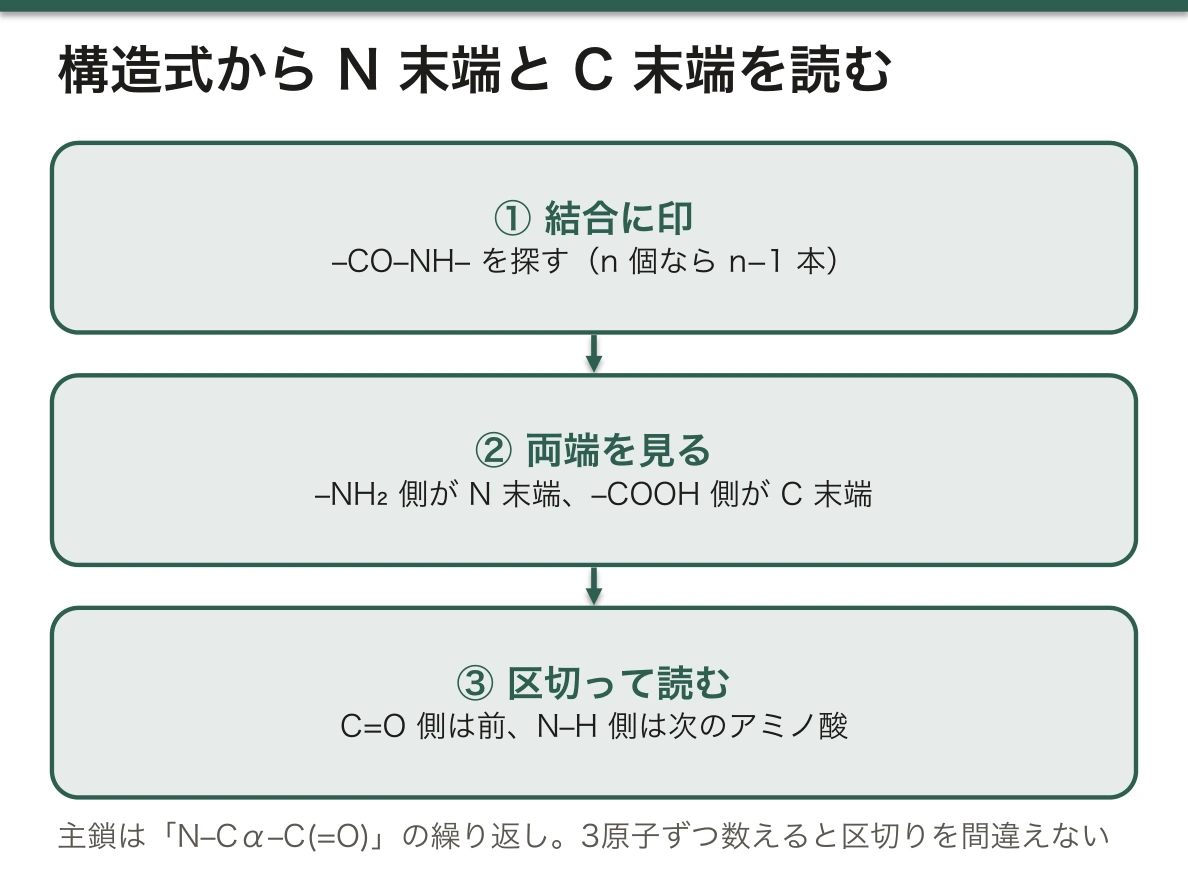
構造式から N 末端と C 末端を読む手順:①–CO–NH– を探してペプチド結合に印を付ける(n 個のアミノ酸なら n−1 本)。②両端を見て、–NH₂(–NH₃⁺)が付いている端が N 末端、–COOH(–COO⁻)が付いている端が C 末端。③ペプチド結合の C=O 側が前のアミノ酸、N–H 側が次のアミノ酸に属する、と区切って側鎖を読む。主鎖は「N–Cα–C(=O)」の繰り返しになっているので、この3原子ずつ数えると区切りを間違えない。
ペプチド結合は、C=O と C–N の間で電子が共有される(共鳴する)ので、二重結合に近い性格を持ち、回転できず平面になる。主鎖が回転できるのは Cα の両側の結合だけで、この制限が αヘリックスや β シートの形を決めている。水の中では加水分解のほうが有利だが、酵素が無ければ非常にゆっくりとしか切れないので、タンパク質は安定に存在できる。
English version
Core
A peptide bond is the amide bond (–CO–NH–) formed by condensation (loss of water) between the –COOH of one amino acid and the –NH₂ of the next.
Forming Ala-Phe: H₂N–CH(CH₃)–COOH + H₂N–CH(CH₂C₆H₅)–COOH → H₂N–CH(CH₃)–CO–NH–CH(CH₂C₆H₅)–COOH + H₂O
The end with a free amino group is the N-terminus; the end with a free carboxyl group is the C-terminus. Sequences are always written from N to C, so Ala-Phe and Phe-Ala are different molecules. In Ala-Phe, Ala is N-terminal and Phe C-terminal; in Phe-Ala it is the opposite, and the –COOH of Phe, not Ala, forms the peptide bond.
Going further
Reading the ends from a structure: (1) find each –CO–NH– and mark it (n residues, n−1 bonds); (2) at the ends, –NH₂ (–NH₃⁺) marks the N-terminus and –COOH (–COO⁻) the C-terminus; (3) the C=O side of a peptide bond belongs to the earlier residue and the N–H side to the next. The backbone repeats N–Cα–C(=O); count in threes and you will not cut in the wrong place.
Electrons are shared between C=O and C–N (resonance), so the peptide bond is partly double, cannot rotate and is planar. Only the bonds on each side of Cα can rotate, and this limit shapes α-helices and β-sheets. Hydrolysis is favoured in water, but without enzymes it is very slow, so proteins are stable.
血漿アミノ酸分析 — 数字の並びから、どの経路が詰まったかを読む
血漿と尿のアミノ酸を一度に定量する検査。先天代謝異常症の診断と、栄養状態の評価に使う。
| 所見 | 読み方 |
|---|---|
| 1つだけが極端に高い | その手前で詰まっている(フェニルアラニン → フェニルケトン尿症) |
| グルタミン・アラニンが高い | アンモニアの負荷(尿素回路異常症) |
| シトルリン | 低ければ OTC・CPS1 欠損、高ければシトルリン血症——尿素回路の分岐点になる |
| 分枝鎖アミノ酸が高い | メープルシロップ尿症 |
| 全体に低い | 低栄養・重症肝疾患・吸収不良 |
尿のアミノ酸が全体に高ければ、腎尿細管の再吸収障害(Fanconi 症候群)を疑う。
「1つだけ高いのか、まとまって高いのか」で、話がまったく変わる。単独の高値は酵素の詰まり、まとまった高値は輸送か全身の代謝の問題になる(先天代謝異常症の総論の章)——「パターンとして読む検査になる」。
シトルリンの値が、尿素回路のどこで止まったかを示す(尿素回路の項)——「シトルリンはミトコンドリアから細胞質へ出る分子なので、その前後で挙動が逆になる」。
尿の所見と組で読むことで、腎の問題と代謝の問題を分けられる(尿を読む章)——「血中が高くて尿にも出る(溢流性)のか、血中は正常なのに尿に出る(腎性)のか」。
シスチン尿症は、後者の代表になる(結石の化学の章)——「シスチン・オルニチン・リジン・アルギニンの4つが尿に出る。結石はシスチンだけが作る」。
採血の条件が結果を大きく変える(検査の性能の章)——「食後・輸液中・発作の合間では、異常が出ない。急性期の検体を保存しておく理由になる」。
English version
Core
A test quantifying the amino acids of plasma and urine at once, used to diagnose inborn errors and to assess nutrition.
| Finding | Read as |
|---|---|
| One alone extremely high | A block just before it (phenylalanine → phenylketonuria) |
| Glutamine and alanine high | An ammonia load (a urea cycle disorder) |
| Citrulline | Low = OTC or CPS1 deficiency; high = citrullinaemia — the branch point of the urea cycle |
| Branched-chain amino acids high | Maple syrup urine disease |
| All low | Malnutrition, severe liver disease, malabsorption |
If the urinary amino acids are all raised, suspect a defect of tubular reabsorption (Fanconi syndrome).
Going further
Whether one alone is high or a group is high changes the story entirely. A single high value means a blocked enzyme; a group means a problem of transport or of the whole metabolism — a test to be read as a pattern.
The citrulline value shows where in the urea cycle it stopped — citrulline is the molecule that leaves the mitochondrion for the cytosol, so its behaviour is opposite on either side of that step.
Read with the urine, it separates a renal from a metabolic problem — is it high in the blood and therefore spilling over, or normal in the blood and appearing in the urine (renal)?
Cystinuria is the classic example of the latter — cystine, ornithine, lysine and arginine all appear in the urine; only cystine makes stones.
The conditions of collection greatly change the result — after a meal, during an infusion, or between attacks, the abnormality does not show. This is why a specimen from the acute episode is preserved.
まとめ
| 側鎖で分けるアミノ酸 — 覚えるべきは例外だけ | 必須アミノ酸9種——バリン・ロイシン・イソロイシン・トレオニン・メチオニン・リジン・フェニルアラニン・トリプトファン・ヒスチジン。 |
|---|---|
| 蛋白質を分ける — 電気泳動が臨床検査になるとき | 血清蛋白分画(セルロースアセテート膜電気泳動)——アルブミン・α₁・α₂・β・γ の5分画。 |
| アミノ酸の分類 — 側鎖ひとつで、性質がすべて決まる | 20種のアミノ酸は骨格が同じ(アミノ基・カルボキシ基・水素・側鎖がα炭素に付く)。 |
| 等電点と緩衝 — アミノ酸が、pH を支えている | アミノ酸は、同じ分子の中に酸(-COOH)と塩基(-NH₃⁺)を持つ(両性電解質)。 |
| 両性イオンの反応式 — アミノ酸は酸とも塩基とも反応する | アミノ酸は、水の中では –COOH が H⁺ を放し、–NH₂ が H⁺ を受け取った両性イオン(H₃N⁺–CHR–COO⁻)として存在する。 |
| ペプチド結合の作り方と、N 末端・C 末端の見分け方 | ペプチド結合は、1つ目のアミノ酸の –COOH と2つ目のアミノ酸の –NH₂ が脱水縮合してできるアミド結合(–CO–NH–)である。 |
| 血漿アミノ酸分析 — 数字の並びから、どの経路が詰まったかを読む | 血漿と尿のアミノ酸を一度に定量する検査。 |
このノートは医学生が授業の内容を自分の言葉でまとめたもので、診療の判断に使うものではありません。誤りに気づいたらお知らせください。